In the presence of air, most of the energy produced by eukaryotic cells is generated by mitochondrial oxidative phosphorylation. This process is driven by a respiratory chain composed of a number of complex membrane proteins that act in sequence to transfer electrons from reduced substrates through a series of oxidation–reduction reactions to oxygen. The net result of this process is the generation of proton and ion gradients across the inner mitochondrial membrane; these gradients are used by ATP synthase to drive ATP synthesis. Several previous studies have demonstrated that the respiratory chain itself regulates the rate of oxidative phosphorylation and have identified cytochrome c oxidase, its terminal member, as a key enzyme in this regulation (Erecinska and Wilson, 1982; Wilson, 1982; Kadenbach, 1986; Poyton et al. 1988; Poyton and McEwen, 1996; Villani and Attardi, 1997). Moreover, it is now clear that many OXPHOS diseases (i.e. fatal and benign infantile myopathies, Leigh’s syndrome, ischemic heart disease,
Alzheimer’s disease and Parkinson’s disease) have cytochrome c oxidase deficiencies (Wallace, 1992; Robinson, 1994). The reduced levels of cytochrome c oxidase present in patients with these diseases may be causally connected to their reduced ability to produce cellular energy. Cytochrome c oxidase is a complex multimeric membrane protein composed of six metal centers (two hemes, two coppers, one zinc and one magnesium) as well as polypeptide subunits encoded by both nuclear and mitochondrial genes (Poyton and McEwen, 1996). To understand how cytochrome c oxidase functions to regulate energy metabolism and its role in OXPHOS diseases, it is essential to examine both the function and expression of its subunit polypeptides. It is also important to understand the functional crosstalk that takes place between those subunit polypeptides encoded by nuclear genes and those subunit polypeptides encoded by mitochondrial genes. These issues are discussed here.
JEB1454
Eukaryotic cytochrome c oxidases are complex oligomeric membrane proteins composed of subunit polypeptides encoded by both nuclear and mitochondrial genomes. While the mitochondrially encoded subunits are encoded by unique genes, some of the nuclear-encoded subunits are encoded by multigene families. The isoforms produced by these multigene families are tissue-specific and/or developmentally regulated in mammals and environmentally regulated in lower eukaryotes. Isoforms for one of the subunits, V, in the yeast Saccharomyces cerevisiae and one of the subunits, VII, in the slime mold Dictyostelium discoideum are regulated differentially by oxygen concentration. Extensive studies with the yeast subunit V isoforms have revealed that the genes for these proteins are switched on or off at very low oxygen concentrations (0.5–1µmol l−1O
2) and that they affect the catalytic properties of holocytochrome c oxidase differentially. By altering an internal step in electron transfer between heme a and the binuclear reaction center (composed of heme a3 and CuB), the ‘hypoxic’ isoform, Vb, enhances the catalytic constant three- to
fourfold relative to the ‘aerobic’ isoform, Va. Modeling studies suggest that this occurs by an interaction between transmembrane helix VII of subunit I and the transmembrane helix in subunit V. The inverse regulation of these two isoforms allows cells to assemble different types of holoenzyme isoenzymes in response to oxygen concentration. Oxygen also regulates the level of transcription of the genes for the other nuclear-coded subunits of yeast cytochrome c oxidase and affects the level of two of the mitochondrially encoded subunits (I and II) post-transcriptionally. Thus, the level of cytochrome c oxidase activity that is produced at different oxygen tensions in yeast is determined in part by the number of holoenzyme molecules that are assembled and in part by the oxygen-regulated isoforms of subunit V. The possibility that this type of control exists in other organisms is considered.
Key words: cytochrome c oxidase, isoforms, yeast, oxygen, respiration, structure, function, gene expression.
Summary
Introduction
STRUCTURE/FUNCTION OF OXYGEN-REGULATED ISOFORMS IN CYTOCHROME c
OXIDASE
PATRICIA V. BURKE ANDROBERT O. POYTON*
Department of Molecular, Cellular, and Developmental Biology, University of Colorado, Boulder, CO 80309-0347, USA
*Author for correspondence (e-mail: [email protected])
Structure/function of eukaryotic cytochrome c oxidases Structure of eukaryotic cytochrome c oxidases
Most of the structural, functional, biosynthetic and genetic data on eukaryotic cytochrome c oxidases have come from studies with mammalian (i.e. bovine) and yeast (i.e.
Saccharomyces cerevisiae) cytochrome c oxidases (Capaldi et al. 1995; Poyton et al. 1995; Tsukihara et al. 1995, 1996;
Poyton and McEwen, 1996). The yeast enzyme has been especially amenable to studies on subunit function and expression, while the mammalian enzyme has been especially amenable to physiochemical studies. Both cytochrome c oxidases catalyze the concerted transfer of four electrons from ferrocytochrome c to molecular oxygen, with the simultaneous pumping of protons across the inner mitochondrial membrane from the matrix to the cytoplasmic side (Babcock and Wikstrom, 1992). Both enzymes contain four redox-active metal centers (heme a, heme a3, CuA, CuB) that participate in electron transfer. Heme a3and CuBare bridged in the resting fully oxidized form and constitute the binuclear reaction center. Electrons pass from cytochrome c, via CuAand heme
a, to the binuclear reaction center, where dioxygen (O2) is reduced to water. The reduction of dioxygen to water is coupled to the translocation of one proton per electron across the inner mitochondrial membrane. This creates a pH and voltage gradient across the membrane.
The protein matrix surrounding the metal centers in eukaryotic cytochrome c oxidases consists of several different polypeptide subunits. The three largest polypeptide subunits (I, II and III) are encoded by mitochondrial genes. These polypeptides are conserved in mammalian and yeast enzymes and they have primary sequence homology to three of the subunit polypeptides of prokaryotic cytochrome oxidases (Saraste, 1990). In addition to the mitochondrially encoded subunits, these eukaryotic cytochrome c oxidases contain several polypeptide subunits that are encoded in the nucleus. The mammalian enzyme contains 10 different subunit polypeptides encoded by nuclear genes (Capaldi et al. 1995; Tsukihara et al. 1996), while the yeast enzyme contains six or eight different polypeptide subunits encoded by nuclear genes, depending on how it is isolated (Power et al. 1984; Taanman
et al. 1992; Gier et al. 1995; Poyton et al. 1995; Poyton and
McEwen, 1996). The simpler yeast enzyme thus contains nine subunit polypeptides, while the more complex yeast enzyme contains 11 subunit polypeptides. The original claim that this preparation contained 12 different polypeptides (Taanman et
al. 1992) has been shown to be incorrect (Gier et al. 1995).
The putative twelfth polypeptide turned out to be a dimer of subunit VIII. The nine-subunit yeast enzyme contains subunits IV, Va or Vb, VI, VII, VIIa and VIII (Poyton et al. 1995), while the 11-subunit yeast enzyme contains subunits IV, Va or Vb, VI, VII, VIIA, VIII, VIa and VIb (Taanman et al. 1992; Gier
et al. 1995). Both forms of the enzyme are fully active,
suggesting that the two ‘extra’ subunits (VIa and VIb) in the 11-subunit enzyme do not affect catalysis. Although the nomenclature used for subunits in the mammalian and yeast enzymes is different, it is obvious from their primary sequences that homologs for all of the yeast subunit polypeptides exist in mammalian cytochrome c oxidases (Patterson et al. 1987; Capaldi et al. 1995) (Table 1).
Recently, the high-resolution crystal structure of the oxidized bovine heart enzyme was published (Tsukihara et al. 1995, 1996). The structure of the six metal centers as well as the atomic coordinates of the amino acids of all 13 polypeptide subunits have been determined. This enzyme is an asymmetric dimer with a transmembrane region that contains 28 α-helices per monomer. All but three of its subunit polypeptides have at least one hydrophobic transmembrane helix. Subunit I has 12 transmembrane helices (numbered I–XII), subunit II has two transmembrane helices and subunit III has seven transmembrane helices. The remaining transmembrane domains reside in the nuclear-coded subunits. Subunit II binds CuAin a large extramembrane domain that lies on the cytosolic side of the inner membrane. Subunit I binds the heme a, heme a3and CuBredox centers; both hemes are perpendicular to the plane of the membrane at an intraplanar angle of 104 ° to one another. They associate with amino acid side-chains from four different transmembrane helices of subunit I: heme a associates with helices XI and XII; heme a3associates with helices VI and VIII; and CuBis close to heme a3and associates with an amino acid side-chain from helix VI as well as two other amino acids.
Redox-sensitive conformers
The means by which electron transfer is coupled to proton translocation in cytochrome c oxidase is unclear, but it has been suggested by many laboratories that the enzyme has distinct redox-sensitive conformational states at its heme–copper binuclear center and that these states couple electron and proton movement (Chan and Li, 1990; Woodruff, 1993; Wikstrom et al. 1994; Iwata et al. 1995; Wittung and Malmstrom, 1996). There is ample evidence that both the oxidized and reduced forms of the enzyme may exist in multiple conformational states and that these are sensitive to pH. For the oxidized enzyme, these are called ‘fast’ and ‘slow’ states (Moody, 1996). They can be distinguished by their spectral signatures. The ‘fast’ form is considered to be a fully oxidized active enzyme, while the ‘slow’ form is considered to be an artifact that arises during the purification and/or storage of the enzyme. The ‘fast’ form converts to the ‘slow’ form at low pH, and the ‘slow’ form may be converted back to the ‘fast’ form by a cycle of reduction and reoxidation. Neither the nature of these pH-induced changes nor their relevance to catalysis is known. For the reduced enzyme, conformers are observable by using Fourier transform infrared Table 1. Correspondence between yeast and mammalian
(bovine) cytochrome c oxidase subunits
Yeast I II III IV Va/Vb VI VII VIIa VIII VIa VIb Bovine I II III Vb IV Va VIIa VIc VIIc VIa VIb
(FTIR) spectroscopy and resonance Raman analysis of carbon monoxide ligated to the fully reduced enzyme (Caughey et al. 1988; Sherman et al. 1991; Wang et al. 1995; Dodson et al. 1996). FTIR spectroscopy of carbon-monoxide-bound reduced cytochrome c oxidase has revealed the presence of two separate populations of CO vibrators for purified bovine, yeast and bacterial cytochrome c oxidases (Allen et al. 1995; Caughey et
al. 1988; Fiamingo et al. 1986; Shapleigh et al. 1992; Mitchell et al. 1996), suggesting that the binuclear reaction centers in
these enzymes have two different conformations. The absorption maxima of these CO stretch bands are similar in the mammalian and yeast enzymes (Allen et al. 1995). This suggests that the binuclear reaction center is in a similar environment in both enzymes. The first report that these conformers are intraconvertible by pH was published recently (Mitchell et al. 1996) for a prokaryotic cytochrome c oxidase. It is not known yet whether this pH-dependent intraconvertibility is also operative in eukaryotic cytochrome c oxidases, which have a much more complex polypeptide subunit structure.
Second-derivative absorption spectroscopy of mammalian or bacterial cytochrome c oxidases has revealed that ferrous cytochrome a also has two absorption maxima when carbon monoxide is bound. This suggests that ferrous cytochrome a, like ferrous cytochrome a3, can adopt two different conformations (Sherman et al. 1991; Copeland, 1991). Recently, Dodson et al. (1996) were able to monitor absorbance simultaneously in both the infrared and visible Soret regions as a function of the redox potential of the solution. They found a slight shift in spectral maxima with redox potential for both cytochromes a and a3. They also found that the redox-dependent shift for cytochrome a3 correlated quantitatively with that for cytochrome a, suggesting that the CO stretching frequency for cytochrome a3 responds to the oxidation state of heme a through some form of allosteric effect mediated by the intervening helices of subunit I. This type of coupling between cytochrome a and cytochrome a3 may occur through helix X in subunit I.
Subunit function in cytochrome c oxidase
From the crystal structure of bovine cytochrome c oxidase (Tsukihara et al. 1995, 1996) and genetic studies with yeast (Poyton and McEwen, 1996; Meunier and Colson, 1994; Meunier et al. 1995), it is clear that mitochondrially encoded subunits I and II of eukaryotic cytochrome c oxidases perform the electron transport functions of the holoenzyme. Subunit III contains no metal centers. It may modulate the proton-pumping activities of the holoenzyme, play a role in the assembly or stability of subunits I and II, or modulate the access of oxygen to the binuclear reaction center (Brunori et al. 1987; Haltia et
al. 1991; Riistama et al. 1996). Together, subunits I, II and III
form the catalytic core of the enzyme. This catalytic core is conserved in all members of the heme/Cu oxidase superfamily (Moody, 1996). Hence, it is appears likely that all of the catalytic functions of eukaryotic cytochrome c oxidases are performed by the mitochondrially encoded subunits.
If the mitochondrially encoded subunits are sufficient for catalysis, what are the functions of the nucleus-encoded subunits? This question has been most readily addressed with yeast cytochrome c oxidase. Genetic studies with yeast suggest that the nucleus-encoded subunits have at least two functions (Poyton and McEwen, 1996). Some of these subunits (yeast V and VIII) modulate catalysis, while other subunits (yeast IV, VI, VII and VIIa) are required for the stability of the catalytic subunits and/or stable assembly of the holoenzyme. The conclusion that some nucleus-coded subunits modulate catalysis is also supported by the discovery of isoforms for some of these subunits in a variety of organisms. So far, these isoforms have been found in several species of the yeast
Saccharomyces (Cumsky et al. 1985, 1987; Trueblood and
Poyton, 1987), in the slime mold Dictyostelium discoideum (Bisson and Schiavo, 1986) and in a variety of mammals including bovine, pig, rat and humans (Kadenbach et al. 1982, 1983; Kuhn-Nentwig and Kadenbach, 1985; Lomax and Grossman, 1989). In yeast and Dictyostelium, these isoforms are expressed in an oxygen-dependent manner (Poyton et al. 1988; Trueblood et al. 1988; Hodge et al. 1989; Sandona et al. 1995; Bunn and Poyton, 1996; Burke et al. 1997), and in mammals these isoforms are tissue-specific (Lomax and Grossman, 1989) and are developmentally regulated (Kadenbach and Reinmann, 1992; Bonne et al. 1993). Their number varies from mammal to mammal. In bovine mammals, there are two isoforms for subunits VIa, VIIa and VIII. One of these (the H type) is expressed in heart and skeletal muscle; the other (the L type) is expressed in liver. In rat, both types of isoform are found for subunits VIa and VIII but only the L isoform of subunit VIIa is expressed. And in humans, subunit VIII occurs as only the L isoform, but both isoforms of subunits VIa and VIIa are expressed. In addition, different isoforms for subunit IV may be expressed in heart and skeletal muscle.
The functions of these isoforms have been studied most intensively using yeast and bovine heart cytochrome c oxidases. Studies with both systems have revealed that isoforms can act as regulators of holoenzyme activity and may do so in more than one way. For example, studies with bovine cytochrome c oxidase have suggested that the subunit VIa isoforms can mediate the regulation of cytochrome c oxidase activity in response to allosteric effectors, such as ATP and other nucleotides (Anthony et al. 1993; Rohdich and Kadenbach, 1993; Frank and Kadenbach, 1996), while studies with the yeast subunit V isoforms have shown a direct effect of these polypeptides on the binuclear reaction center and the kinetics of interaction with cytochrome c. The genes for these latter isoforms are inversely regulated by oxygen concentration.
Yeast subunit V isoforms: structure and function
holocytochrome c oxidase function. The yeast Saccharomyces
cerevisiae is ideal for such studies for several reasons. First, in
contrast to mammals, S. cerevisiae possesses only one nuclear-coded subunit (V) for which isoforms exist (Poyton et al. 1995; Poyton and McEwen, 1996). Second, these isoforms are encoded by single-copy genes, COX5a and COX5b, which can be deleted easily to produce strains that carry one isoform or the other (Cumsky et al. 1987; Trueblood and Poyton, 1987). And third, the catalytic functions of yeast cytochrome c oxidase can be assayed in vivo within whole cells (Waterland
et al. 1991), thereby avoiding purification-related artifacts.
The subunit V isoforms, Va and Vb, have 66 % primary sequence homology (Fig. 1) and differ in length by one amino acid (Cumsky et al. 1987). Both are integral proteins of the inner membrane and possess one hydrophobic transmembrane helix. Moreover, both polypeptides have similar secondary structures and amphipathicities. Some regions of these polypeptides are highly conserved while others are not.
Differential function(s) of subunits Va and Vb
Functional differences between Va and Vb have been studied
in vivo by examining the catalytic properties of cytochrome c
oxidase isoenzymes in mutant strains that contain either Va or Vb. These studies revealed that the isoforms of subunit V affect the turnover number of holocytochrome c oxidase and do so by altering the rates of intramolecular transfer between heme a and the binuclear reaction center (Waterland et al. 1991). Intramolecular electron transfer from heme a to the binuclear reaction center is three- to fourfold faster in the Vb isoenzyme than in the Va isoenzyme, while the activation energy of the reaction in both enzymes is the same. This finding was taken as evidence that the subunit V isoforms function allosterically to alter the conformation of the protein environment around the binuclear reaction center, within subunit I, so as to limit the accessibility of heme a3to electrons without altering the barrier height of the electron transfer reaction itself. Recently, Allen et
al. (1995) examined the effects of the subunit V isoforms on
the interaction of holocytochrome c oxidase with cytochrome
c. As expected, they found that the subunit V isoforms do not
affect the Km for cytochrome c binding to cytochrome c oxidase, but do alter the maximum turnover number (TNmax) of the holoenzyme.
To test the hypothesis that the subunit V isoforms affect the binuclear reaction center, Allen et al. (1995) used FTIR spectroscopy of Va and Vb isoenzymes liganded with carbon monoxide. Carbon monoxide is an excellent probe because of its high affinity for cytochrome c oxidase and its strong infrared absorbance. Moreover, because carbon monoxide binds heme a3 in the binuclear reaction center and the frequencies and bandwidths of C–O infrared stretch bands are highly sensitive to the bonding environment between heme iron and CO and to the environment around the CO ligand, these bands provide a sensitive way of assessing the effects of isoforms or mutant subunits on the binuclear reaction center. These studies, performed using purified cytochrome c oxidase isoenzymes containing Va or Vb, revealed a fundamental difference between the Va and Vb isoenzymes. Two separate populations of CO vibrators, with peaks at 1965 cm−1 and 1961.7 cm−1, could be detected in the Va isoenzyme, while only a single population of vibrators, with a peak at 1965 cm−1, could be detected in the Vb isoenzyme. These were designated CII and CI to correspond to similar bands that have been observed in bovine heart cytochrome c oxidase. The two separate populations of vibrators in the Va isoenzyme, with peaks shifted by 3 cm−1, probably result from the existence of two protein conformers within the population. The band corresponding to the CII conformer has the greatest intensity, indicating that it is the more stable conformer. These findings provide direct support for the conclusion that subunit V affects ligand binding around the binuclear reaction center and alters the environment around heme a3. In addition, the finding that both the bandwidths and wave numbers for the Va isoenzyme are similar to those from bovine heart suggests that the environment around the ligand site of heme a3in the binuclear reaction center is remarkably similar in mammalian and yeast cytochrome c oxidases.
To fit the above findings with the observed differences in turnover numbers for Va and Vb isoenzymes, Allen et al. (1995) proposed that the CII conformer is productive for electron transfer between heme a and the binuclear reaction center, and that the CI conformer is less productive. Moreover, they proposed a model in which these conformers are intraconvertible and the overall rate of electron transfer from heme a to a3is determined by the ratio of CII to CI. For the
A Q T H A L S N A A V M D L Q S R W E N M P S T E Q Q D I V S K L S E R Q K L P W A Q L T
V Q T K A L S K A T L T D L P E R W E N M P N L E Q K E I A D N L T E R Q K L P W K T L N
E P E K Q A V W Y I S Y G E W G P R R P V L N K G D S S F I A K G V A A G L L F S V G L F
N E E I K A A W Y I S Y G E W G P R R P V H G K G D V A F I T K G V F L G L G I S F G L F
A V V R M A G G Q D A - K T M N K E W Q L K S D E Y L K S K N A N P W G G Y S Q V Q S K
G L V R L L A N P E T P K T M N R E W Q L K S D E Y L K S K N A N P W G G Y S Q V Q S K Va H2
N-Vb H
2
N--COOH
-COOH
Vb isoenzyme, the binuclear reaction center has only one conformation; this corresponds to the productive conformer, CII. Hence, the enhanced rate observed with the Vb isoenzyme results from the presence of a productive conformer that does not intraconvert to a less productive one. So, according to this model, Vb would lock the binuclear reaction center in the CII conformer. This model is supported by the recent finding that the two conformers (a and b) of a prokaryotic cytochrome c oxidase intraconvert in a pH-dependent fashion, and that the turnover number of the enzyme is related to the a/b ratio (Mitchell et al. 1996).
Conserved function(s) of Va and Vb
Previous studies have revealed that yeast cells require a subunit V isoform for a functional holocytochrome c oxidase and that Va and Vb are interchangeable (Trueblood and Poyton, 1987); that is, either isoform can function in the holoenzyme. These findings suggest that, in addition to the differential functions mentioned above, these two isoforms have conserved functions as well. From a comparison of their primary sequences, it is clear that there are regions of perfect homology in these two polypeptides. One of these is at their carboxyl termini. Recently, we have analyzed several point mutations in COX5a (P. V. Burke and R. O. Poyton, in preparation). One of these, cox5a-1, is a nonsense mutation at residue 124 in the carboxyl-terminal domain; this deletes the ten carboxyl-terminal amino acids of the protein and leads to complete loss of activity. Another is a revertant that replaces the stop codon at residue 124 with a serine and, in so doing, restores partial activity. This revertant, with Ser124 replacing Trp124, supports approximately half the level of activity of the wild-type protein. Together, these two mutants establish that the ten carboxyl-terminal amino acids, which are completely conserved between Va and Vb, are essential.
Structural considerations
In order to understand the molecular bases for the differential and conserved functions of the subunit V isoforms, it is useful to analyze the spatial relationships between subunit V and the two subunits, I and II, that carry the redox-sensitive prosthetic groups. The recently published high-resolution crystal structure of bovine cytochrome c oxidase (Tsukihara et
al. 1996) together with spatial constraint molecular modeling
software (Sali and Blundell, 1993) has allowed us to model the three-dimensional structures of yeast subunits Va, I and II. Our approach to modeling these subunits has been first to align the yeast subunits with their bovine counterparts, by taking into account primary sequence, predicted secondary structure, hydropathy analysis (to identify transmembrane domains), residues that are conserved among many species and the locations of the amino and carboxyl termini on the determined crystal structure of bovine cytochrome c oxidase. Once aligned, we then carry out comparative modeling using the program Modeller (Sali and Blundell, 1993). This program uses our alignment to overlay the backbone of the target sequence (i.e. the yeast subunit) on that of the known structural
template (the bovine subunit) and constructs a model taking into account the different spatial constraints for the two sequences by making use of a large database of constraints derived from known structures. It then optimizes the initial model and uses molecular dynamic simulations and annealing to give the best structure.
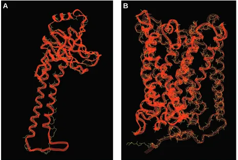
For these studies, we have used bovine subunit IV as the structural template for yeast subunit Va, bovine subunit I as the structural template for yeast subunit I, and bovine subunit II as the structural template for yeast subunit II. Comparisons of the structural template for each bovine subunit and the modeled structure for their yeast counterparts are shown in Figs 2 and 3. In each case, the three-dimensional model of the yeast subunit aligns well with its bovine counterpart. In each case, the fit (i.e. the root mean square displacement) between the bovine template and the predicted yeast structure is better than 0.086 nm.
[image:5.609.330.556.74.379.2]A
B
Fig. 3. Comparison of the crystal structure determined for bovine cytochrome c oxidase subunits I and II with the predicted three-dimensional structure of yeast cytochrome c oxidase subunits I and II. The primary sequences of yeast subunits I (B) and II (A) were aligned with the primary sequences of bovine subunits I and II, respectively; they were then modeled on the three-dimensional structure determined for the bovine subunits. Each bovine subunit is a wire-frame image in yellow, and each yeast subunit is a ribbon in red. Subunit I was modeled in two parts (indicated by three-strand and six-strand ribbons) and then combined for alignment. The top of each panel faces the intermembrane space side of the inner membrane; the bottom faces the matrix.
Fig. 4. A cross section through a yeast cytochrome c oxidase monomer in the inner mitochondrial membrane. This cross section was taken in the hydrophobic core of the membrane but close to the intermembrane space side and is redrawn for yeast from a similar cross section through oxidized bovine heart cytochrome c oxidase (Tsukihara et al. 1996). The locations of transmembrane α-helixes (designated by circles) in their subunit polypeptides and heme a, heme a3and CuBare shown. The helices from subunit I are shown in
aqua and are numbered from I to XII; the helices from subunit II are shown in green and are labeled I and II; and the helices from subunit III are shown in yellow and are labeled I to VII. The helix in subunit Va (or Vb) is shown in orange. Unlabelled circles (pink) represent the transmembrane α-helices from other nuclear-coded subunits. Heme a and heme a3 are shown as red bars, and CuBis shown as
small orange dot close to helix VI of subunit I. The binuclear reaction center (heme a3 and CuB) is shown between helices VI,
VIII, IX and X of subunit I, and heme a is shown near helices X and XII of subunit I. The entire monomer is outlined with a dotted line. Interior dotted lines denote semicircles observed in the arrangement of the subunit I helices.
I
II
III
III
I
I
II
II
IV
Va
V
V
VI
VI
VII
VII
VIII
IX
X
XI
XII
[image:6.609.53.281.512.697.2]subunits are integral membrane proteins. Subunit Va has a single transmembrane domain (amino acids 69–100) which contains an α-helix (amino acids 73–96) and which crosses the membrane at an angle (see Figs 5, 7). In addition, it has a hydrophilic amino-terminal domain (amino acids 1–68) on the matrix side of the inner membrane and a hydrophilic carboxyl-terminal domain (amino acids 97–133) on the cytosolic side of the membrane. Subunit II has two transmembrane helices and a large hydrophilic domain, which contains the CuA site, on the cytosolic side of the membrane. This hydrophilic domain resembles a horse’s head. Subunit I has 12 transmembrane α -helices and is largely embedded in the membrane. Heme a, heme a3and CuBare located within this subunit. The spatial relationships between these prosthetic groups and the transmembrane α-helices of subunits I, II and Va are most easily visualized in a cross-sectional view through the hydrophobic core of the membrane, close to its cytosolic surface (Fig. 4). From this cross-sectional view, it is clear that
the helices of subunit I surround heme a, heme a3 and CuB. Heme a and a3are perpendicular to the plane of the membrane at an intraplanar angle of 104 ° to one another. It is also clear that the α-helix of subunit V is adjacent to helix XII of subunit I, and that the α-helices of subunit II are adjacent to helices IX and VIII of subunit I.
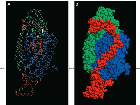
[image:7.609.65.546.308.669.2]The spatial relationships in three dimensions between yeast or bovine subunits I and II, and between yeast Va and bovine IV, are shown in Fig. 5. The transmembrane domain of subunit V crosses the membrane, immediately adjacent to subunit I, while its carboxyl-terminal hydrophilic domain lies along the side of the hydrophilic carboxyl-terminal (‘horse’s head’) domain of subunit II. The α-helix within this hydrophilic domain lies along a groove in subunit II formed by the jaw of the ‘horse’s head’. From these spatial considerations, it appears that the transmembrane domain of subunit V interacts with subunit I, while its carboxyl-terminal domain interacts with subunit II. The close physical
Fig. 5. Spatial relationships between subunits I, II and V of cytochrome c oxidase viewed within the plane of the inner mitochondrial membrane. Using the atomic coordinates determined by Tsukihara (1996), we have drawn a model for three of the subunits of cytochrome c oxidase. The relative location of the inner mitochondrial membrane is shown by the dashed lines on the sides of each panel. The top of the figure faces the intermembrane space side of the membrane, while the bottom faces the mitochondrial matrix. (A) Alpha carbon backbone showing prosthetic groups and their relationship to subunit Va (in red), subunit II (in green) and subunit I (in blue). Heme groups are shown in red, CuBis shown in yellow and the CuAcluster (two atoms) in subunit II is shown in white. (B) Space-filling version of the image shown in A.
association of subunits I, II and Va suggested by this model is supported by the finding that cox5a mutants have reduced amounts of subunits I, II and III (McEwen et al. 1986; L. E. Farrell and C. E. Trueblood, unpublished observations) and, conversely, mutants lacking subunit I contain substoichiometric amounts of subunit Va relative to the other subunits (McEwen et al. 1986).
A molecular model for subunit V function
The structure predicted for subunit Va, together with the genetic and functional studies mentioned above, suggests that subunit V and its isoforms have at least two important functional domains: (1) a carboxyl-terminal domain which is conserved between both isoforms and which may be essential for the transfer of electrons from cytochrome c to heme a and/or for the exit of protons from the enzyme, and (2) one or more differential domains that account for the differential effects of Va and Vb on the electron-transport activities of the holoenzyme.
Carboxyl-terminal domain
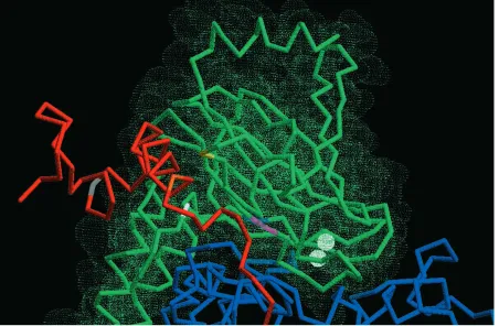
On the basis of the structural models in Fig. 5, it appears that the conserved carboxyl-terminal domain of yeast subunit V lies in a cleft between subunits I and II, close to the jaw in the ‘horse’s head’ region of subunit II (Fig. 6). The importance
of this region can be deduced from the following findings. A mutation that removes the last 10 amino acids of subunit V knocks out cytochrome c oxidase activity. A mutation that converts Trp124 to Ser124 has decreased activity (P. V. Burke and R. O. Poyton, in preparation). Glu 107 (yeast Va nomenclature) is conserved in bovine subunit IV, both isoforms of yeast subunit V and homologous subunits in other mammals and fungi (Lin et al. 1993). And Gly156 in subunit II, a near neighbor of Glu107 in subunit Va, is also conserved in many species. In addition, mutations in the region of subunit II that lines the cleft also lead to the lack of cytochrome c oxidase activity (Fig. 6). One of these is a Gly to Glu mutation at residue 156, and another is a double mutation that converts Ala220 to Val and Ala189 to Val (Meunier and Colson, 1994). It has been proposed that this cleft acts as an exit pathway or channel for protons or water (Iwata et al. 1995; Tsukihara et
al. 1996). If this channel functions in the exit of protons or
water, one would expect it to be regulated (i.e. gated) so as to prevent backflow. Perhaps the ten C-terminal amino acids in Va and Vb, which are essential and extend beyond the surface of the cleft, function in this gating.
A putative differential domain
[image:8.609.78.530.395.691.2]In considering possible candidates for the region of subunit V that confers its differential functions, it is important to note
Fig. 6. Enlarged region of Fig. 5A showing the ‘horse’s head’ region of subunit II and its relationship to the carboxyl terminus of subunit Va and to subunit I. Subunit I is in blue, subunit II is in green, subunit Va is in red and the CuAcluster (two atoms) is in white. Several mutations
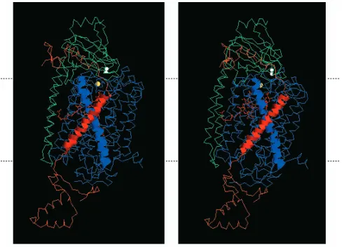
that the subunit V isoforms affect the TNmaxbut not the Kmfor cytochrome c binding and that they effect an internal step in electron transfer between heme a and the binuclear reaction center. This can be understood in terms of the models for yeast subunits Va, I and II described above. The region of subunit V that is closest to the binuclear reaction center is its transmembrane domain, especially its transmembrane α-helix. The transmembrane α-helix in subunit V lies adjacent to transmembrane α-helix XII of subunit I (Fig. 4), crossing it near the site at which heme a is liganded at an angle of 50 ° (Fig. 7). The region of subunit V that crosses helix XII is between amino acids 80 and 90. The amino acids within this region are all within 1 nm of the junction between helix XII of subunit I and the transmembrane helix of subunit V. They are also close to the farnesyl tail of heme a. Five out of the 11 residues in this region are not conserved. One or more of the non-conserved amino acid side-chains may alter the relative positions of both helices (i.e. the transmembrane helix of subunit V and helix XII of subunit I). This could change the relative orientation of hemes a and a3 (which have an
intraplanar angle of 104 ° in the oxidized bovine enzyme) and, in turn, affect the electron transfer rates between these two prosthetic groups. Studies are currently under way to test this hypothesis.
Yeast subunit V isoforms: differential regulation by O2 concentration
As discussed by Kwast et al. (1998), the genes COX5a and
COX5b, which encode Va and Vb, are differentially regulated
by oxygen and heme. The ‘aerobic’ isoform, Va, is expressed in aerobic cells and in cells grown at oxygen concentrations down to 1µmol l−1O
2. In contrast, the ‘hypoxic’ isoform, Vb, is expressed in anaerobic cells and in cells grown at oxygen concentrations up to 1µmol l−1 O
[image:9.609.64.555.71.423.2]2. Oxygen regulates the expression of both isoforms at the level of the transcription of their genes. In both cases, a heme-dependent transcription factor is involved (Poyton and Burke, 1992; Bunn and Poyton, 1996; Kwast et al. 1998). In air, the COX5a gene is activated by the Hap2/3/4/5p transcription factor complex, while the Fig. 7. A stereo pair showing the relationships between subunits I, II and Va. The transmembrane α-helix of subunit Va is shown as a red ribbon, and helix XII of subunit I is shown as a blue helix. The relative location of the inner mitochondrial membrane is shown by the dashed lines on the sides of each panel. The top of the figure faces the intermembrane space side of the membrane, while the bottom faces the mitochondrial matrix. Alpha carbon backbones are shown for the rest of subunit I (blue), for subunit Va (red) and for subunit II (green). Hemes
COX5b gene is repressed by the Rox1(Reo1)p transcription
factor. The expression of Rox1(Reo1)p is mediated directly by heme and Hap1p. Because the biosynthesis of heme requires oxygen, cellular heme levels have been proposed to provide a gauge of oxygen availability. Hence, in the presence of oxygen, heme levels are sufficiently high to activate Hap2/3/4/5p-dependent transcription of COX5a and Hap1p-dependent transcription of Rox1(Reo1)p, which represses expression of COX5b. This leads to the preferential expression of the Va isoform. In the absence of oxygen, heme is not made; hence, the COX5a gene can no longer be activated and the
COX5b gene can no longer be repressed. Consequently, in the
absence of oxygen, COX5a is down-regulated while COX5b is up-regulated. This leads to the preferential expression of the Vb isoform.
Oxygen affects cytochrome c oxidase levels in two different ways
The regulation of cytochrome c oxidase levels in yeast cells is affected by oxygen in at least two different ways. First, as discussed above, oxygen differentially affects the expression of COX5a and COX5b. The inverse regulation of these two genes allows cells to assemble different types of holoenzyme isoenzymes in response to oxygen concentration. Cells grown at low oxygen concentrations (i.e. below 0.5µmol l−1 O
2) express Vb, which enhances the TNmax of holocytochrome c oxidase, whereas cells grown at oxygen concentrations above 0.5µmol l−1 O
2express Va, which reduces the TNmax of the holoenzyme (Fig. 8).
Second, oxygen concentration also determines the number of holoenzyme molecules that are assembled. Evidence for this comes from studies on both the oxygen-dependent expression of COX genes and from measurements of cytochrome c oxidase levels and turnover numbers in cells grown at different oxygen concentrations. The level of expression of the nuclear COX genes (COX4, COX6, COX7, COX8 and COX9) in yeast
is determined by oxygen concentration per se and not merely by the presence or absence of oxygen. The expression of each of these genes has a low threshold for oxygen (0.5–1.0µmol l−1 O2). For these genes, there is a gradual decline in expression between 200µmol l−1 O
2 (air) and their oxygen threshold. Below this threshold, expression drops precipitously. For
COX5a, the level of expression is nearly constant between
200µmol l−1O
2and 0.5µmol l−1O2and then drops off rapidly. The level of expression of two of the mitochondrially encoded subunits, I and II, is also affected by oxygen. This effect occurs post-transcriptionally (Poyton and McEwen, 1996). These findings imply that the levels of cytochrome c oxidase decrease with decreasing oxygen concentration and fall off rapidly at oxygen concentrations below 0.5µmol l−1 O
2. Moreover, because the expression of the subunit isoforms Va and Vb switches at an oxygen concentration that is near 0.5µmol l−1 O2, these findings imply that cells assemble holocytochrome c oxidase molecules with higher TNmax values at low oxygen concentrations. These predictions are supported by the finding that both cytochrome c oxidase activity and intracellular levels of cytochromes aa3are undetectable in cells grown at oxygen concentrations below 0.1µmol l−1, increase in cells grown at oxygen concentrations between 0.1 and 1µmol l−1, and stay nearly constant at oxygen concentrations between 1 and 200µmol l−1 (Rogers and Stewart, 1973a,b). Moreover, as predicted from the expression of COX5a and COX5b, the
TNmaxof cytochrome c oxidase was higher in cells grown at oxygen concentrations below 0.2µmol l−1O
2.
Considered together, these results suggest that the effects of oxygen concentration on the expression of both the nuclear-encoded and mitochondrially nuclear-encoded subunits are sufficient to account for the regulation of intracellular cytochrome c oxidase levels in response to oxygen.
Extrapolation to other organisms
As discussed above, isoforms of the nuclear-coded subunits of cytochrome c oxidase are present in both microbes and mammals. This leads to the question of whether some of these isoforms are oxygen-regulated and whether they function like subunits Va and Vb. Here, we will consider this question
vis-a-vis the isoforms in Dictyostelium discoideum and mammals.
The subunit VII isoforms in Dictyostelium
So far, oxygen-regulated isoforms of the nuclear-encoded subunits of cytochrome c oxidase have only been observed in
Saccharomyces cerevisiae, as discussed above, and in the
slime mold Dictyostelium discoideum. Interestingly, the oxygen-regulated isoforms in Dictyostelium are for subunit VII, the homolog of yeast subunit VIIa (Bisson et al. 1997), and not the homolog of yeast subunit V. As in Saccharomyces
cerevisiae, one of the two isoforms (VIIe) of subunit VII is
expressed in aerobic vegetative cells; the other, VIIs, is induced by hypoxia. However, the oxygen threshold for induction of the hypoxic isoform of subunit VII in vegetative cells of Dictyostelium is considerably higher, 100µmol l−1O
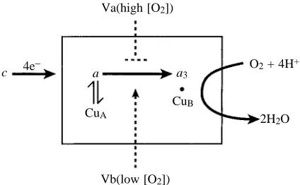
[image:10.609.62.274.536.667.2]2, Fig. 8. Subunit V isoforms in yeast regulate cytochrome c oxidase
activity. In the presence of air or high oxygen concentrations, yeast cells express the Va isoform; at low oxygen concentrations, they express subunit Vb. These two isoforms affect the rate of intramolecular electron transfer between heme a and a3: Vb
enhances the rate, while Va slows it down. Va(high [O2])
a a3
O2+ 4H+
2H2O c 4e−
CuA
CuB
than that for the induction of the hypoxic isoform of yeast subunit V. The control of VIIe expression by oxygen is apparently overriden after vegetative cells aggregate and begin to differentiate (Sandona et al. 1995). Thus, it is possible that some developmental program set in place at the time of cellular aggregation also controls isoform switching. Currently, it is not clear whether, or how, these isoforms of subunit VII affect the function(s) of Dictyostelium cytochrome
c oxidase. However, because they are not structural homologs
of yeast subunit V, it seems unlikely that they function like the yeast isoforms.
Do mammals possess isoforms analogous to yeast Va and Vb?
It is not yet known whether the mammalian subunit IV, the counterpart of yeast subunit V, has an oxygen-regulated isoform that functions to affect the binuclear reaction center. However, considering that this subunit has not been completely sequenced from different tissues and that the cDNA libraries that have been used to isolate its gene were not made from cells or tissues that were grown at low oxygen concentration, it is quite possible that a mammalian subunit IV isoform has gone undetected thus far. Currently, there are several pieces of suggestive evidence that hint at the presence of a hypoxic isoform of mammalian subunit IV. First, immunological differences have been reported between subunit IV from fetal and adult rats, suggesting the occurrence of fetal and adult isoforms of subunit IV (Kuhn-Nentwig and Kadenbach, 1985). Second, immunohistochemical studies suggest that fiber-specific isoforms of subunit IV exist in human skeletal muscle (Romero et al. 1990). Third, purification studies have revealed a putative isoform in human heart cytochrome c oxidase. This isoform has a blocked N terminus (Van Kuilenburg et al. 1992). Finally, it is well established that cytochrome synthesis in cultured mammalian cells is repressed by growth at low oxygen tensions (Pious, 1979) and that the cytochrome contents of tissues that are exposed to low oxygen levels, e.g. fetal tissue, are low (Hallman, 1971; Wilson, 1972). In an intriguing study performed several years ago, it was demonstrated that the cytochrome aa3content of newborn puppies increases during the first 5 days of life, but that the effective turnover rate of cytochrome c oxidase decreases during this period (Mela et al. 1975). These changes were attributed to an increase in arterial PO∑ after
birth and suggest that holocytochrome c oxidase assembled at low oxygen tensions (in the fetus) has a higher turnover rate than holocytochrome c oxidase assembled at atmospheric oxygen tensions (in the adult). These findings are especially interesting because they parallel what we currently know about the expression and function of yeast subunits Va and Vb.
In summary, it is clear that the isoforms of a nuclear-encoded subunit of cytochrome c oxidase can affect the catalytic functions of a mitochondrially encoded subunit. They also serve to illustrate the intricate interplay between oxygen concentration and holoenzyme function.
This work was supported in part by Public Health Service Grant 30228 from the National Institutes of Health.
References
ALLEN, L. A., ZHAO, X.-J., CAUGHEY, W. ANDPOYTON, R. O. (1995).
Isoforms of yeast cytochrome c oxidase subunit V affect the binuclear reaction center and alter the kinetics of interaction with the isoforms of yeast cytochrome c. J. biol. Chem. 270, 110–118. ANTHONY, G., REIMANN, A. AND KADENBACH, B. (1993).
Tissue-specific regulation of bovine heart cytochrome c oxidase activity by ADP via interaction with subunit VIa. Proc. natn. Acad. Sci.
U.S.A. 90, 1652–1656.
BABCOCK, G. T. ANDWIKSTROM, M. (1992). Oxygen activation and
the conservation of energy in cell respiration. Nature 356, 301–309. BISSON, R. AND SCHIAVO, G. (1986). Two different forms of
cytochrome c oxidase can be purified from the slime mold
Dictyostelium discoidium. J. biol. Chem. 261, 4373–4376.
BISSON, R., VETTORE, S., ARATRI, E. AND SANDONNA, D. (1997). Subunit change in cytochrome c oxidase: identification of the oxygen switch in Dictyostelium. EMBO J. 16, 739–749.
BONNE, G., SEIBEL, P., POSSEKEL, S., MARSAC, C. ANDKADENBACH,
B. (1993). Expression of human cytochrome c oxidase subunits during fetal development. Eur. J. Biochem. 217, 1099–1107. BRUNORI, M., ANTONINI, G., MALETESTA, F., SARTI, P. ANDWILSON,
M. T. (1987). Cytochrome c oxidase – Subunit structure and proton pumping. Eur. J. Biochem. 169, 1–8.
BUNN, H. F. AND POYTON, R. O. (1996). Oxygen sensing and
molecular adaptation to hypoxia. Physiol. Rev. 76, 839–885. BURKE, P. V., RAITT, D. C., ALLEN, L. A., KELLOGG, E. A. AND
POYTON, R. O. (1997). Effects of oxygen concentration on the expression of cytochrome c and cytochrome c oxidase genes in yeast. J. biol. Chem. 272, 14705–14712.
CAPALDI, R. A., MARUSHICH, M. F. AND TAANMAN, J.-W. (1995).
Mammalian cytochrome c oxidase: Characterization of enzyme and immunological detection of subunits in tissue extracts and whole cells. Meth. Enzymol. 260, 117–132.
CAUGHEY, W. S., DONG, A., SAMPATH, V., YOSHIKAWA, S. ANDZHAO,
X. J. (1988). Probing heart cytochrome c oxidase structure and function by infrared spectroscopy. J. Bioenerg. Biomembr. 25, 81–91.
CHAN, S. I. AND LI, P. M. (1990). Cytochrome c oxidase:
Understanding nature’s design of a proton pump. Biochemistry 29, 1–12.
COPELAND, R. A. (1991). Conformational switching at cytochrome a during steady state turnover of cytochrome c oxidase. Proc. natn.
Acad. Sci. U.S.A. 88, 7281–7283.
CUMSKY, M. G., KO, C., TRUEBLOOD, C. E. ANDPOYTON, R. O. (1985).
Two non-identical forms of subunit V are functional in yeast cytochrome c oxidase. Proc. natn. Acad. Sci. U.S.A. 82, 2235–2239.
CUMSKY, M. G., TRUEBLOOD, C. E., KO, C. ANDPOYTON, R. O. (1987).
Structural analysis of two genes encoding divergent forms of yeast cytochrome c oxidase subunit V. Molec. cell. Biol. 7, 3511–3519. DODSON, E. D., ZHAO, X.-J., CAUGHEY, W. S. ANDELLIOTT, C. M.
(1996). Redox dependent interactions of the metal sites in carbon-monoxide-bound cytochrome c oxidase monitored by infrared and UV/Visible spectroelectrochemical methods. Biochemistry 35, 444–452.
ERECINSKA, M. AND WILSON, D. F. (1982). Regulation of cellular
FIAMINGO, F. G., ALTSCHULD, R. A. ANDALBEN, J. O. (1986). Alpha and beta forms of cytochrome c oxidase observed in rat heart myocytes by low temperature Fourier transform infrared spectroscopy. J. biol. Chem. 261, 12976–12987.
FRANK, V. AND KADENBACH, B. (1996). Regulation of the H+/e−
stoichiometry of cytochrome c oxidase from bovine heart by intramitochondrial ATP/ADP ratios. FEBS Lett. 382, 121–124. GIER, B., SCHAGGER, H., ORTWEIN, C., LINK, T. A., HAGEN, W. R.,
BRANDT, U. ANDVONJAGOW, G. (1995). Kinetic properties and ligand binding of the eleven-subunit cytochrome c oxidase from
Saccharomyces cerevisiae isolated with a novel large-scale
purification method. Eur. J. Biochem. 227, 296–302.
HALLMAN, M. (1971). Changes in mitochondrial respiratory chain proteins during perinatal development. Evidence for the importance of environmental oxygen. Biochim. biophys. Acta
253, 360–372.
HALTIA, T., SARASTE, M. ANDWIKSTROM, M. (1991). Subunit III of cytochrome c oxidase is not involved in proton translocation: a site-directed mutagenesis study. EMBO J. 10, 2015–2021.
HODGE, M., KIM, G., SINGH, K. ANDCUMSKY, M. G. (1989). Inverse
regulation of the yeast COX5 genes by oxygen and heme. Molec.
cell. Biol. 9, 1958–1964.
IWATA, S., OSTERMEIER, C., LUDWIG, B. AND MICHEL, H. (1995). Structure at 2.8 Å resolution of cytochrome c oxidase from
Paracoccus denitrificans. Nature 376, 660–669.
KADENBACH, B. (1986). Regulation of respiration and ATP synthesis
in higher organisms: hypothesis. J. Bioenerg. Biomembr. 18, 39–54.
KADENBACH, B., HARTMAN, R., GLANVILLE, R. ANDBUSE, G. (1982). Tissue-specific genes code for polypeptide VIa of bovine liver and heart cytochrome c oxidase. FEBS Lett. 138, 236–238.
KADENBACH, B. ANDREINMANN, A. (1992). Cytochrome c oxidase:
tissue-specific expression of isoforms and regulation of activity. In
Molecular Mechanisms in Bioenergetics (ed. L. Ernster), pp.
241–263. Amsterdam: Elsevier Press.
KADENBACH, B., UNGIBAUER, M., JANAUSCH, J., BUGE, U. ANDKUHN
-NENTWIG, L. (1983). The complexity of respiratory complexes.
Trends biochem. Sci. 8, 398–400.
KUHN-NENTWIG, L. AND KADENBACH, B. (1985). Isolation and properties of cytochrome c oxidase from rat liver and quantification of immunological differences between isozymes from various rat tissues with subunit-specific antisera. Eur. J. Biochem. 149, 147–158.
KWAST, K. E., BURKE, P. V. AND POYTON, R. O. (1998). Oxygen
sensing and transcriptional regulation of oxygen-responsive genes in yeast. J. exp. Biol. 201, 1177–1195.
LIN, J., PAN, L. P. ANDCHAN, S. I. (1993). The subunit location of magnesium in cytochrome c oxidase. J. biol. Chem. 268, 22210–22214.
LOMAX, M. ANDGROSSMAN, L. I. (1989). Tissue-specific genes for
respiratory proteins. Trends biochem. Sci. 14, 501–503.
MCEWEN, J. E., KO, C., KLOECKENER-GRUISSEM, B. ANDPOYTON, R.
O. (1986). Nuclear functions required for cytochrome c oxidase biogenesis in Saccharomyces cerevisiae. J. biol. Chem. 261, 11872–11879.
MELA, L., GOODWIN, C. W. ANDMILLER, L. D. (1975). Correlation of
mitochondrial cytochrome content and activity to oxygen availability in the newborn. Biochem. biophys. Res. Commun. 64, 384–390.
MEUNIER, B. AND COLSON, A. M. (1994). Random deficiency
mutations and reversions in cytochrome c oxidase subunits I, II and
III of Saccharomyces cerevisiae. Biochim. biophys. Acta 1187, 112–115.
MEUNIER, B., COLSON, A. M. ANDRICH, P. R. (1995). The topology of CuA in relation to other metal centers in cytochrome c oxidase of Saccharomyces cerevisiae as determined by analysis of second-site revertants. Biochim. biophys. Acta 1253, 13–15.
MITCHELL, D. M., SHAPLEIGH, J. P., ARCHER, A. M., ALBEN, J. O. AND
GENNIS, R. B. (1996). A pH-dependent polarity change at the
binuclear reaction center of reduced cytochrome c oxidase detected by FTIR difference spectroscopy of the CO adduct. Biochemistry
35, 9446–9450.
MOODY, A. J. (1996). ‘As prepared’ forms of fully oxidized haem/Cu
terminal oxidases. Biochim. biophys. Acta 1276, 6–20.
PATTERSON, T. E., TRUEBLOOD, C. E., WRIGHT, R. M. ANDPOYTON,
R. O. (1987). Polypeptide subunits encoded by nuclear genes are essential components of eukaryotic cytochrome c oxidase. In
Cytochrome Systems: Molecular Biology and Bioenergetics (ed. S.
Papa, B. Chance and L. Ernster), pp. 253–260. New York: Plenum Press.
PIOUS, D. (1979). Induction of cytochromes in human cells by oxygen. Proc. natn. Acad. Sci. U.S.A. 65, 1001–1008.
POWER, S. D., LOCHRIE, M. A., SEVARINO, K. A., PATTERSON, T. E. AND
POYTON, R. O. (1984). The nuclear-coded subunits of yeast cytochrome c oxidase. I. Fractionation of the holoenzyme into chemically pure polypeptides and the identification of two new subunits using solvent extraction and reversed phase high performance liquid chromatography. J. biol. Chem. 259, 6564–6570. POYTON, R. O. AND BURKE, P. V. (1992). Oxygen regulated
transcription of cytochrome c and cytochrome c oxidase genes in yeast. Biochim. biophys. Acta 1101, 252–256.
POYTON, R. O., GOEHRING, B., DROSTE, M., SEVARINO, K. A., ALLEN, L. A. ANDZHAO, X.-J. (1995). Cytochrome c oxidase (Complex IV)
from Saccharomyces cerevisiae. Meth. Enzymol. 260, 97–116. POYTON, R. O. ANDMCEWEN, J. E. (1996). Crosstalk between nuclear
and mitochondrial genomes. A. Rev. Biochem. 65, 563–607. POYTON, R. O., TRUEBLOOD, C. E., WRIGHT, R. M. ANDFARRELL, L.
E. (1988). Expression and function of cytochrome c oxidase isologues. Modulators of cellular energy production? Ann. N.Y.
Acad. Sci. 550, 289–307.
RIISTAMA, S., PUUSTINEN, A., GARCAI-HORSMAN, A., IWATA, S.,
MICHEL, H. ANDWIKSTROM, M. (1996). Channelling of dioxygen into a respiratory enzyme. Biochim. biophys. Acta 1275, 1–4. ROBINSON, B. H. (1994). MtDNA and nuclear mutations affecting
oxidative phosphorylation: correlating severity of clinical defect with extent of bioenergetic compromise. J. Bioenerg. Biomembr.
26, 311–317.
ROGERS, P. J. AND STEWART, P. R. (1973a). Mitochondrial and peroxisomal contributions to the energy metabolism of
Saccharomyces cerevisiae in continuous culture. J. gen. Microbiol.
79, 205–217.
ROGERS, P. J. ANDSTEWART, P. R. (1973b). Respiratory development in Saccharomyces cerevisiae grown at controlled oxygen tension.
J. Bacteriol. 115, 88–97.
ROHDICH, F. ANDKADENBACH, B. (1993). Tissue-specific regulation
of cytochrome c oxidase efficiency by nucleotides. Biochemistry
32, 8499–8503.
ROMERO, N., MARSAC, C., FARDEAU, M., DROSTE, M., SCHNEYDER, B.
ANDKADENBACH, B. (1990). Immunohistochemical demonstration
of fiber-type specific isozymes of cytochrome c oxidase in human skeletal muscle. Histochemistry 94, 211–215.
modelling by satisfaction of spatial restraints. J. molec. Biol. 234, 779–815.
SANDONA, D., GASTALDELLO, S., RIZZUTO, R. ANDBISSON, R. (1995). Expression of cytochrome c oxidase during growth and
development of Dictyostelium. J. biol. Chem. 270, 5587–5593. SARASTE, M. (1990). Structural features of cytochrome oxidase. Q.
Rev. Biophys. 23, 331–66.
SHAPLEIGH, J. P., HILL, J. J., ALBEN, J. O. ANDGENNIS, R. B. (1992).
Spectroscopic and genetic evidence for two heme-Cu-containing oxidases in Rhodobacter sphaeroides. J. Bacteriol. 174,
2338–2343.
SHERMAN, D., KOTAKE, S., ISHIBE, N. ANDCOPELAND, R. A. (1991).
Resolution of the electronic transitions of cytochrome c oxidase: evidence for two conformational states of ferrous cytochrome a.
Proc. natn. Acad. Sci. U.S.A. 88, 4265–4269.
TAANMAN, J. W. ANDCAPALDI, R. A. (1992). Purification of yeast
cytochrome c oxidase with a subunit composition resembling the mammalian enzyme. J. biol. Chem. 267, 22481–22485.
TRUEBLOOD, C. E. AND POYTON, R. O. (1987). Differential effectiveness of yeast cytochrome c oxidase subunit V genes results from differences in expression not function. Molec. cell. Biol. 7, 3520–3526.
TRUEBLOOD, C. E., WRIGHT, R. M. AND POYTON, R. O. (1988). Differential regulation of the two genes encoding Saccharomyces
cerevisiae cytochrome c oxidase subunit V by heme and the HAP2
and REO1 genes. Molec. cell. Biol. 8, 4537–4540.
TSUKIHARA, T., AOYAMA, H., YAMASHITA, E., TOMIZAKI, T., YAMAGUCHI, H., SHINZAWA-ITOH, K., NAKASHIMA, R., YAONO, R. ANDYOSHIKAWA, S. (1995). Structures of metal sites of oxidized bovine heart cytochrome c oxidase at 2 Å. Science 269, 1069–1072.
TSUKIHARA, T., AOYAMA, H., YAMASHITA, E., TOMIZAKI, T.,
YAMAGUCHI, H., SHINZAWA-ITOH, K., NAKASHIMA, R., YAONO, R.
ANDYOSHIKAWA, S. (1996). The whole structure of the 13-subunit
oxidized cytochrome c oxidase at 2 Å. Science 272, 1136–1144. VAN KUILENBURG, A., VAN BEEUMAN, J., DEMOL, H., VAN DEN
BOGERT, C., SCHOUTEN, I. ANDMUIJERS, A. O. (1992). Subunit IV of human cytochrome c oxidase, polymorphism and a putative isoform. Biochim. biophys. Acta 1119, 218–224.
VILLANI, G. ANDATTARDI, G. (1997). In vivo control of respiration by
cytochrome c oxidase in wild-type and mitochondrial DNA mutation-carrying human cells. Proc. natn. Acad. Sci. U.S.A. 94, 1166–1171.
WALLACE, D. C. (1992). Mitochondrial genetics: a paradigm for aging
and degenerative diseases? Science 256, 628–632.
WANG, J., TAKAHASHI, S., HOSLER, J. P., MITCHELL, D. M., FERGUSON
-MILLER, S., GENNIS, R. B. AND ROUSSEAU, D. L. (1995). Two conformations of the catalytic site in the aa3-type cytochrome c
oxidase form Rhodobacter sphaeroides. Biochemistry 34,
9819–9825.
WATERLAND, R. A., BASU, A., CHANCE, B. ANDPOYTON, R. O. (1991). The isoforms of yeast cytochrome c oxidase subunit V alter the in
vivo kinetic properties of the holoenzyme. J. biol. Chem. 266,
4180–4186.
WIKSTROM, M., BOGACHEV, A., FINEL, M., MORGAN, J. E., PUUSTINEN, A., RAITIO, M., VERKHOVSKAYA, M. AND VERKHOVSKY, M. I.
(1994). Mechanism of proton translocation by the respiratory oxidases. The histidine cycle. Biochim. biophys. Acta 1187, 106–111.
WILSON, D. F. (1982). Regulation of in vivo mitochondrial oxidative
phsophorylation. In Membranes and Transport, vol. 1 (ed. A. Martinosi), pp. 349–355. New York: Plenum Press.
WILSON, J. E. (1972). The relationship between glycolytic and mitochondrial enzymes in the developing rat brain. J. Neurochem.
19, 223–227.
WITTUNG, P AND MALMSTROM, B. G. (1996). Redox-linked
conformational changes in cytochrome c oxidase. FEBS Lett. 388, 47–49.
WOODRUFF, W. H. (1993). Coordination dynamics of heme-copper oxidases. The ligand shuttle and the control and coupling of electron transfer and proton translocation. J. Bioenerg. Biomembr.