Intracranial dural arteriovenous fistulas with spinal venous drainage: relation between clinical presentation and angiographic findings
Full text
Figure

Related documents
This study was designed to identify the added clinical value of ASL for the diagnosis of small AVMs or DAVFs. We ex- cluded AVMs ⬎ 2 cm to better capture patients in whom di- agnosis
The aim of this article is to describe, in the largest series of patients so far from 2 different hospitals, the venous reopening technique to treat dural AV shunts via a
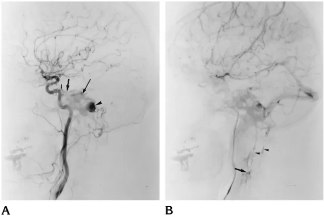
A , Arterial phase of a right external carotid angiogram shows a DAVF at the right TSS ( large arrow ) with retrograde venous drainage into the superior sagittal sinus and
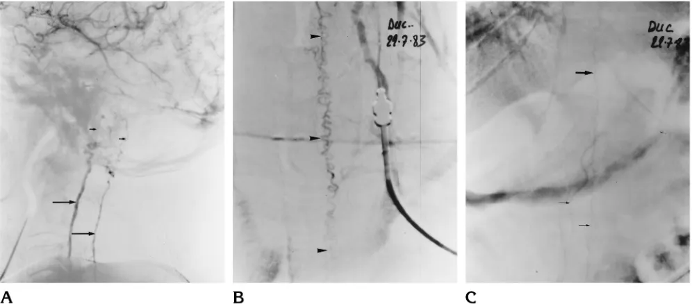
In 6 patients, a prolonged (17- to 29-minute) single middle meningeal artery injection was sufficient to occlude the venous site of the fistula with retrograde occlusion of
Box and whisker plot show mean relative cerebral blood volume (rCBV) ratio in control subjects and in patients with dural arteriovenous fistulas (DAVF) with retrograde cortical
MR imaging findings included flow void cluster, engorged ophthalmic vein/proptosis, white matter hyperintensity, intracranial hemor- rhage, dilated leptomeningeal or medullary
BACKGROUND AND PURPOSE: Tortuous, engorged veins can be identified on the venous phase of the brain circulation in patients with venous congestion related to an intracranial
It is believed that long-standing enhancement of the spinal cord in patients with dural arteriovenous fistula probably results from chronic progressive venous ischemia, which
