With 5 text-figures Printed in Great Britain
DIRECTIONAL SENSITIVITY OF SACCULAR MICROPHONIC
POTENTIALS IN THE HADDOCK
BY P. S. ENGER, A. D. HAWKINS*, O. SAND AND C. J. CHAPMAN*
Institute of Zoophysiology, University of Oslo, Oslo, Norway, and
Marine Laboratory, Aberdeen, Scotland
{Received 12 March 1973)
INTRODUCTION
The function of the swimbladder in fish hearing is well established for the ostario-physine species, which possess a chain of ossicles connecting the swimbladder mechanically to the perilymph of the labyrinth (cf. von Frisch, 1936; Poggendorf, 1952). However, the auditory function of the swimbladder in fish lacking a mechanical connexion between the swimbladder and the ear is less clear. Van Bergeijk (1964) and Alexander (1966), from hypothetical considerations, have pointed out the possible widespread use of the swimbladder in sound reception. They suggested that the swimbladder might function as a pressure/displacement transformer, even in the absence of a mechanical linkage, acting as a secondary sound-source re-radiating near-field particle displacements which stimulate the auditory receptors. However, experimental evidence supporting this theory has been sparse. Several authors have compared the hearing of species with and without swimbladders under the same acoustic conditions and concluded that the swimbladder is involved in sound recep-tion (Enger & Andersen, 1967; Iversen, 1969; Chapman & Sand, 1973). Recently a more direct approach has been employed by Sand & Enger (1973), who showed that in cod kept at 6 m depth the saccular microphonic potentials were drastically reduced when the swimbladder was emptied by sucking the gas out of it through a hypodermic syringe.
The advantage of utilizing the swimbladder pulsations in hearing is a lowering of the auditory threshold. A concomitant disadvantage, however, is a possible loss of ability to determine the direction of the sound source (van Bergeijk, 1964). This follows from the assumption that the re-radiated sound from the swimbladder will mask any differences in arrival time, phase and intensity of the incident sound at the two ears. Earlier behavioural studies supported this view by indicating that fish are unable to determine the direction of a sound source except at close range, where the lateral line organs probably are involved (von Frisch & Dijkgraf, 1935). Sharks are known to have directional hearing at long range (Nelson & Gruber, 1963; Myrberg, Banner & Richard, 1969), but recent investigations by Olsen (1969) on herring and by Schuijf, Baretta & Wildschut (1972) on wrasse (Labrus berggylta), undertaken under much better acoustic conditions than earlier studies, have shown that fish with swimbladders can also distinguish between different sound-source directions. Herring were reported to be able to determine direction within at least 45 °, probably
426 P. S. ENGER, A. D. HAWKINS, O. SAND AND C. J. CHAPMAN
better, for frequencies from 20 to 6000 Hz, whereas the wrasse at 115 Hz (the onlj frequency tested) showed a directional resolution better than 70 ° and possibly as good as io°.
This experimental evidence thus conflicts with any theory which supposes that directional hearing is impaired in fish possessing a swimbladder. However, as stressed by Schuijf et al. (1972), the excitation of the maculae in the pars inferior by shearing forces makes this receptor inherently directionally sensitive. It is therefore possible that stimulation of the maculae by re-radiated sound from the swimbladder is avoided by a proper orientation of the hair-cells, i.e. some or all of the hair-cells could be so arranged that they are insensitive to radial displacements emanating from the swim-bladder. Wersall, Flock & Lundquist (1965) have examined the orientation of the hair-cells in the sacculus of a gadoid fish, the burbot (Lota lota). From the figure they present it would appear that the hair-cells are arranged in two opposing directions, giving maximal sensitivity to vertical or near-vertical displacement of the otolith. However, the macula has a complex curved shape, which suggests that taken as a whole it is not exclusively sensitive to any single displacement direction. The direc-tional response of the otolith organ is determined not only by the orientation of the hair-cells but also by the nature of the otolith suspension. The manner in which the otolith is suspended or mounted may restrict its motion to particular directions. Little information is available on this question, however.
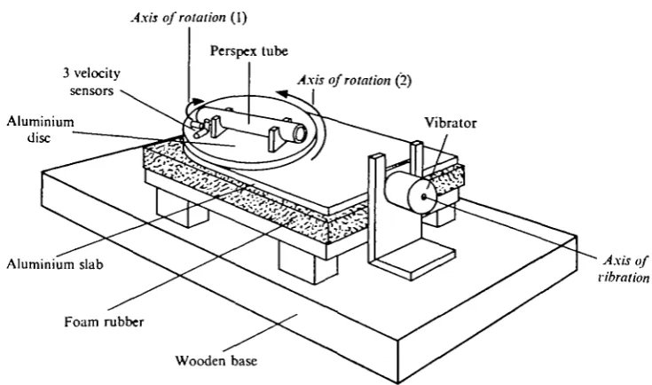
Clearly, experimental evidence is needed to establish whether fish with an un-specialized swimbladder are sensitive to displacements radiated from the bladder. Furthermore, it must be established whether separate groups of hair-cells are sensitive to other vibration directions, thus providing the fish with directional sensitivity. To clarify these questions we have therefore measured saccular microphonic potentials in the haddock (Melanogrammus aeglefinus) as a function of the direction of vibration. To obtain well-defined directional stimuli the experiments were performed by mounting the fish on a rotatable vibration table.
MATERIALS AND METHODS
Directional sensitivity of saccular micropkonic potentials 427
Axis of rotation (1)
Perspex tube
3 velocity
sensors Axis of rotation (2)
Aluminium disc
Vibrator
Aluminium slab Axis of
vibration
Foam rubber
[image:3.451.44.412.64.281.2]Wooden base
Fig. i. Vibration table. Perspex tube with fish is rotatable around its long axis and in the horizontal plane. Further explanation in text.
those produced by gill movements. The electrodes were bent at right angles with the proximal part of the electrode resting on the skull, where it was secured by dental cement. The thin flexible electrode leads were attached to the skin by sutures at two points, just behind the incision. A short piece of stainless-steel wire inserted into the back muscles served as the ground electrode.
With the electrodes implanted, the fish was placed in a perspex tube 30 cm long and 6 cm in diameter. A pair of stainless-steel bars clamped the fish skull firmly, just above the eyes. The tube was then placed on the vibration table.
Stimulating and recording equipment. The vibration table, shown schematically in
428 P. S. ENGER, A. D. HAWKINS, O. SAND AND C. J. CHAPMAN
60
40
1 o
x 20
e
I 6
e
X) > 4
90° 180°
Vibration angle
270° 360°
Fig. 2. Vibration amplitude at 200 Hz of the rota table, experimental table, versus azimuth angle of the fiah holder, measured with three transducers. Two are placed in the horizontal plane, parallel to the long axis of the fish (•), and at the right angles to the long axis of the fish (O), and one placed in the vertical axis (A).
(rms) were recalculated to displacement values. One series of measurements, as the fish was rotated stepwise from o° to 360°, is given in Fig. 2 for 200 Hz. It can be seen that the two horizontal components were 90° out of phase, as expected, and that the ratio between the horizontal vibration component parallel to the driving force and the horizontal component perpendicular to this direction was as high as 30. The horizon-tal movement of the disc was thus essentially linear. The vertical vibrations varied with the angular position of the fish holder and in some positions were unfortunately rather high. The maximum amplitude was about five times the minimum, reaching values of about one-third of the horizontal vibration amplitude. The table responded in much the same way to the other frequencies tested.
Microphonic potentials from the fish were amplified by a pre-amplifier (Tektronix 122) and displayed on a storage oscilloscope (Tektronix 564). The signal was also fed into a logarithmic level recorder (Bruel & Kjaer, 2305) for measurement, after being amplified, and in some cases filtered through a frequency analyser (Bruel & Kjaer, 2107). Microphonic potentials are in this paper given in dB re i/iVrms.
RESULTS
Directional sensitivity of saccular microphonic potentials 429
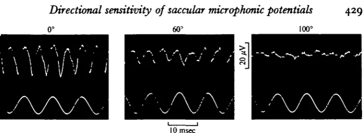
[image:5.451.39.414.48.185.2]10 msec
Fig. 3. Saccular microphonic potentials (upper trace) recorded during ioo Hz vibration of 1 "2 x io~* cm amplitude (lower trace) at three different angles of azimuth.
found when recording from the fish sacculus during sound stimulation (Zotterman, 1943; Enger & Andersen, 1967). The amplitude of the potentials was a function both of the stimulus strength and of the direction of vibration. In this and most other cases the fish was subjected to vibration in the normal upright position (zero tilt). However, some experiments were performed on fish which were tilted about the long axis, as mentioned below.
In Fig. 3 the maximum microphonic amplitude was recorded at an azimuth angle of o° (i.e. when the fish was subjected to vibration along its axis). A minimum was recorded at an azimuth angle of ioo° (i.e. when the fish was subjected to vibration from the side). The microphonic potential amplitudes recorded from two different fish at 100 and 200 Hz, respectively, are plotted versus the angle of azimuth in Fig. 4A and B. The amplitude minima were rather sharp at 900 and 2700 in A, and at ioo° and 2800 in B. In most other fish tested corresponding minimum values were obtained at azimuth angles in the range 85-105° and 260-2800. Maximum amplitudes in these cases were recorded over a wide sector of 60-700, centred around o° and 1800. A comparison between Figs. 2 and 3 confirms that the microphonic potential ampli-tude reached a maximum when the horizontal vibration component along the long axis of the fish was maximal (i.e. at azimuth angles 0-180°). At the angle of minimum microphonic response the horizontal vibration component transverse to the long axis was maximal and the vertical vibration component close to maximal. Therefore, it must be concluded that the sensory cells which produced the recorded responses were relatively insensitive to vibration in these directions.
The above description applied to most recordings, but one of the notable exceptions is presented in Fig. 5. This figure shows that the maximum microphonic potential amplitude was obtained from this particular fish when it was subjected to vibration in a direction at right angles to the long axis (i.e. at azimuth angles of 90-270°). This result indicates that the sensory cells contributing to the recorded potentials at this electrode position are mainly sensitive to transverse vibration of the fish. How-ever, in this particular case the amplitude of the vertical vibration component was relatively large (though not at a maximum) with the fish in this position. We therefore cannot eliminate the possibility that a sensitivity to vertical vibrations might have con-tributed to the maximal response.
430 P. S. ENGER, A. D. HAWKINS, O. SAND AND C. J. CHAPMAN
90-270
Fig. 4. Polar diagrams, showing rms-values of saccular microphonic potential at different angles of azimuth. Note that maximal response is obtained to vibration along the long axis of the fish (azimuth of o and 1800 angles). A. Vibration frequency 100 Hz, amplitude i-2X io~l cm. B. Vibration frequency 300 Hz, amplitude 6x io~*cm (another fish).
less (4-6 dB), and the variation pattern did not seem to follow any of the vibration components given in Fig. 1. One reason for this might be that we were simultaneously recording from cells of different directional sensitivity. Alternatively the cells may have been mainly sensitive to the vertical vibration component (which varied much less than the horizontal components).
We must therefore stress that though our data indicate that in the majority of fish examined the parts of the sensory macula examined showed greatest sensitivity to vibration along the long axis of the animal, in other preparations greatest sensitivity was shown to vibration in other directions.
In some cases saccular microphonic potentials were also recorded while the fish was tilted stepwise around its long axis. If this was done at azimuth angles giving maximal microphonic potentials, the microphonic potentials decreased in amplitude when the fish was rotated from its normal upright position towards a position with the dorsal side down. Furthermore, if the fish was quickly returned to the upright position, several seconds elapsed before the microphonic potential amplitude regained its previous maximal amplitude. This indicates that the position of the otoliths changes only slowly when the fish is tilted, and that the movement of the otolith must therefore be opposed by relatively strong visco-elastic forces.
DISCUSSION
particu-Directional sensitivity of saccular microphonic potentials 431
ftar axis of highest sensitivity to vibrational stimuli. This has a bearing on two aspects of fish hearing, namely (1) directional hearing, i.e. the ability of the fish to determine the direction of the sound source, and (2) the role of the swimbladder in the hearing process. The latter aspect becomes clear when bearing in mind that the swimbladder may act as a pressure/displacement transformer and thus produce displacements which stimulate the ear in a direction along the long axis of the fish.
At first sight our method of subjecting the fish to vibration in order to simulate auditory stimulation may seem unusual and unnatural, since it only produces particle displacement on the experimental table and not sound pressure. However, we believe that this method is justified since it now seems to be well established that particle displacement, and not sound pressure, is the adequate stimulus for the auditory re-ceptors. To use pure vibrational stimuli is advantageous in that any effects arising from the swimbladder are eliminated. In addition, the vibration direction can be readily determined and thus yield information about the possible sensitivity axes of the sensory cells. The vibration amplitude of the vibration table reached a maximum of 1-2 x io~5 cm at 100 Hz. In comparison, threshold values for particle displacement in dab (Chapman & Sand, 1973) are around 3 x io~9 cm in the optimal frequency range, thus giving a difference of 72 dB. An auditory stimulus intensity of 72 dB above thres-hold cannot be considered abnormally high, and artifacts caused by overdriving the hearing system probably did not play a role in our experiments.
The position of the electrode in all the experiments performed was found to be in the middle and anterior half of the saccular sensory epithelium, but no detailed and systematic mapping of the recording loci was performed. Generally, it must be assumed that the greatest contribution to the amplitude of the microphonic potentials comes from the cells closest to the electrode. From this rather safe assumption it is quite clear that a certain proportion of saccular sensory cells have their axes of highest sensitivity to vibratory stimuli orientated along the long axis of the fish. It seems therefore justified to conclude that a definite prerequisite for the participation of the swimbladder in fish hearing, namely that the otolith/hair cell system is sensitive to vibrations issuing radially from the swimbladder, is fulfilled.
There is an apparent discrepancy between this finding and the morphological indication of a predominantly dorso-ventral sensitivity axis in the macula (Wersall
et al. 1965). This difference must be sought either in a species difference or, more
likely, in that the suspension of the otoliths causes complicated otolith movements when the fish is subjected to vibration. However, the vibration pattern of the otoliths in response to auditory or vibrational stimuli is not known. We suggest that this is a field which is definitely worth further study.
432 P. S. ENGER, A. D. HAWKINS, O. SAND AND C. J. CHAPMAN
270-Fig. 5. As for 270-Fig. 4, but note that maximal response is obtained to vibration at right angles to the long axis of the fish (azimuth angles of 90 and 270°). Vibration frequency 175 Hz, amplitude 6-9 x io"'cm.
transversal sensitivity maximum in the horizontal plane has been presented in Fig. 5, and other recordings indicate sensitivity axes in other directions. Although further studies are required for a complete description of the response patterns of the sensory cells, the results presented provide the first step towards a neurological understanding of the behaviouristically observed ability of fish to detect the direction of the sound source (Olsen, 1969; Schuijf et al. 1972).
We suggest that one possible way in which a fish could detect the direction of a sound source is by possessing different groups of hair cells in the ear, having different axes of maximal sensitivity. While some of these cells may be sensitive to particle displacements re-radiated from the swimbladder, other cells are rather insensitive to displacements in this direction. The latter cells may thus detect the particle displace-ments of the incident sound, without being affected by the amplified displacedisplace-ments from the swimbladder. If such a mechanism is operating, one would expect to find a considerably higher threshold for detection of the direction of a sound source than for simply detecting the sound itself.
SUMMARY
1. Microphonic potentials from the sacculus in the haddock have been recorded by implanted electrodes during horizontal vibration of the fish in air. This gives a good simulation of sound stimulation in water.
2. The microphonic potential amplitude was a function of the vibration angle, and from most recording loci maximal amplitudes were obtained for vibration directions parallel to the long axis of the fish. The sensory cells contributing to this response are therefore most sensitive to displacements in the same direction as sound-induced swimbladder pulsations would produce. This result thus supports the theory of an accessory role of the swimbladder in sound reception.
Directional sensitivity of saccular tmcrophonic potentials 433
p the fish has been obtained from other recording loci. One example of highest sensitivity to a vibration direction at right angle to the long axis of the fish is presented. 4. The findings that different sensory cells appear to have different axes of maximal sensitivity to vibration provides one possible neurological explanation for the ability of fish to detect the direction to a sound source.
REFERENCES
ALEXANDER, R. M C N . (1966). Physical aspects of swimbladder function. Biol. Rev. 41, 141-76. VAN BERGEIJK, W. A. (1964). Directional and nondirectional hearing in fish. In Marine Bio-Acoustics
(ed. W. N. Tavolga), pp. 269-80. Pergamon Press.
CHAPMAN, C. J. & SAND, O. A field study of hearing in two species of flatfish, Pleuronectes platessa (L.) andZ-i manda limanda (L.) (family Pleuronectidae). Comp. Biochem. Physiol. (In the Press.) ENGER, P. S. & ANDERSEN, R. (1967). An electrophysiological field study of hearing in fish. Comp.
Biochem. Physiol. 33, 517-25.
VON FRISCH, K. (1936). Uber den GehOrsinn der Fische. Biol. Rev. 11, 210-46.
VON FRISCH, K. & DIJKGRAAF, S. (1935). Konnen die Fische die Schallrichtung wahrnehmen? Z. vergl. Physiol. 33, 641-55.
IVERSEN, R. T. B. (1969). Auditory thresholds of the scombrid fish, Euthymxus affinis, with comments on the use of sound in tuna fishing. Fish. Rep. FAO no. 62, 3, 849-59.
LOWENSTELN, O. (1971). The labyrinth. In Fish Physiology, vol. v (ed. W. S. Hoar and D. J. Randall), pp. 207-40. Academic Press.
MYRBERO, A. A. JR., BANNER, A. & RICHARD, J. D. (1969). Shark attraction using a video-acoustic system. Marine Biol. 3, 264—76.
NELSON, D. R. & GRUBER, S. H. (1963). Sharks: Attraction by low-frequency sounds. Science, N.Y. M*.
975-7-OLSEN, K. (1969). Directional responses in herring to sound and noise stimuli. Int. Coun. Explor. Sea., 1969/B20.
POGGENDORF, D. (1952). Die absoluten H6rschwellen des Zwergwelses (Amiurus nebulosus) und Beitrfige zur Physik des Weberschen Apparates der Ostariophysen. Z. vergl. Physiol. 34, 222-57.
SAND, O. & ENGER, P. S. Evidence for an auditory function of the swimbladder in the cod. J. Exp. Biol. 59.
4°5-i4-SCHUIJF, A., BARETTA, J. W. & WILDSCHUT, J. T. (1072). A field investigation on the discrimination of sound direction in Labrus berggylta (Pisces: Perciformes). Neth. J. Zool. 22, 81-104.
WERSALL, J., FLOCK, A. & LUNDQUIST, P. G. (1965). Structural basis for directional sensitivity in cochlear and vestibular sensory receptors. Cold Spring Harbor Symp. quant. Biol. 30, 115-32. ZOTTERMAN, Y. (1943). The microphonic effect of teleost labyrinths and its biological significance.