Edward R. Noble, Wendy R. K. Smoker, and Nitya R. Ghatak
Summary: We present two cases of unusually large skull base paragangliomas. The first tumor was accompanied by marked bony destruction of the central skull base and multiple associ-ated cysts. The second tumor arose along the petrous ridge, with a large intracranial component. The CT, MR imaging, angio-graphic, histologic, and electron microscopic findings of these unusual lesions are described.
Index terms: Paraganglioma; Skull, neoplasms
Paragangliomas are tumors that arise from
paraganglion cells of the parasympathetic
sys-tem, cells of neural crest origin. Paraganglioma
of the head and neck was reported in 1862 by
Von Luschka, who described a carotid body
tu-mor (1). In the head and neck, the most
com-mon locations for paragangliomas are the
ca-rotid bifurcation, the jugular bulb, and the
middle ear. Paragangliomas have been described
in many locations in which paraganglionic tissue
is not normally located, including the sella and
parasellar regions (1–3). The first case we
de-scribe is unusual because of the extensive
in-volvement of the central skull base and its
heter-ogeneous cystic/solid composition. The second
case is that of a very large tumor arising along the
petrous ridge and extending intracranially.
Case Reports
Case 1A 71-year-old man began to notice a change in his peripheral vision when fixing his boat dock. On physical examination he was found to have bilateral optic atrophy, bitemporal hemianopsia, and anosmia. The remainder of his cranial nerves were intact and no endocrinologic dis-turbance was detected. A computed tomographic (CT) scan obtained at an outside hospital showed an enhancing mass with extensive destruction of the central skull base and clivus. A biopsy of the mass was done, and the initial pathologic report was meningioma.
The patient came to our institution for additional eval-uation. Arteriography showed a vascular mass involving the region of the sella, encasement of both internal carotid arteries, displacement of the basilar artery, and tumor blush in the region of the central skull base and right orbital apex (Fig 1A–C). A repeat CT study and magnetic reso-nance (MR) imaging were performed, at which time the mass was seen to replace all of the central and a portion of the anterior skull base, severely compressing the brain stem and displaying heterogeneous enhancement (Fig 1D–I). Multiple associated cysts were seen at the margins of the mass, some with the intensity of cerebro-spinal fluid and others with shortened T1 and prolonged T2 relaxation times. The diagnosis of meningioma was ques-tioned.
A right frontotemporoparietal craniotomy was per-formed. Several large cysts containing “crankcase-type” fluid were encountered during resection of the temporal tip. The mass was found to encase major vessels, the optic nerves, and the optic chiasm. A partial resection of the mass was performed to relieve compression of the optic nerves, chiasm, and brain stem.
The tumor was studied by using various staining meth-ods, including immunohistochemistry for chromogranin, S-100 protein, cytokeratin, and the following pituitary hor-mones: prolactin, growth hormone, corticotropin, thyro-tropin, follicle-stimulating hormone, and luteinizing hor-mone. In some areas of the tumor, the cells were arranged in clusters best seen in reticulin-stained sections (Fig 1J). In other areas, they were arranged in a linear or trabecular fashion. Most tumor cells displayed strong reactivity for chromogranin. A variable number of supporting cells throughout the tumor showed positive reaction for S-100 protein. The tumor cells showed no reaction for any pitu-itary hormones or cytokeratin.
Case 2
The second patient, a previously healthy 14-year-old boy, had a 1-month history of headaches, vomiting, un-steady gait, and personality changes. The headaches were right-sided and associated with right-sided facial pain. Physical examination revealed a palsy of right cranial nerve VI, mild right-sided hearing loss, and unsteady gait.
Received August 24, 1995; accepted after revision October 4, 1996.
From the Departments of Radiology (E.R.N., W.R.K.S.) and Pathology (N.R.G.), Medical College of Virginia, Richmond.
Address reprint requests to W. R. K. Smoker, MD, Department of Radiology, Medical College of Virginia, 1200 E Marshall St, Box 980615, Richmond, VA 23298-0615.
AJNR 18:986–990, May 1997 0195-6108/97/1805–0986©American Society of Neuroradiology
Fig 1. A 71-year-old man with enhancing mass accompanied by extensive destruction of the central skull base and clivus. Arterial (A) and venous (B) phase lateral views from a left internal carotid artery angiogram show a vascular mass supplied by branches of the meningohypophysial trunk (white arrowhead, A). The lesion appears to encase the carotid artery (black arrowheads, A). A homogeneous blush persists late into the venous phase (arrowheads, B).
C, Lateral view from a left vertebral arteriogram shows marked posterior displacement of the basilar artery (arrows). The sella, dorsum sella, and clivus cannot be defined.
D, Axial CT scan obtained after the angiographic study reveals a huge, centrally solid, faintly enhancing mass with peripheral cystic components involving the central skull base, destroying the clivus and sella, extending into both cavernous sinuses, encasing the internal carotid arteries (black arrowheads) and orbital apices, and compressing and displacing the midbrain and basilar artery posteriorly (white arrowhead).
The remainder of his physical examination was normal, although he was somewhat lethargic.
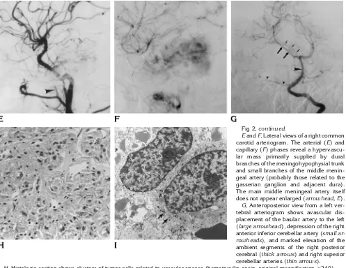
CT and MR studies revealed a large, enhancing mass along the right petrous ridge. The mass extended intracra-nially, compressing the brain stem and showing intense, homogeneous enhancement (Fig 2A–D). Angiography re-vealed early and persistent tumor blush over the right petrous ridge with marked displacement of posterior cir-culation vascular structures, although the tumor was sup-plied by the right anterior circulation (Fig 2E–G).
[image:3.587.90.250.83.262.2]After placement of a right frontal ventriculostomy, a right suboccipital craniectomy was performed. The sev-enth and eighth cranial nerves were adherent to the infero-posterior capsule of the tumor, which, when incised, was found to be quite vascular. The bulk of the tumor was removed with suction and the capsule collapsed on itself. The tumor was also adherent to the brain stem medially and to the cerebellum inferiorly. Part of the capsule was removed and the cavity was cauterized and filled with biofibular collagen.
Fig 2. A 14-year-old boy with a 1-month history of headaches, vomiting, unsteady gait, and personality changes.
AandB, Axial CT scans before (A) and after (B) contrast enhancement reveal a slightly hyperdense lesion that enhances dramatically and homogeneously. Note the marked displacement of the basilar ar-tery (arrowhead, B).
C and D, Contrast-enhanced T1-weighted (700/20/2) MR images in the ax-ial (C) and coronal (D) planes also reveal a solid, fairly homogeneously enhancing mass centered along the right petrous apex with extension into the posteroinfero-medial aspect of the right middle cranial fossa, accounting for the slight asymmetry and elevation of the right temporal horn (arrowhead, D).Figure continues.
[image:3.587.216.548.333.729.2]In addition to the staining methods used in case 1, we also used electron microscopy to study this tumor. In most areas, the tumor cells were arranged in clusters, closely associated with vascular spaces (Fig 2H). A relatively small number of tumor cells showed a positive reaction for chromogranin. The supporting cells, especially at the pe-riphery of the cell clusters, showed a positive staining reaction for S-100 protein. Electron microscopy showed typical membrane-bound secretory granules within the tu-mor cells (Fig 2I). The tutu-mor was negative for pituitary hormones.
Discussion
Classically, paragangliomas are highly
vas-cular lesions that can be very destructive. They
typically exhibit flow voids on MR images,
pro-ducing a characteristic “salt-and-pepper”
ap-pearance, and are highly vascular on
angio-graphic studies, classically supplied by the
ascending pharyngeal artery. Flow voids were
not observed on MR studies in either of our
cases, which we attribute to their being supplied
predominately by the small dorsal clival
men-ingeal branches of the meningohypophysial
trunk rather than by the larger vessels that
sup-ply the more common jugular paragangliomas
(ie, ascending pharyngeal artery).
Rarely, paragangliomas have been reported
to involve the sella and parasellar regions: one
reported case seems to have shown evidence of
lytic destruction of the central skull base (2– 4).
Our first case is distinctly unusual because of
the associated cysts. The second case is
un-usual by virtue of its location along the petrous
ridge with its associated large intracranial
com-ponent. Otherwise, this tumor had features
more typical of classic paragangliomas.
The diagnosis of paraganglioma in both
Fig 2,continued.
EandF, Lateral views of a right common carotid arteriogram. The arterial (E) and capillary (F) phases reveal a hypervascu-lar mass primarily supplied by dural branches of the meningohypophysial trunk and small branches of the middle menin-geal artery (probably those related to the gasserian ganglion and adjacent dura). The main middle meningeal artery itself does not appear enlarged (arrowhead, E).
G, Anteroposterior view from a left ver-tebral arteriogram shows avascular dis-placement of the basilar artery to the left (large arrowhead), depression of the right anterior inferior cerebellar artery (small ar-rowheads), and marked elevation of the ambient segments of the right posterior cerebral (thick arrows) and right superior cerebellar arteries (thin arrows).
H, Histologic section shows clusters of tumor cells related to vascular spaces (hematoxylin-eosin, original magnification3240).
[image:4.587.52.549.342.726.2]cases was based on their histologic
appear-ance, characterized by the arrangement of
tu-mor cells in clusters (zellballen) and their
im-munoreactivity for chromogranin. Although
only a small number of tumor cells showed a
positive reaction for chromogranin in case 2,
electron
microscopy
revealed
membrane-bound
secretory
granules
characteristically
seen in paragangliomas. Because of the
para-sellar location, we considered the possibility
that these tumors might represent aggressive
pituitary adenomas, which may sometimes
re-semble paragangliomas. However, we believe
that the tumors in our patients can be
distin-guished from pituitary adenomas by the total
absence of immunoreactivity for major pituitary
hormones and, perhaps more important, by the
positive staining reaction of the supporting cells
for S-100 protein.
These lesions would most certainly be
classi-fied as “paragangliomas of uncertain cell origin”
(5). Although paraganglionic cells have not
been found in the region of the central skull
base, Bilbao et al (2) proposed that neural crest
tissue may be involved in the development of
the pituitary gland and that a nest of
paragan-glionic tissue may remain in the region of the
adenohypophysis. Others have suggested that
such tumors may arise from abnormally
mi-grated paraganglionic tissue in the fetus or
ne-onate (6).
References
1. Von Luschka H. Uber de drusenartige natur des sogenanten gan-glion intercaroticum.Arch Anat Physiol1862:405
2. Bilbao JM, Horvath E, Kovacs K, Singer W, Hudson AR. Intrasellar paraganglioma associated with hypopituitarism.Arch Pathol Lab Med1978;102:95–98
3. Steel TR, Dailey AT, Born D, Berger MS, Mayberg MR. Paragan-gliomas of the sellar region: report of two cases.Neurosurgery 1993;32:844 – 847
4. Flint EW, Claassen D, Pang D, Hirsch WL. Intrasellar and supra-sellar paraganglioma: CT and MR findings.AJNR Am J Neurora-diol1993;14:1191–1193
5. Russell DS, Rubenstein LJ.Pathology of Tumors of the Nervous System.Baltimore, Md: Williams & Wilkins, 1989:692– 697 6. Ho KC, Meyer G, Garancis J, Hanna J. Chemodectoma involving


