CARDIOVASCULAR RESPONSES TO SCYLIORHININ I AND II
IN THE RAINBOW TROUT, ONCORHYNCHUS MYKISS, IN VIVO
AND IN VITRO
JENS KÅGSTRÖM, MICHAEL AXELSSON ANDSUSANNE HOLMGREN
Comparative Neuroscience Unit, Department of Zoophysiology, University of Göteborg, Medicinaregatan 18, S-413 90 Göteborg, Sweden
Accepted 7 February 1994
Summary
Changes in cardiac output, heart rate, dorsal aortic blood pressure and coeliac artery blood flow were measured in unrestrained rainbow trout, Oncorhynchus mykiss, following injections of the elasmobranch tachykinins scyliorhinin I and II. The resistance in the coeliac vascular bed and the total systemic vasculature were calculated from blood pressure and flow. In addition, isolated tails were perfused to investigate the effect of the peptides on the somatic vasculature. Scyliorhinin I (SCY I) produced a biphasic change in the coeliac vascular resistance: an initial decrease was followed by an increase. The decrease in coeliac vascular resistance was accompanied by a decrease in the total systemic vascular resistance, leading to an increased cardiac output. The ensuing increase in coeliac vascular resistance caused a slight increase in blood pressure. In the perfused tail, SCY I produced a marked increase in the somatic vascular resistance. Scyliorhinin II (SCY II) decreased the systemic vascular resistance, causing an increase in cardiac output. SCY II also caused a late increase in the coeliac vascular resistance, which led to hypertension and bradycardia. In vitro, SCY II produced a biphasic response in which an initial decrease in the somatic resistance was followed by a larger increase. The results demonstrate that exogenous SCY I and II are vasoactive peptides that act by different mechanisms in the rainbow trout cardiovascular system. Their actions also differ from the actions of substance P previously observed in the cod, Gadus morhua, and possibly involve a neural reflex.
Introduction
The autonomic nervous system has a considerable capacity to regulate the cardiovascular system and thus to control the blood distribution. In addition to the classical adrenergic and cholinergic neurotransmitters, cardiovascular control involves peptidergic, purinergic and serotonergic neurotransmitters, some of them sometimes co-existing in the same neurones (Burnstock and Griffith, 1988; Nilsson and Holmgren, 1992). Intestinal blood flow is regulated by perivascular nerves, but also by other factors, such as circulating hormones, metabolic products and extravascular compression (Fara,
1984). Immunohistochemical studies clearly show that nerve terminals innervating the gut and cardiovascular system of fish contain several non-adrenergic, non-cholinergic transmitters, including tachykinin-like peptides (Holmgren et al. 1982; Jensen and Holmgren, 1985; Holmgren and Jönsson, 1988; Jensen et al. 1987; Bjenning et al. 1989; Burkhart-Holm and Holmgren, 1989; Thorndyke and Holmgren, 1990).
The peptides scyliorhinin I and II were originally isolated from the intestine of the common dogfish (Scyliorhinus canicula) by Conlon et al. (1986), and recently scyliorhinin I together with a substance-P-like peptide were isolated from the brain of the same species (Waugh et al. 1993). Scyliorhinin I and II belong to the tachykinin family of polypeptides and share the C-terminal amino acid sequence -Phe-X-Gly-Leu-Met-NH2
with, amongst others, substance P (SP; X=Phe), neurokinin A (NKA; X=Val) and neurokinin B (NKB; X=Val). The C terminus is considered to be of primary importance in the interaction of the peptide with its receptor (Regoli et al. 1989). Scyliorhinin I (SCY I; X=Tyr) is a linear decapeptide most closely resembling the tachykinin physalaemin, whereas scyliorhinin II (SCY II; X=Val) is a cyclic 18 amino acid peptide most similar to Glu2-Pro5-kassinin.
The only histochemical study, to our knowledge, of scyliorhinin reactivity in fish gut has been made on Scyliorhinus canicula, where SCY I and II immunoreactivity was found predominantly in endocrine-like cells in the gastric and intestinal mucosa (Van Giersbergen et al. 1991). In the brain of the goldfish Carrassius auratus, SCY-I-like immunoreactivity has also been found (Conlon et al. 1991). Substance-P-like immunoreactivity in the gut of the rainbow trout has been demonstrated in endocrine cells in the mucosa of the stomach and proximal intestine, and in nerves mainly concentrated in the myenteric plexus throughout the gut and, to some extent, in the smooth muscle layers of the intestine (Holmgren et al. 1982). No perivascular fibres immunoreactive to SP or any other tachykinin have been found along the coeliac or mesenteric artery (Jensen, 1990). Recently, SP and NKA have been isolated and sequenced from the intestine and brain of rainbow trout and from brain of the Atlantic cod, Gadus morhua (Jensen and Conlon, 1992; Jensen et al. 1993).
The effects of the tachykinin peptides on vessels and gastrointestinal smooth muscles have been thoroughly investigated, mainly in mammals (for reviews, see Pernow, 1983; Barthó and Holzer, 1985; Otsuka and Yoshioka, 1993), but also to some extent in non-mammalian species (see Jensen, 1990; Holmgren and Nilsson, 1991). In many species of fish, various tachykinins cause gastrointestinal smooth muscles to contract in a dose-dependent manner (Jensen et al. 1987, 1994; Jensen and Holmgren, 1991). SP has a direct action on smooth muscles and, in most species, an additional indirect pathway of action
via enteric cholinergic and/or serotonergic excitatory neurones (Holmgren et al. 1985;
The only work on the cardiovascular regulatory role of scyliorhinin I and II in vivo or in
vitro was performed on the common dogfish, where no change in heart rate could be
demonstrated after SCY I injection, and only a small increase in blood pressure occurred at higher doses (Waugh et al. 1993).
This has prompted us to investigate the effect of the fish peptides scyliorhinin I and II on gastrointestinal blood flow and cardiac performance in the rainbow trout,
Oncorhynchus mykiss, in vivo, with emphasis on coeliac artery blood flow.
Materials and methods
Rainbow trout, [Oncorhynchus mykiss (Walbaum)], of either sex and with a body mass of 900–1300 g, were used in this study. The fish, bought from a local hatchery, were kept unfed in aerated recirculating fresh water at 10 ˚C and used within 2 weeks. The experiments were performed in February–May.
Surgical procedure for in vivo studies
The fish were anaesthetized in MS 222 (tricaine methane sulphonate, 120 mg l21,
Sigma) until breathing movements ceased, and then transferred to the operating table, where aerated fresh water containing MS 222 (100 mg l21) was passed over the gills
throughout the operation. For drug injection and for recording of blood pressure (PDA), a polyethylene cannula (PE 50) filled with heparinized (100 i.u. ml21) 0.9 % NaCl was
inserted into the dorsal aorta through the roof of the mouth, using a trocar method described by Aldman et al. (1992). The cannula was tunnelled through the snout and secured with sutures in the roof of the mouth and on the back of the fish.
If a rainbow trout is placed on its left side, the ventral aorta is visible under the operculum beneath the skin and connective tissue ventral to the gill arches. In order to measure cardiac output (Q˙), the ventral aorta was exposed caudal to the fourth afferent branchial artery, freed from surrounding tissue and then fitted with a Doppler flow probe (2.0–3.0 mm i.d., single-crystal, Titronics Medical Instruments). To measure blood flow in the coeliac artery (q˙CoA), an incision approximately 4 cm long was made
dorsal to the pectoral fin starting 2 cm posterior to the edge of the operculum. The coeliac artery was dissected free, a Doppler flow probe (1.5–2.0 mm i.d.) was placed around the vessel and the lead was tunnelled to the outside. Both leads were secured with skin sutures.
After surgery, the fish was placed in the experimental chamber and left to recover for at least 22 h. During this time, the effects of anaesthesia and handling wore off and the cardiovascular variables stabilized (Smith et al. 1985).
The Doppler flow probes were connected to a Doppler flow meter (Iowa University). The cannula was attached to a Statham P23 pressure transducer, which was calibrated against a static column of water. The flow probe signals and the pressure signal were amplified and displayed on a Grass Polygraph recorder system (model 79 D). Heart rate (fH) was derived from the pulsatile blood flow signal (Q˙) using a Grass 7P44 tachograph unit and expressed as beats per minute. PDAis expressed in kPa, while Q˙ and q˙CoA are
resistances of the coeliac (RCoA) and systemic (RSys) vascular beds were calculated from PDA/q˙CoAand PDA/Q˙, respectively (Axelsson and Nilsson, 1986).
The method used in the present study does not give absolute values of flows, but the method is widely accepted and gives reliable information on changes in flow. The directional pulsed-Doppler flowmeter accurately measures blood flow velocity in the vessels and displays this velocity in kHz Doppler shift. Previous work by Axelsson and Fritsche (1991) has demonstrated a high degree of linear correlation between the Doppler signal and mean volume flow, and we are therefore confident that the percentage changes in Doppler shift recorded in this experiment are directly correlated to changes in blood flow in the arteries.
This study was performed with a permit from Gothenburg Animal Ethical Committee, Dnr 299 (1992-10-07) valid for 3 years.
Experimental protocol
Drugs were injected in boluses of 0.1 ml kg21body mass. After each injection, the
cannula was immediately flushed with 0.2 ml of 0.9 % NaCl solution. The different drugs were injected in random order, and between drug injections, the cannula was flushed with 0.5 ml of 0.9 % NaCl solution. There was an interlude of at least 15 min between each injection of drug, preventing intermingling effects from the previous injection. An injection of adrenaline or noradrenaline (1 nmol kg21body mass) always initiated each
experiment as a test of the reactivity of the cardiovascular system.
Tail perfusion
Tail perfusion was performed according to the method of Wahlqvist and Nilsson (1981). The fish was injected with heparin (0.4 ml; 5000 i.u. ml21) into the dorsal aorta
through the roof of the mouth, and after 1 min killed by a sharp blow to the head. The tail was cut off behind the kidney and cannulated through the dorsal aorta (PE 50) and caudal vein (PE 90) for the inflow and outflow, respectively, of a salmon Ringer’s solution containing (g l21); NaCl, 7.63; KCl, 0.36; CaCl2.2H2O, 0.22; MgSO4.7H2O, 0.3;
NaHCO3, 2.0; NaH2PO4.2H2O, 0.43; glucose, 1.0 (Holmgren, 1983, modified from
Lockwood, 1961), and also 531029mol l21 adrenaline to imitate the physiological
condition in the fish (Milligan et al. 1989). The Ringer’s solution was bubbled with a gas mixture of O2(97 %) and CO2(3 %). The tail was immersed in saline in an organ bath
(8–9 ˚C) and the inflow catheter connected to a constant-flow peristaltic pump. Inflow pressure was measured with a Statham pressure transducer connected to the inflow catheter and a Grass polygraph recorder, as described above. The perfusion flow was adjusted to give a perfusion pressure of approximately 4 kPa, corresponding to mean dorsal aortic blood pressure levels in unexercised rainbow trout (Smith, 1978).
As perfusion flow was constant, changes in somatic vascular resistance induced by drugs were directly proportional to the measured changes in inflow pressure in the dorsal aorta. The outflow pressure was kept at zero. The drugs were injected in boluses of 0.1 ml into the inflow catheter.
Chemicals
The following drugs were used: L-adrenaline bitartrate (Sigma), L-noradrenaline
bitartrate (Sigma), scyliorhinin I (Peninsula) and scyliorhinin II (Peninsula). The drugs were dissolved in stock solutions of 0.9 % NaCl, containing 0.002 % merthiolate and 1 mg ml21bovine serum albumin, and subsequently diluted in 0.9 % NaCl.
Calculations
In addition to the Grass polygraph recordings, a data-acquisition software package (AD/DATA; P. Thorén, University of Göteborg) was used to transfer all data into an IBM-compatible computer. Data are presented as means ± standard error of mean (S.E.M.). Wilcoxon signed-ranks tests for paired samples (two-tailed) were used for
statistical evaluation of the results, and a modified Bonferoni procedure was used to reduce the risk of discarding a true null hypothesis when repeated tests were made (Holm, 1979). Differences where P<0.05 were regarded as statistically significant.
Results
Resting values for heart rate (fH; 56.8±1.8 beats min21; N=8) and dorsal aortic pressure
(PDA; 3.7±0.2 kPa; N=8) agree well with previously reported values in the rainbow trout
(Smith, 1978). Initial testing was made to determine which doses were suitable for further studies, i.e. doses that produced consistent responses which declined to resting values within 15 min. The experiments started with an injection of adrenaline (1 nmol kg21body mass) or noradrenaline (1 nmol kg21body mass), which gave
essentially the same effect: an elevation of PDAleading to a decrease in fHdue to the barostatic reflex and an initial small increase in coeliac artery blood flow followed by a large decrease in flow. Adrenaline increased cardiac output (Q˙), while noradrenaline only occasionally increased Q˙.
In vivo effects of scyliorhinin I and II
Injection of SCY I (0.1 nmol kg21body mass; N=7; Fig. 1) caused an initial decrease in
coeliac artery vascular resistance, RCoA(20.5±3.7 %), leading to an increase in blood flow
(q˙CoA, 23.0±3.6 %) through the vascular bed. Simultaneously, the systemic vascular
resistance, RSys, was decreased by 20.0±1.8 % coinciding with an elevation of Q˙ by
22.1±3.5 %. About 9 min after the injection of SCY I, a small increase in PDA (from
3.8±0.3 to 4.2±0.3 kPa) and a small reduction in fH (from
56.8±5.2–52.5±5.6 beats min21) developed. This was correlated with an increase in RCoA
(15.3±3.8 %). The vascular effects of SCY I started within 2 min of the injection and lasted 10–12 min.
SCY II (0.1 nmol kg21body mass; N=7; Fig. 2) decreased RSys by 27.4±8.6 % and
raised Q˙ by 25.4±6.1 %. Q˙ returned to the resting level within 10–12 min. There was no significant change in q˙CoA. A tendency for an initial decrease in RCoAwas noted, but this
a prolonged significant increase in PDA (from 3.8±0.4 to 4.5±0.7 kPa) lasting at least
8 min. There was also a delayed decrease in fH, 8–9 min after injection (from 55.6±4.2 to 49.8±5.9 beats min21). By the time of maximum blood flow produced by SCY II
(2–4 min after injection), RSysand RCoAhad begun to increase and RCoAwas significantly
raised above the control level (32.6±12.5 %) and remained high for at least 6 min.
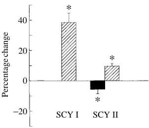
Perfused tail
In the tail preparations (Fig. 3), SCY I (0.1 nmol; N=11) produced a marked increase in perfusion pressure due to an increase in the somatic vascular resistance (38.5±6.1 %). The pressure usually returned to control level within 2 min. SCY II (0.1 nmol; N=9) caused a biphasic response, in which an initial decrease in resistance (5.7±2.9 %) was followed by a prolonged increase (9.7±1.7 %). In two of the nine preparations, only the increase was seen. Occasionally the response to SCY II was more long-lasting than that to SCY I, with the second phase lasting for several minutes.
80 70 60 50 40 30 20 8
6
4
2
160
140
120
100
80 140
120
100
80
60 0
Resistance (%
change)
Flow
(%
change)
P
DA
(kPa)
f
H
(beats
min
−
1)
2 4 6 8 10 12
Time (min)
*
*
*
*
*
*
*
Q. q.CoA [image:6.595.220.379.149.448.2]RSys RCoA
80 70 60 50 40 30 20 8
6
4
2
160
160 140
120
100
80
140 120 100 80 60 0
Resistance
Flow
(%
change)
P
DA
(kPa)
f
H
(beats
min
−
1)
2 4 6 8 10 12
Time (min)
*
*
*
*
*
Q. q.CoA [image:7.595.216.374.150.451.2]RSys RCoA
Fig. 2. The cardiovascular responses to arterial injection of scyliorhinin II (0.1 nmol kg21body mass) in the rainbow trout; mean values ±S.E.M.; N=7. Other details as in Fig. 1.
40
20
0
SCY I SCY II
−20
Percentage
change
*
*
*
[image:7.595.220.371.511.641.2]Discussion
In addition to the rapid control of the arteries by perivascular nerves, chromaffin tissues and endocrine cells exert a longer-lasting influence on vascular tonus by releasing different substances into the bloodstream. These substances may act directly on neuronal or smooth muscle receptors or indirectly through endothelial receptors that induce the release of an endothelium-derived factor that, in turn, affects smooth muscle cells. In the present study, the drugs were injected into the bloodstream and therefore acted like circulating hormones rather than like synaptic transmitters released from nerve endings. Whether tachykinin receptors in the fish circulatory system are normally stimulated by neuronal input or react only to circulating tachykinins is unknown. In the rabbit, nerve stimulation induces tachykinin-mediated vasodilatation in skeletal muscle (Persson et al. 1991).
In this study, in vivo, SCY I produces a decrease in the total systemic resistance, leading to an elevation of cardiac output whereby the blood pressure is maintained. Since the heart rate is unchanged, the elevation of cardiac output is mainly due to an increased stroke volume. In addition, there is an increase in blood flow through the coeliac artery vascular bed because of a reduction in the coeliac vascular resistance. By the time the hyperaemia wears off, the tonus of the vascular beds (and the coeliac vasculature in particular) has increased, leading to an increase in vascular resistance. This, in turn, causes the observed late increase in blood pressure. Thus, SCY I induces a biphasic response in the coeliac vascular resistance. In the cod, SP generates a triphasic response in coeliac artery blood flow, in which an initial increase in flow is followed by a rapid decrease and a subsequent increase (Jensen et al. 1991). This is caused by an overall reduction in both systemic and coeliac vascular resistances similar to that produced by SCY I in the rainbow trout, except for the intermediate phase where RCoA increases
temporarily because of a local cholinergic mechanism.
In the study by Waugh et al. (1993), SCY I produced a potent hypotension in the rat cardiovascular system, while no effect was obtained in the common dogfish Scyliorhinus
canicula. However, the only variable measured was aortic blood pressure, and regional
variations in blood flow can occur without any marked changes in aortic pressure, as has been demonstrated in the present study (see Figs 1 and 2). SP injected in vivo in the spiny dogfish, Squalus acanthias, has effects on the coeliac vascular bed similar to those of SCY I in rainbow trout, but only small effects on cardiac output (Holmgren et al. 1992).
Axelsson and Fritsche (1991) demonstrated a postprandial increase in coeliac and mesenteric blood flow in the cod. At the same time, there was a decrease in coeliac and systemic vascular resistances and an increase in cardiac output. The mechanisms behind this postprandial increase in gut blood flow are not known. However, the fish tachykinin SCY I produces an increase in coeliac vascular blood flow in the rainbow trout and, along with SP, is the only putative neurotransmitter or hormone so far shown consistently to cause hyperaemia in the gut.
there. The action of the tachykinins might be directly on the vascular smooth muscles or
via the endothelium. Another possibility is an interaction with adrenergic neurones.
Systemic blood pressure in trout is controlled by tonically active adrenergic nerves acting on systemic vessels viaa-adrenoceptors (Smith, 1978). In comparison, in the perfused tail of the spiny dogfish Squalus acanthias, SP causes a vasodilatation (Holmgren et al. 1992). The reason for the difference in vascular responses between the results in vivo and in
vitro in rainbow trout is not known, but a neuronal reflex stimulation in vivo (centrally or
peripherally mediated) that overrides the constriction seen in vitro is possible. Tachykinins have been isolated from fish brain (Conlon et al. 1991; Jensen and Conlon, 1992; Waugh et al. 1993) and, in addition, a novel type of neurokinin (NK) receptor has been found in the brain and stomach of the dogfish Scyliorhinus canicula, where SCY I and II are the most potent ligands (Van Giersbergen et al. 1991).
In mammals, three types of tachykinin receptors are known: NK-1, with a preference for binding to SP; NK-2, with a preference for NKA; and NK-3, as a receptor for NKB (see Lee et al. 1986; Regoli et al. 1989; Maggi et al. 1993). In fish tissue, the presence of at least NK-1-like receptors has been suggested (Holmgren et al. 1985; Kitazawa et al. 1988; Van Giersbergen et al. 1991; Jensen et al. 1994). In mammalian tissues, SCY I shows high affinity for both NK-1 and NK-2 binding sites, and a low affinity for NK-3 binding sites (Buck and Krstenansky, 1987; Beaujouan et al. 1988). SCY II, in contrast, is a NK-3-selective tachykinin which is more selective for NK-3 than is NKB (Buck and Krstenansky, 1987; Beaujouan et al. 1988). The rainbow trout cardiovascular system may contain different tachykinin receptor subtypes affected by SCY I and SCY II in a dissimilar manner, or a common receptor type with different affinities for SCY I and SCY II. However, taking into account the disparate responses to SCY I and SCY II demonstrated in our study, and considering their different affinities for mammalian NK receptors, it is very likely that there is more than one type of NK receptor in fish as well.
Injection of SCY II in vivo leads to a decrease in systemic vascular resistance, resulting in an increased venous return and therefore an increase in cardiac output. In most cases, the blood pressure tends to decrease for 1–2 min before it subsequently increases to a level significantly higher than control. By this time, there is a marked vasoconstriction in the coeliac vascular bed and also a bradycardia. It has been shown that in the exercising trout cardiac output increases while the systemic resistance decreases, leading to a redistribution of blood to the working muscles at the expense of visceral structures (Randall and Daxboeck, 1982). Whether SCY II has a regulatory function in this context in fish is not known. The experiments were performed using a low water velocity in the experimental chamber, thus keeping the fish in a non-exercising condition. The late increase in RCoA induced by SCY I and SCY II in vivo could reflect extravascular
compression through the tachykinin receptors known to be located on enteric cholinergic nerves and intestinal smooth muscle cells in fish (Holmgren et al. 1985; Jensen and Holmgren, 1991; Kitazawa, 1991) or could depend on a reflex overcompensation after the previous inhibition of the smooth muscle.
vivo response where RSys is diminished. This may imply that there are NK-3-like receptors in the systemic circulation in the trout. The secondary constriction by SCY II may be produced by an action on the same receptors that mediate the SCY I constriction. However, the effect is weak compared with that of SCY I, which may reflect a lower affinity for the same receptor. The tachykinin receptor present on smooth muscle cells from the carp intestinal bulb is responsive to both SCY I and SCY II, and here also SCY I is the most potent tachykinin (Kitazawa, 1991). Further studies with specific receptor antagonists are needed to reveal the different mechanisms of action of these peptides.
It is clear from this study that the fish scyliorhinins are capable of altering the vascular resistances of both the systemic and gastrointestinal circuits, a property which can be useful to the animal during different physiological conditions, such as feeding or exercise. Besides SP, SCY I is the only substance found that consistently reduces the resistance of gut blood vessels in fish. This regulatory function of tachykinin peptides on the circulatory system is also found in mammals, which suggests a conserved role throughout the evolution.
This work was supported by the Swedish Forestry and Agricultural Science Research Council, the Swedish Natural Science Research Foundation, the Hierta-Retzius Foundation, the Wilhelm and Martina Lundgren Research Foundation and the Helge Ax:son Johnson Foundation. We are grateful to Mrs B. Blomberg for help with experimental animals.
References
ALDMAN, G., GROVE, D. J. ANDHOLMGREN, S. (1992). Duodenal acidification and intraarterial injection of CCK8 increase gallbladder motility in the rainbow trout Oncorhynchus mykiss. Gen. comp.
Endocr. 86, 20–25.
AXELSSON, M. ANDFRITSCHE, R. (1991). Effects of exercise, hypoxia and feeding on the gastrointestinal blood flow in the Atlantic cod, Gadus morhua. J. exp. Biol. 158, 181–198.
AXELSSON, M. AND NILSSON, S. (1986). Blood pressure control during exercise in the Atlantic cod, Gadus morhua. J. exp. Biol. 126, 225–236.
BARTHO, L. ANDHOLZER, P. (1985). Commentary. Search for a physiological role of substance P in gastrointestinal motility. Neuroscience 16, 1–32.
BEAUJOUAN, J. C., SAFFROY, M., PETITET, F., TORRENS, Y. ANDGLOWINSKI, J. (1988). Neuropeptide K, scyliorhinin I and II: new tools in the tachykinin receptor field. Eur. J. Pharmac. 151, 353–354. BJENNING, C., DRIEDZIC, W. ANDHOLMGREN, S. (1989). Neuropeptide Y-like immunoreactivity in the
cardiovascular nerve plexus of the elasmobranchs Raja erinacea and Raja radiata. Cell Tissue Res. 255, 481–486.
BUCK, S. H. ANDKRSTENANSKY, J. L. (1987). The dogfish peptides scyliorhinin I and scyliorhinin II bind with differential selectivity to mammalian tachykinin receptors. Eur. J. Pharmac. 144, 109–111. BURKHARDT-HOLM, P. ANDHOLMGREN, S. (1989). A comparative study of neuropeptides in the intestine
of two stomachless teleosts (Poecilia reticulata, Leuciscus idus melanotus) under conditions of feeding and starvation. Cell Tissue Res. 255, 245–254.
BURNSTOCK, G. ANDGRIFFITH, S. G. (1988). Nonadrenergic Innervation of Blood Vessels, vols I and II. Boca Raton, FL: CRC Press Inc.
CONLON, J. M., DEACON, C. F., O’TOOLE, L. ANDTHIM, L. (1986). Scyliorhinin I and II: two novel tachykinins from the dogfish gut. FEBS Lett. 200, 111–116.
FARA, J. W. (1984). Postprandial mesenteric hyperemia. In Physiology of the Intestinal Circulation (ed. A. Shepherd and D. N. Granger), pp. 99–119. New York: Raven Press.
HOLM, S. (1979). A simple sequentially rejective multiple test procedure. Scand. J. Statist. 6, 65–70. HOLMGREN, S. (1983). The effects of putative non-adrenergic, non-cholinergic autonomic transmitters
on isolated strips from the stomach of rainbow trout, Salmo gairdneri. Comp. Biochem. Physiol. 74C, 299–238.
HOLMGREN, S., AXELSSON, M. ANDFARRELL, A. P. (1992). The effect of catecholamines, substance P and VIP on blood flow to the gut in the dogfish, Squalus acanthias. J. exp. Biol. 168, 161–175.
HOLMGREN, S., GROVE, D. J. AND NILSSON, S. (1985). Substance P acts by releasing 5-hydroxytryptamine from enteric neurons in the stomach of the rainbow trout, Salmo gairdneri.
Neuroscience 14, 683–693.
HOLMGREN, S. AND JÖNSSON, A. C. (1988). Occurrence and effects on motility of bombesin-related peptides in the gastrointestinal tract of the Atlantic cod, Gadus morhua. Comp. Biochem. Physiol. 89C, 249–256.
HOLMGREN, S. ANDNILSSON, S. (1991). Novel neurotransmitters in the autonomic nervous systems of non-mammalian vertebrates. In Novel Peripheral Neurotransmitters. Section 135 of International
Encyclopedia of Pharmacology and Therapeutics (ed. C. Bell), pp. 293–328. New York: Pergamon
Press.
HOLMGREN, S., VAILLANT, C. ANDDIMALINE, R. (1982). VIP-, substance P-, gastrin/CCK-, bombesin-, somatostatin- and glucagon-like immunoreactivities in the gut of the rainbow trout, Salmo gairdneri.
Cell Tissue Res. 223, 141–153.
JENSEN, J. (1990). Tachykinins and other regulatory peptides in the gastrointestinal canal of fish. PhD thesis, University of Göteborg, Göteborg.
JENSEN, J., AXELSSON, M. ANDHOLMGREN, S. (1991). Effects of substance P and vasoactive intestinal polypeptide on gastrointestinal blood flow in the Atlantic cod Gadus morhua. J. exp. Biol. 156, 361–373.
JENSEN, J. ANDCONLON, J. M. (1992). Substance P-related and neurokinin A-related peptides from the brain of the cod and trout. Eur. J. Biochem. 206, 659–664.
JENSEN, J. ANDHOLMGREN, S. (1985). Neurotransmitters in the intestine of the Atlantic cod, Gadus
morhua. Comp. Biochem. Physiol. 82C, 81–89.
JENSEN, J. ANDHOLMGREN, S. (1991). Tachykinins and intestinal motility in different fish groups. Gen.
comp. Endocr. 83, 388–396.
JENSEN, J. ANDHOLMGREN, S. (1992). Release of substance P-like immunoreactive material from the stomach of the rainbow trout. J. comp. Physiol. B 162, 184–188.
JENSEN, J., HOLMGREN, S. ANDJÖNSSON, A. C. (1987). Substance P-like immunoreactivity and the effects of tachykinins in the intestine of the Atlantic cod, Gadus morhua. J. Autonomic Nervous System 20, 25–33.
JENSEN, J., OLSON, K. R. ANDCONLON, J. M. (1993). Primary structures and effects on gastrointestinal motility of tachykinins from the rainbow trout. Am. J. Physiol. 256, R804–R810.
JENSEN, J., OLSON, K. R. ANDCONLON, J. M. (1994). Primary structures and effects on gastrointestinal motility of tachykinins from the rainbow trout. Journal name (in press).
KITAZAWA, T. (1991). Exitatory responses to scyliorhinins I and II in smooth muscle strips isolated from the carp intestinal bulb. Naunyn-Schmiedeberg’s Arch. Pharmac. 343, 525–531.
KITAZAWA, T., KIMURA, A., FURUHASHI, H., TEMMA, K. ANDKONDO, H. (1988). Contractile response to substance P in isolated smooth muscle strips from the intestinal bulb of the carp (Cyprinus carpio).
Comp. Biochem. Physiol. 89C, 277–285.
LEE, C. M., CAMPBELL, N. J., WILLIAMS, B. J. ANDIVERSEN, L. L. (1986). Multiple tachykinin binding sites in peripheral tissues and in brain. Eur. J. Pharmac. 130, 209–217.
LOCKWOOD, A. P. M. (1961). Ringer solutions and some notes on the physiological basis of their ionic composition. Comp. Biochem. Physiol. 2, 241–289.
MAGGI, C. A., PATACCHINI, R., ROVERO, P. AND GIACHETTI, A. (1993). Tachykinin receptors and tachykinin receptor antagonists. J. auton. Pharmac. 13, 23–93.
MILLIGAN, C. L., GRAHAM, M. S. AND FARRELL, A. P. (1989). The response of trout red cells to adrenaline during seasonal acclimation and changes in temperature. J. Fish Biol. 35, 229–236. NILSSON, S. ANDHOLMGREN, S. (1992). Cardiovascular control by purines, 5-hydroxytryptamine and
OTSUKA, M. ANDYOSHIOKA, K. (1993). Neurotransmitter functions of mammalian tachykinins. Physiol.
Rev. 73, 229–308.
PERNOW, B. (1983). Substance P. Pharmac. Rev. 35, 85–141.
PERSSON, M. G., HEDQVIST, P. ANDGUSTAFSSON, L. E. (1991). Nerve-induced tachykinin-mediated vasodilatation in skeletal muscle is dependent on nitric oxide formation. Eur. J. Pharmac. 205, 295–301.
RANDALL, D. J. AND DAXBOECK, C. (1982). Cardiovascular changes in the rainbow trout (Salmo
gairdneri Richardson) during exercise. Can. J. Zool. 60, 1135–1140.
REGOLI, D., DRAPEAU, G., DION, S. ANDD’ORLEANS-JUSTE, P. (1989). Receptors for SP and related neurokinins. Pharmacology 38, 1–15.
SMITH, D. G. (1978). Neural regulation of blood pressure in rainbow trout, Salmo gairdneri. Can. J.
Zool. 56, 1678–1683.
SMITH, D. G., NILSSON, S., WAHLQVIST, I. ANDERIKSSON, B.-M. (1985). Nervous control of the blood pressure in the Atlantic cod, Gadus morhua. J. exp. Biol. 117, 335–347.
THORNDYKE, M. ANDHOLMGREN, S. (1990). Bombesin potentiates the effect of acetylcholine on isolated strips of fish stomach. Regulatory Peptides 30, 125–135.
VANGIERSBERGEN, P. L. M., CONLON, J. M. AND BUCK, S. H. (1991). Binding sites for tachykinin peptides in the brain and stomach of the dogfish, Scyliorhinus canicula. Peptides 12, 1161–1163. WAHLQVIST, I. ANDNILSSON, S. (1981). Sympathetic nervous control of the vasculature in the tail of the
Atlantic cod, Gadus morhua. J. comp. Physiol. 144, 153–156.
WAUGH, D., WANG, Y., HAZON, N., BALMENT, R. J. ANDCONLON, J. M. (1993). Primary structures and biological activities of substance P-related peptides from the brain of the dogfish, Scyliorhinus