CONTROL OF CENTRAL PATTERN GENERATORS BY AN
IDENTIFIED NEURONE IN CRUSTACEA: ACTIVATION OF
THE GASTRIC MILL MOTOR PATTERN BY A NEURONE
KNOWN TO MODULATE THE PYLORIC NETWORK
BY PATSY S. DICKINSON*, FREDERIC NAGY AND MAURICE MOULINS
Laboratoire de Neurobiologie et Physiologie Comparees, Place du Dr Peyneau, 33120 Arcachon, France and Department of Biology, Bowdoin College,
Brunswick, ME 04011, USA
Accepted 16 December 1987
Summary
In the red lobster (Palinurus vulgaris), an identified neurone, the anterior pyloric modulator neurone (APM), which has previously been shown to modulate the output of the pyloric central pattern generator, was shown to modulate the output of the gastric mill central pattern generator. APM activity induced a rhythm when the network was silent and increased rhythmic activity when the network was already active. Rhythmic activity was induced whether APM fired in single bursts, tonically or in repetitive bursts. A single burst in APM induced a rhythm which considerably outlasted the burst, whereas repetitive bursts effec-tively entrained the gastric oscillator.
These modulations involved two major mechanisms. (1) APM induced or enhanced plateau properties in some of the gastric mill neurones. (2) APM activated the extrinsic inputs to the network, thus increasing the excitatory synaptic drive to most of the neurones of the network. As a result, when APM was active, all the neurones of the pattern generator actively participated in the rhythmic activity. By its actions on two separate but behaviourally related neural networks, the APM neurone may be able to control an entire concert of related types of behaviour.
Introduction
Several studies in recent years have shown that the output of central pattern generators (CPGs) can be modulated, either by the experimental application of neurotransmitters or hormones (Grillner, 1973; Willard, 1981; Kristan & Weeks, 1983; Truman & Weeks, 1983; Hooper & Marder, 1984; O'Shea & Schaffer, 1985; Harris-Warrick, 1987; Marder, Hooper & Siwicki, 1986; Marder, Calabrese,
'Present address: Department of Biology, Bowdoin College, Brunswick, ME 04011, USA.
Nusbaum & Trimmer, 1987) or by the activity of modulatory neurones (Nagy & Dickinson, 1983; Chiel, Weiss & Kupfermann, 1986; Weiss, Chiel, Koch & Kupfermann, 1986a). This helps provide the flexibility of motor output needed for a CPG to subserve its functions under changing internal or external conditions. Such modulatory elements can provoke long-lasting alterations of pre-existing nervous activity; for example, there can be changes in the levels of activity in individual neurones, in the number of neurones actively participating in pattern generation and even in the phase relationships between the members of a CPG (Calabrese & Arbas, 1985; Kupfermann, 1979; Marder, 1987; Nagy & Dickinson, 1983; Nagy & Moulins, 1987).
The stomatogastric nervous system of decapod crustaceans includes several CPGs, two of which, because the circuitry underlying the pattern generation has been extensively studied, have frequently served as model CPGs (Miller & Selverston, 1985; Russell, 1985ft; Selverston, Russell, Miller & King, 1976; Selverston, Miller & Wadepuhl, 1983; Selverston & Moulins, 1985; Miller, 1987; Mulloney, 1987; Selverston, 1987). The output of one of these, the pyloric pattern generator, controlling the filtering movements of the pyloric stomach, is dramati-cally altered by the activity of an identified modulatory interneurone, the anterior pyloric modulator (APM). APM modulates the pyloric network largely via its effects on the regenerative membrane properties of the neurones which constitute the pyloric CPG. APM is thus able both to turn on or activate the pyloric motor pattern and to control and alter the patterned output of an already active pyloric network (Dickinson & Nagy, 1983; Nagy & Dickinson, 1983).
The pyloric network is also modulated by a variety of other inputs. For example, two further modulatory neurones have recently been identified. The modulatory proctolin neurone (MPN) activates the pyloric motor pattern, increasing its frequency and the intensity of discharges in some of the neurones (Nusbaum & Marder, 1987). The pyloric suppressor neurone (PS) interrupts the pyloric rhythm by suppressing regenerative properties in pyloric neurones (Cazalets, Nagy & Moulins, 1987). In addition, a number of transmitters which are present in the system significantly alter the pyloric pattern when experimentally applied to the network (Flamm & Harris-Warrick, 1986a,b; Hooper & Marder, 1984; Marder & Hooper, 1985; Marder et al. 1986; Nusbaum & Marder, 1988). This suggests that the pyloric CPG is subject to parallel modulation from a number of pathways.
The mechanisms for generating patterned output in the other stomatogastric CPG, the gastric mill network, which controls the 'chewing' movements of the gastric mill teeth, differ somewhat from those used by the pyloric network. Historically, the gastric network was considered as a 'network oscillator', in which the rhythmic output resulted from synaptic interactions and not from intrinsic regenerative membrane properties or endogenous bursting (Mulloney & Selver-ston, 1974a,6; Selverston & Mulloney, 1974; Selverston et al. 1976; Russell,
I985a,b). Recent studies have shown that synaptic relationships within the
(Hartline & Russell, 1984; Russell & Hartline, 1984). In addition, rhythmic extrinsic inputs play a role in generating the gastric rhythm (Robertson & Moulins, 1981, 1984).
In the present study, we show that APM, the modulatory neurone which activates the pyloric network, also activates the gastric mill network. It can induce long-lasting activity in a silent network and can control the level of rhythmic activity in an active network. We also show that these effects take place via modulations of cellular properties of some neurones in the network, as is the case for the pyloric network, and that, in addition, APM affects the gastric rhythm by increasing the level of rhythmic excitation coming from extrinsic inputs to the network.
Thus, modulation of central pattern generators shows not only convergence, in which several parallel modulatory inputs impinge upon a single network, but also divergence, in which a single modulatory neurone can influence more than one CPG. Such a modulatory neurone may be in a position to influence an entire behavioural sequence requiring the participation of multiple pattern generators.
Materials and methods
Experiments were conducted on male and female red lobsters, Palinurus
vulgaris, ranging in mass from 300 to 1000 g. Lobsters were maintained in aerated,
running sea water for 1-4 weeks before use.
The stomach was removed from the animal and the stomatogastric nervous system, consisting of the motor nerves of the gastric mill, together with the stomatogastric ganglion (STG), the paired commissural ganglia (COGs), the oesophageal ganglion (OG) and the connecting nerves (Fig. 1A), was dissected out. The nerves innervating the gastric muscles have been identified (Maynard & Dando, 1974; see also Selverston etal. 1976; Claiborne & Ayers, 1987) and the activities of the motor neurones were recorded extracellularly from them (see Table 1). Gastric mill neurones recorded intracellularly were identified by the presence of action potentials in the appropriate nerves and by the synaptic interactions between the various neurones (Selverston et al. 1976; Mulloney, 1987). The anterior pyloric modulator neurone (APM), located in the oesophageal ganglion, was identified by the presence of its axons in the inferior oesophageal nerve (ion), the superior oesophageal nerve (son) and the stomatogastric nerve (stn) as well as by its characteristic modulatory effects on the pyloric motor output (Nagy & Dickinson, 1983).
The isolated stomatogastric nervous system was pinned in a Sylgard-covered Petri dish and superfused with oxygenated saline (composition, in mmolF1): NaCl, 479-12; KC1, 12-74; MgSO4, 10-0; Na2SO4, 3-91; CaCl2, 13-67; Hepes, 5-0;
dlvn
Return stroke
Interneurone
Commissural
stn
amn dgn aln
lgn mgn lpgn
Interneurone
Medial tooth Medial tooth Medial tooth
Lateral teeth Lateral teeth Lateral teeth
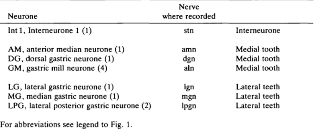
Table 1. Neurones of the gastric mill network, indicating interneurones, medial and
lateral tooth motor neurones, the number (in parentheses) of each neuronal type in the circuit, and the nerves in which the action potentials of the various neurones were
recorded extracellularly in these experiments
Nerve Neurone where recorded Intl, Interneurone 1 (1)
AM, anterior median neurone (1) DG, dorsal gastric neurone (1) GM, gastric mill neurone (4)
LG, lateral gastric neurone (1) MG, median gastric neurone (1)
LPG, lateral posterior gastric neurone (2)
For abbreviations see legend to Fig. 1.
placed around the COGs to allow separate supervision. Synaptic activity in the COGs was blocked by superfusing them with saline in which Ca2+ was replaced with Mg2*, and 2 4 m m o i r1 Co2+ or Mn2+ was added.
Intracellular recordings were made from neuronal somata using glass microelec-trodes (resistances 10-20MQ with thin-walled glass) filled with 3moll"1 KC1 or 2moll"1 potassium acetate. Current was injected into neurones through the recording electrodes via a bridge circuit in the World Precision Instruments M707 amplifier. Extracellular recordings were made using platinum wire electrodes, as previously described (see Moulins & Nagy, 1981). Data were recorded directly onto a Gould ES1000 recorder or photographed from the stored image on a Tektronix 5113 oscilloscope screen.
Results
The gastric mill pattern generator in Palinurus vulgaris
The neuronal network in the stomatogastric nervous system which controls the teeth of the gastric mill has been extensively studied in the spiny lobster Panulirus
interruptus (Mulloney & Selverston, 1974a,£>; Selverston & Mulloney, 1974;
Hartline & Maynard, 1975; Selverston et al. 1976; Russell, 1985a,b). Recent studies (e.g. Hartline & Russell, 1984; Russell & Hartline, 1984) have shown that most of the neurones in the gastric mill network possess, at least to some extent, membrane properties which allow them to produce 'plateau potentials' and thus give them a certain amount of 'burstiness'. Nonetheless, the synaptic relationships within the network [particularly the reciprocal inhibitions, notably between the lateral gastric (LG) neurone and interneurone 1 (Int 1); Fig. IB] appear to be of paramount importance in determining the rhythm. Although the network in the stomatogastric ganglion (STG) alone may be capable of producing a gastric rhythm, rhythmic inputs from neurones in the commissural ganglia (COGs) are also important in generation of the gastric rhythm. In the red lobster, Palinurus
vulgaris, the pattern of motor output from the gastric mill network (Fig. 2), the
synaptic relationships within the network (Fig. IB), the extrinsic inputs to the network (Fig. 1C) and the intrinsic properties of the gastric neurones appear, with a few exceptions, to be quite similar to those previously described in Panulirus
interruptus. It must, however, be noted that none of the synaptic connections has
been extensively tested for monosynapticity; the synaptic relationships shown are based solely on responses observed in the various neurones in normal saline.
The gastric mill network can be considered as two subsystems of motor neurones; one controls the two lateral teeth, the other controls the single medial tooth of the gastric mill (Fig. IB). The primary linkage between the two subsystems is the single interneurone, I n t l , although electrical coupling between the LG and GM (gastric mill) motor neurones also plays a role.
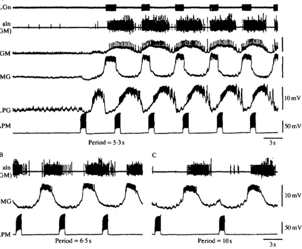
We have observed a number of variants of the gastric rhythm in Palinurus, the most common of which is shown in Fig. 2. The overall period of the rhythm was approximately 5-6 s. The 'power stroke' and 'return stroke' neurones in each subsystem discharged alternately and the two subsystems were slightly out of phase with one another. Activity in the power stroke neurones of the lateral teeth (MG and LG) was in antiphase with activity in the two lateral posterior gastric (LPG) neurones, which control the return stroke of the lateral teeth (see Fig. 2A,
LGn
DGn
AMn
dlvn (LPG-GM)
DG
LPG
10 mV
10 mV
10 mv
Ai
LPG-Aii
000*1
10 mV
2mV
0-2s
Bi
2mV
LG-
11U
10 mV0-2 s
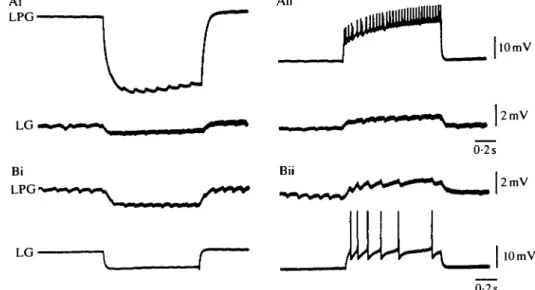
Fig. 3. LPG and LG are electrically coupled. Injecting hyperpolarizing (Ai) or depolarizing (Aii) current in LPG causes hyperpolarization or depolarization, respectively, of LG. Conversely, hyperpolarizing (Bi) or depolarizing (Bii) current injected in LG causes hyperpolarization or depolarization, respectively, of LPG. Action potentials in LG also provoke discrete IPSPs in LPG (Bii).
LG vs LPG and Fig. 2B, LG and MG vs LPG). Although the activity in LG and MG was nearly in phase (Fig. 2B) as a result of their electrical coupling (Fig. IB), the LG motor neurone began its discharge slightly (about 0-5 s) before the MG motor neurone. This contrasts with the case in Panulirus, in which MG generally begins firing slightly before LG (Mulloney & Selverston, 1974a; Russell, 1985a; Selverston, 1987). It should be noted, however, that the phase of LG with respect to all the other neurones of the gastric mill network is somewhat variable and can be controlled by modulatory inputs (Nagy, Dickinson & Moulins, 1987). The alternation of LG-MG and LPG resulted primarily from reciprocal inhibitory synapses between the power stroke and return stroke motor neurones (Fig. IB; see also Fig. 3Bii). In addition, however, the LG-LPG synapse is mixed; these two neurones are also electrically coupled, as can be seen in Fig. 3A,B. This electrical coupling has not been reported in Panulirus (Mulloney, 1987).
Discharges in the neurones of the two subsystems were slightly out of phase, with lateral tooth activity preceding medial tooth activity by several hundred milliseconds. This can be seen clearly by comparing the power stroke neurones LG and GM (Fig. 2A,B), as well as those in the return stroke, in which LPG leads DG-AM (Fig. 2A,B). This delay results, at least in part, from the complex interactions of the two subsystems with I n t l (Fig. IB; Selverston etal. 1976; Russell, 19856; see also Nagy etal. 1987).
In addition, input from two types of interneurones in the commissural ganglia (COGs) plays an important role in the overall determination of the gastric rhythm (Fig. 1C). First, the P cells (Selverston etal. 1976; Selverston & Miller, 1980) synapse onto the return stroke motor neurones of both systems (i.e. DG, AM, LPG; see Fig. 1C). The EPSPs from the P cells follow the pyloric rhythm and are one of the elements responsible for the pyloric modulation of the gastric rhythm in
Panulirus (Selverston etal. 1976), a modulation which is also seen in Palinurus.
Second, the E neurones in Panulirus (Russell, 1976; Selverston etal. 1976) excite the GM, LG, MG and LPG neurones and are in turn inhibited by Int 1; their EPSP volleys therefore follow the pyloric rhythm itself. The CG neurones in Homarus (Robertson & Moulins, 1981, 1984) are very similar. They synapse onto the same neurones; in addition, they appear to be endogenous bursters forming part of the commissural gastric oscillator, which is implicated in the generation of the gastric rhythm (Robertson & Moulins, 1981). It has been suggested that the CG and E cells are in fact homologous (Nagy & Moulins, 1987). In Palinurus, the GM, MG, LG and LPG neurones receive volleys of simultaneous EPSPs, which come from commissural neurones. The projections of these commissural neurones are thus identical to those of the CG neurones in Homarus. Moreover, these EPSPs, like those from the CG neurones, can be silenced by the firing of Intl. Although we have not recorded directly from such commissural neurones in Palinurus, we shall assume that the cells responsible for the rhythmic EPSP volleys in this species are also the CG neurones, as shown in Fig. 1C. An additional line of evidence suggests that this is the case. In Homarus, spikes in the CG neurones, and corresponding EPSPs in the gastric motor neurones, are triggered one-for-one by spikes in the anterior gastric receptor neurone (AGR), a sensory neurone located in the STG (Simmers, 1987; Simmers & Moulins, 1987). AGR also exists in Palinurus. As in
Homarus, each AGR spike was found to provoke the commissural-derived EPSP
in the GM, LG and LPG neurones (MG not tested; F. Nagy, unpublished observations), corroborating the suggestion that the neurones reponsible for these EPSPs are also the CG neurones.
> E
5?
s
stomatogastric ganglion (Dickinson & Nagy, 1983; Nagy & Dickinson, 1983). The previously demonstrated modulatory effects of APM, in particular an increase in the frequency of the pyloric rhythm and in the intensity of activity in the pyloric neurones, especially the constrictor motor neurones, can be seen in the vlvn recording in Fig. 4B. This modulation began after a delay of a few seconds (see Fig. 4B) and lasted for up to a minute after a 2-5 s discharge of APM. The time course of APM's modulations of the two networks, pyloric and gastric, can be seen clearly in Fig. 4B (compare vlvn with aln and LGn).
Modulation of the gastric mill CPG by the anterior pyloric modulator neurone (APM)
Induction and activation of the gastric rhythm
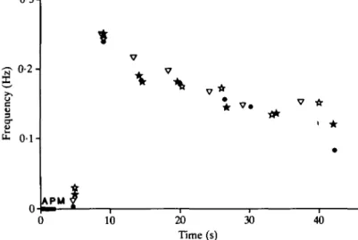
When the gastric mill network was silent, a brief (2-6 s) discharge of APM could turn on the entire gastric rhythm (Fig. 5A,B). This effect had a relatively long latency, as the first neurone to be activated (LPG) did not begin firing for approximately 2 s after the onset of APM activity, while APM's action potentials reached the STG within 100 ms (see Fig. 11). The duration of the activity induced by APM was considerably longer than the duration of the APM discharge; here a 2-s discharge (Figs 5B, 6) provoked 40-45 s of gastric rhythm. The time course of the increased gastric rhythm is shown graphically in Fig. 6, in which the frequency of the gastric rhythm is plotted over time after a 2-s discharge of APM. Both the long latency and the long duration of APM's induction of the gastric rhythm are evident. Furthermore, the gastric rhythm induced by APM was complete: all the neurones of the network were activated (Fig. 5A,B; MG and I n t l not shown).
When a gastric rhythm was present before an APM discharge, APM further activated the system (Fig. 5C). The overall frequency of the rhythm increased, the amplitude of oscillations increased in most of the neurones (e.g. see DG, GM in Fig. 5C) resulting in an increased intensity of the bursts in these motor neurones (again, see DG and GM in Fig. 5C), and neurones which were previously silent began to fire (see LG in Fig. 5C). In the case shown here, the network was relatively inactive before the APM discharge. In cases in which it was more active, similar changes were seen, although the evolution of action potential frequency in LG was unusual and could in some cases decrease. This phenomenon is considered more fully elsewhere (Nagy etal. 1987).
APM 10 mV 2s
dlvn (GM-LPG)
AMn
LG*w
APM
1 I
GM
A P M
2s
-10 mV
Fig. 5
0-3-
0-2-u
3
o-i H
10 20
Time (s)
30 40
Fig. 6. APM's activation of the gastric rhythm is long-lasting. Frequency of the gastric rhythm is plotted as a function of time after a 2s APM discharge (bar; elicited by current injection) for four trials in the same preparation (four symbols on graph). Frequency was calculated as the inverse of the period of bursts in the GM neurone; each value for frequency is plotted at the time of the start of the second burst used in that calculation. The frequency of the first burst, which occurred at nearly the same time after the APM burst in all four trials, is therefore zero. In all cases, the gastric rhythm continued for 40s after the APM burst.
Control by tonic and rhythmic activity of APM
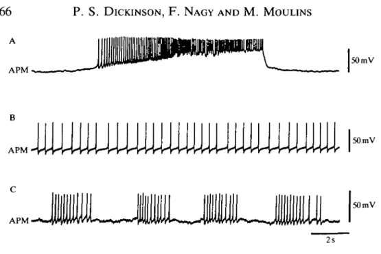
APM could spontaneously fire in bursts similar to those seen in Fig. 5 (Fig. 7A) or it could fire tonically (Fig. 7B). In addition, it could fire spontaneously in repeated bursts (Fig. 7C), thus providing a rhythmic input to the gastric network. All these patterns of spontaneous activity led to increased activity of the gastric mill network.
As is the case with a single burst in APM, tonic activity could induce a gastric rhythm in a previously silent system (Fig. 8). In this case, the intensity of the bursts in the motor neurones was a function of the frequency of firing in APM (shown here for LG and GM, Fig. 8B,C,D). The frequency of the overall rhythm also increased with increased APM frequency, but to a much smaller extent. Here an
APM
50 mV
50mV
APM
50 mV
2s
Fig. 7. Patterns of spontaneous activity in APM. APM can fire (A) in single bursts, (B) tonically or (C) in repetitive short bursts.
increase from 5 to 15 Hz in APM caused the frequency of the gastric bursts to increase by 14 %; i.e. the period decreased from 4-35 s to 3-85 s.
When APM fired spontaneously in repeated bursts or when this firing pattern was simulated by repeated depolarizations, a gastric rhythm was again induced (Figs 9,10A). Fig. 9 shows that when APM was bursting spontaneously (Fig. 9A), silencing it by hyperpolarization caused a cessation of the gastric rhythm (Fig. 9B). This indicates that spontaneous rhythmic activity in APM is, in itself, capable of driving a gastric rhythm. Further, whether APM fired in bursts spontaneously or was driven experimentally, the frequency of the induced gastric rhythm appeared to be the same as that of the APM bursts. To determine whether APM could entrain the gastric rhythm, we depolarized APM at different burst frequencies, and found that the gastric rhythm effectively followed the rhythm of APM bursts (Fig. 10). Furthermore, the phase of the gastric bursts in the period of APM [seen here in MG, for example, and calculated as (latency to the onset of the MG burst)/(period of APM)] varied as a function of that period (for MG, period = 5-3 s, phase = 0-79; period = 6-5 s, phase = 0-57; period = 10s, phase = 0-45), indicating that APM is not simply phasically activating the system, but is truly entraining the gastric oscillator.
CO
o
< -I o
5
0-LG n DG n
'P
'It'l'tH
M
"I
111!
1 In t 1 APM . 2 0 m V 2 s Fig . 9 . Spontaneou s rhythmi c activit y i n AP M ca n driv e th e gastri c rhythm . (A ) Whe n AP M fires rhythmically , a gastri c rhyth m whos e frequenc y matche s tha t o f AP M i s recorde d (her e i n LG , DG , G M an d In t 1) . (B ) Whe n th e spontaneou s activit y i n AP M i s terminate d b y hyperpolarization , th e gastri c rhyth m cease s an d Int l fires tonically .10 mV
50 mV
APM
Period = 6-5 s
Fig. 10. APM can entrain the gastric mill rhythm. (A) Rhythmic bursting activity in APM (with a period of 5-3 s, produced by current injection) initiates and drives activity of the gastric mill network. Gastric bursts follow APM bursts one-for-one. Note that the LPG neurone is the first to begin firing, as is the case when APM is made to fire tonically or in single longer bursts. (B,C) As the period of APM bursts is increased (to 6-5 s in B, 10 s in C), the gastric rhythm follows, retaining one-for-one bursting. However, the phase of the gastric neurones (see MG, GM) in the period of APM is a function of the APM period (for calculations see text).
10 mV
50 mV
Mechanisms underlying APM's modulation of the gastric rhythm Synaptic projections of APM
AM DG GM LG MG LPG
d,n d,n
X
n d
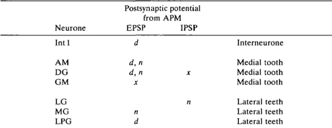
Table 2. Postsynaptic potentials from APM which were recorded in the gastric mill neurones
Postsynaptic potential from APM Neurone EPSP IPSP
Int 1 d Interneurone
Medial tooth x Medial tooth Medial tooth
n Lateral teeth Lateral teeth Lateral teeth
Synapses are indicated as d, for direct (not blocked by blocking COG synapses); n, for not direct (blocked when COG synapses are blocked); and x, for present, but unknown whether direct or not direct.
AM, and with MG, LG and GM starting last, generally after the end of the APM burst (see Fig. 5A,B).
In each of the gastric neurones, it was possible to record discrete synaptic events associated with APM spikes (Table 2). Because APM's axons cross the commissu-ral ganglia (Fig. 11A), it is possible that some of these synaptic events were due to commissural neurones activated by APM. To test this possibility, we experimen-tally blocked synapses in the commissural ganglia by superfusing the COGs with 0 C a2 + + Co2 + saline (see Materials and methods). Some of the synaptic events associated with APM action potentials were clearly not the result of APM synapses on the motor neurones in the STG, because they were blocked by this treatment. However, even these indirect synaptic potentials (marked n, for not direct, in Table 2) followed APM spikes one-for-one. Other synaptic potentials were not blocked when all synaptic activity in the COGs was blocked and are thus
APM
nG Int 1
APM
D DG
LPG
APM
APM
3 Hz
F MG
APM
Giii
5 Hz
Fig. 11
|2mV
20 mV
40 ms
2mV
|20mV 7 Hz
considered to be direct; since no neurones in the STG are known to produce such synaptic potentials, it seems likely that these direct events are in fact monosynap-tic. We cannot, however, rule out the possibility that other neurones in the COGs are activated by APM via electrotonic synapses and that these neurones in turn act on the gastric neurones.
APM provoked a direct EPSP in Intl (Fig. 11B). In this recording, in which synaptic activity in the COGs was blocked, each EPSP was sufficient to cause an action potential in Int 1.
In both of the return stroke neurones of the medial tooth, DG and AM, APM provoked both a direct EPSP and an indirect EPSP. Under normal conditions, an APM spike evoked a large, apparently double EPSP (Fig. 11C); when commissu-ral ganglion synapses were blocked, an EPSP remained, but it was now single and considerably smaller (Fig. 11D). In DG, this synaptic event was even more complex, for it was followed by a slower inhibitory phase. This is not shown in Fig. 11C or D, but it appeared clearly when APM fired repetitively (Fig. 11G). The inhibition was most visible when APM fired at higher frequency (e.g. 7 Hz in the example shown), and thus appears to be a function of spike frequency in APM. It is not yet clear whether this inhibition is direct or indirect. When APM fired a burst at relatively high frequency, such as that shown in Fig. 5B, it was clear that DG was transiently excited and subsequently inhibited, the inhibition lasting until the end of the APM burst.
In GM, the power stroke neurone of the medial tooth, APM also evoked an EPSP (Fig. H E ) . It is not clear whether this EPSP is direct or indirect because, when the COGs were blocked, GM was bombarded by IPSPs from I n t l , which fired tonically under these conditions. These IPSPs were much larger than the EPSPs coming from APM, so we could not tell whether the EPSP was still present. Although GM was excited by APM, it was always amongst the last of the gastric neurones to begin firing after an APM discharge (see Fig. 5A,B). Inhibition from Int 1 (itself activated by APM) apparently was sufficient in this case to mask the excitation from APM.
Although LG and MG are frequently considered together in the gastric circuit and they are both involved in the power stroke of the lateral teeth, APM provoked an IPSP in LG (Fig. 11C) and an EPSP in MG (Fig. 11F). Both of these PSPs were indirect. When APM was excited, LG was inhibited both by Int 1 and by APM itself, and thus fired very late in the sequence. MG, though excited by APM, was also inhibited by Int 1 and by the electrical coupling with LG, and thus it, too, fired only after the end of the APM burst.
The LPG neurone (lateral tooth return stroke) was seen to receive a direct EPSP from APM. This, coupled with the fact that the only neurone within the gastric network which inhibits LPG is LG (which was silent), explains the observation that LPG was always the first to fire when APM induced the gastric rhythm.
long-term activation of rhythmic activity in all the gastric neurones. Instead, this appears to be due to effects of APM on at least two additional factors involved in the production of the gastric rhythm: plateau properties in the gastric neurones and extrinsic excitatory inputs to the gastric network.
Induction of plateau properties in gastric motor neurones
It has been shown that most neurones in the gastric network are capable under some conditions of producing plateau potentials (Russell & Hartline, 1984). We first examined the effects of APM on MG, a neurone known to possess the regenerative membrane properties which underlie plateau potentials. Like the other gastric neurones, MG did not display these regenerative membrane properties when the network was silent. In this case, APM could induce such properties. Before activity was induced in APM, the MG neurone responded passively to a brief pulse of depolarizing current (Fig. 12). When APM fired, the same pulses injected into MG produced long regenerative depolarizations which held its membrane potential above threshold, generating bursts of action potentials (Fig. 12). The APM neurone thus induced plateau properties in MG and thereby increased MG's firing when APM was active.
APM was likewise able to induce plateau properties in the lateral tooth neurone LPG (Fig. 13). Again, when the gastric rhythm and APM were both silent, LPG responded passively to the injection of a depolarizing current pulse (Fig. 13Ai), but produced a plateau potential in response to the same size (or a smaller) current pulse during activity in APM (Fig. 13Aii). (In the case shown, APM was induced to fire in repetitive bursts.) Because the presence of plateau properties in LPG has not previously been demonstrated (see Selverston, 1987, for a review), we examined this phenomenon more thoroughly. Two additional lines of evidence confirmed that the induction of plateau properties by APM was at least partially responsible for the increased rhythmic firing of LPG when APM was active. First, when APM fired, the membrane potential of the previously silent LPG neurone (Fig. 13Bi) began to oscillate rhythmically, producing rhythmic bursts of action potentials (Fig. 13Bii). Hyperpolarization of LPG increased the amplitude and decreased the duration of these depolarizations (Fig. 13Biii,iv), indicating that these were endogenous plateaus (Russell & Hartline, 1984) which had been unmasked by APM. Second, under the same conditions, when APM was firing, it was possible to advance the beginning of these plateaus by the injection of brief depolarizing current pulses (Fig. 13C). Thus, the modulatory neurone APM can unmask or induce regenerative plateau potentials even in the LPG neurone, which does not commonly exhibit such properties.
L
_
/{/•PWI
i
J~
L
J
T
IT
lOm V 2n A APM -I s Fig . 12 . AP M induce s platea u propertie s i n th e M G neurone . Befor e th e AP M discharge , M G respond s passivel y t o a 25 0 m s puls e o f depolarizin g curren t (IMG) Afte r AP M ha s fired a t 1 8 H z fo r 6-8 s (drive n b y cunen t injection) , th e sam e depolarizin g pulse s caus e platea u potential s i n MG . Th e onse t o f thi s inductio n i s slow ; th e puls e i n M G delivere d afte r 2 s o f AP M activit y stil l faile d t o produc e a platea u potential .Ai Aii
10 mV
| 3 n A
A P M '
n
2s
Bi Bii
APM
• • • • • •
10 mV
50 mV
OnA OnA - 0 - 5 nA - l - 5 n A
2s
Fig. 13. APM induces plateau properties in the LPG neurone. (A) Before an APM discharge, LPG responds passively to the injection of a 150 ms depolarizing current pulse (3-2 nA in Ai); during APM activity (repetitive bursts in the case shown) the same or a smaller (1-0 nA in Aii) current pulse provokes a plateau potential in LPG. (B) When the gastric network is silent (Bi), activity in APM leads to rhythmic bursts of firing in LPG (Bii-Biv). The depolarizations which underlie LPG activity during APM activity increase in amplitude and decrease in duration when LPG is progressively hyperpolarized (by 0-5 nA in Biii, by 1-5 nA in Biv). (C) During APM activity (trace not shown), the depolarizing waves in LPG can be advanced by injecting depolarizing current pulses (400 ms, 2 nA) into LPG. The expected times of the start of LPG bursts are marked with triangles.
lOmV
Activation of extrinsic inputs to the gastric network
APM appears to activate excitatory synaptic input to the gastric network from the commissural ganglia; activity in both the P cells and the CG neurones, monitored indirectly via their EPSPs on gastric neurones, was enhanced.
In most cases, the AM neurone was constantly bombarded by trains of EPSPs which followed the pyloric rhythm (Fig. 14). Previous studies (Selverston etal. 1976; Selverston & Miller, 1980) have shown that these come from the P cells in the COGs. A burst of action potentials in APM provoked a strong and long-lasting activation of these EPSPs (Fig. 14A,B). At the beginning of an APM discharge the P cells appeared to be inhibited but, after 1-2 s, the EPSPs from the P cells reappeared in distinct bursts. Both the frequency of the bursts and the frequency
APM
Ci
DG
5mV
|20mV
2s
Cii
APM-Di AMJ
APM'
\
5mV
|20mV
Is
Dii
20mV
of the EPSPs within each burst were considerably increased. It has been shown (Selverston et al. 1976) that the P cell burst frequency is determined largely by feedback from the anterior burster (AB) neurone in the pyloric network. Since the pyloric network is also activated by APM (Nagy & Dickinson, 1983), the increased frequency of P cell bursts is likely to be an indirect effect of this feedback loop. However, the frequency of EPSPs within each P cell burst is not controlled by such feedback, and therefore is probably a more direct effect of APM, one which may occur within the COGs, where the P cells are located.
Activation of the P cells in turn activated the gastric neurones onto which they synapse (i.e. AM, DG and I n t l ; Fig. 14A), as is shown for AM in Fig. 14B and DG in Fig. 14C. When APM was prevented from firing by the injection of hyperpolarizing current, EPSPs derived from the P cells occurred at low frequency (Fig. 14Di). When the hyperpolarization was released and APM was allowed to fire spontaneously in bursts, an overall activation of the P cells was once again seen as an increase in the frequency of the EPSPs (Fig. 14Dii). In this case, the combination of the transient inhibition and the longer-lasting excitation by APM led to a double rhythmicity in the P cells; they followed both the pyloric and gastric rhythms. This, in turn, could divide the gastric bursts of AM and DG into pyloric sub-bursts (Fig. 14Cii; see also Fig. 5B for DG).
The other known commissural inputs to the gastric network, the CG neurones, were also activated by APM. When the gastric network was silent, the GM neurone was constantly bombarded by EPSPs which occurred synchronously with EPSPs in the LG, MG and LPG neurones. As discussed above, the only commissural neurones known to synapse on all these gastric neurones are the CGs (Fig. 15A); we therefore assume that the EPSPs recorded in these gastric neurones can be used as indicators of CG activity. These EPSPs in a silent gastric network could occur either in bursts or tonically (see Fig. 15). When APM fired, either in a single burst or in repetitive bursts, the frequency of the EPSPs from the
B
LPG
GM
MG
Di
APM-Ei
GM'
APM-A
Ci
|2mV
|2mV Cii
GM
|2mV
0-2s LG
Dii
|2mV
|2mV
0 1 s
|2mV
|50mV
Is
|2mV
ULJLJLJ
50mVCG neurones increased considerably (compare Fig. 15Ci and Cii, Di and Dii, Ei and Eii). Furthermore, when APM induced rhythmic activity in the gastric network, the volleys of EPSPs from the CG neurone were also rhythmic (Fig. 15Eii). It is conceivable that the induction of rhythmicity in the CG neurones was indirect, due to inhibitory rhythmic feedback from the gastric network via I n t l (see Fig. IB; see also Russell, 1976). However, the strong increase in firing frequency of the CG neurones (as seen in the high frequency of EPSPs recorded in GM) could not occur via such feedback, but must instead be the result of an effect of APM in the commissural ganglia. This activation of the CG neurones by APM could result in a considerable increase in the activity of the neurones they excite, notably GM.
APM thus induces rhythmic activity in the gastric network using at least three mechanisms: (1) it provokes one-for-one PSPs, either direct or indirect, in all the neurones of the gastric mill network; (2) it induces or unmasks plateau properties in at least two neurones, the MG and LPG motor neurones; (3) it activates the two known extrinsic excitatory inputs to the gastric network, the P cells and the CG neurones. The latter two effects are both long-lasting and apparently underhe the long-term activation of the gastric network by APM.
Discussion
APM modulates the gastric mill central pattern generator
We have shown here that an identified modulatory neurone, known to alter the rhythmic output of a central pattern generator in decapod crustaceans, can also activate and modulate a second pattern generator. This neurone, the anterior pyloric modulator (APM), modulates the output of the pyloric pattern generator of the stomatogastric nervous system by inducing and altering the regenerative membrane properties, or plateau properties, of the neurones which make up the
pattern generator (Dickinson & Nagy, 1983; Nagy & Dickinson, 1983). We have shown here that the same neurone can provoke rhythmic activity in a silent gastric mill network and that it can increase the level of gastric activity in an already oscillating gastric network. Furthermore, input from the APM neurone can entrain the cycling gastric oscillator.
Activation of the gastric mill rhythm by APM occurs with a long latency (several seconds) and considerably outlasts the duration of APM's discharge. Such a slow onset and long time course are typical of neuromodulation (Kupfermann, 1979). Similar 'slow modulating synaptic actions' (Kandel etal. 1987), which have been increasingly documented in recent years (Harris-Warrick, 1987; Kaczmarek & Levitan, 1987), have been shown to play a role in a variety of phenomena, including the presynaptic control of transmitter release underlying certain forms of learning (Kandel etal. 1987), the control of neuronal excitability (Hartzell, 1981; Adams, Brown & Constanti, 1982; Nicoll, 1982), the control of bursting properties in neurones (Barker & Gainer, 1974; Wilson & Wachtel, 1978; Russell & Hartline, 1982; Dickinson & Nagy, 1983; Adams & Benson, 1985) and the control of myogenic activity in muscles (Evans & O'Shea, 1978; Benson, Sullivan, Watson & Augustine, 1981; Calabrese & Maranto, 1984; Calabrese & Arbas, 1985; Meyrand & Moulins, 1986). However, relatively few identified neurones have been shown to modulate the activity of an entire group of neurones involved in motor pattern generation. In the leech, the serotonergic Retzius cells are known to modulate the swim CPG (Kristan & Nusbaum, 1983) and the heartbeat CPG (Calabrese & Arbas, 1985). However, the mechanisms of action at the cellular level are not yet well-established in either of these cases. In Aplysia, neurones involved in the control of feeding are modulated by the metacerebral cells (Weiss et al. 1981) and by the C2 interneurone (Chiel etal. 1986; Weiss etal. 1986a; Weiss, Chiel & Kupfermann, 19866). The mechanisms by which these modulations take place are well-understood, but the organization of the network controlling the behaviour is still relatively unknown. Because the pyloric and gastric mill CPGs are fairly well-understood (see Selverston & Moulins, 1987) and the resulting behaviour patterns can now be analysed in the intact animal (Heinzel, 1987), studies of the modulation of the pyloric and gastric networks by APM may be fruitful. In particular, a comparison of the mechanisms used by APM in modulating the pyloric and gastric rhythms may enable us to shed light on the relationships between the mechanisms of modulation and the functional results of that modulation.
APM simultaneously alters several building blocks underlying gastric mill pattern generation
One way of considering the mechanisms that underlie pattern generation is as building blocks, which can be put together in different ways to generate various motor outputs (Getting, 1987). Several such building blocks are thought to contribute to the generation of the gastric mill motor pattern: (1) the synaptic relationships within the network (Mulloney & Selverston, 1974a,b; Selverston & Mulloney, 1974; Selverston etal. 1976), (2) plateau properties in some of the gastric neurones (Russell, 1985ft; Russell & Hartline, 1978,1984) and (3) rhythmic volleys of EPSPs from the commissural CG neurones (Robertson & Moulins, 1981, 1984). We have shown here that APM directly alters the latter two building blocks. These alterations can, in turn, alter the functional expression of the first (i.e. the synaptic relationships within the network; Nagy et al. 1987). In addition, APM provokes one-for-one EPSPs in all the gastric neurones; these might be considered as another building block involved in generating the gastric rhythm.
The role of the one-for-one PSPs mediated by APM cannot be in long-term modulation of the gastric rhythm, for both the direct and the indirect one-for-one PSPs, unlike other indirect PSPs, occur only during the firing of APM. There remain at least two possibilities as to the function of these PSPs in the overall effects of APM on the gastric mill network. First, together with the synaptic interactions within the network itself, they determine the order in which the gastric neurones start to fire when APM activates the rhythm. This order is constant (see Figs 5,10), with LPG always starting first, followed by DG and AM. The power stroke neurones MG, LG and GM fire at nearly the same time and are last in the sequence. Consequently, whenever the rhythm is turned on by APM, it will start with the same sequence of movements: opening of the lateral teeth (due to firing of LPG) followed by retraction of the medial tooth (due to DG and AM). The return strokes of both the lateral and medial teeth will thus precede any power strokes. Functionally, this may be of considerable importance.
The second possible role of the one-for-one PSPs is in the entrainment of the rhythm by APM. We have shown here that spontaneous bursting in APM does occur in isolated preparations, and that this bursting activity entrains the gastric rhythm. We have not examined this role of the APM-induced PSPs in detail, but have noted, for example, that when APM drives the rhythm at a high frequency its firing can prematurely terminate activity in LG (via the APM-to-LG IPSP). This, together with the activation of I n t l , may contribute to the entrainment process. The other one-for-one PSPs, by providing a discrete temporal signal, are also probably important. In this context, it is interesting to note that APM, which does not appear to produce discrete PSPs in the pyloric neurones (Nagy & Dickinson, 1983), does not entrain that rhythm.
properties were readily inducible in some of the neurones, but were apparently absent in others, suggesting that the membrane properties of certain neurones might allow them a relatively more important role in generating the rhythm (Russell & Hartline, 1984). We have shown in this paper and elsewhere (Nagy
etal. 1987) that APM can selectively control these properties in some gastric
neurones and, therefore, may alter therelative importances of these neurones in determining the final output of the gastric CPG.
APM alters the plateau properties of at least three gastric neurones. First, APM's discharge can induce plateau properties in the MG neurone. Previous studies have shown that plateau properties can be induced in MG by input from premotor centres (Russell & Hartline, 1984); APM appears to be one such input. Second, APM can suppress plateau properties in the LG neurone (Nagy etal. 1987). Previous studies have shown that the plateau properties of LG, like those of MG, can be modulated (Russell & Hartline, 1984). In Palinurus, LG's discharge has two components, one of which is due primarily to LG's plateau properties. Suppression of this endogenous component of LG's discharge by APM leads to extensive and long-lasting changes in the phase relationships of neuronal dis-charges in both the lateral and medial tooth subsystems (Nagy et al. 1987). Such a suppression of plateau properties is not common in the stomatogastric nervous system; it has been reported only in the present case and in the control of bursting pacemaker potentials in three pyloric neurones by the pyloric suppressor neurone (Cazalets etal. 1987). Third, we have shown here that APM induces plateau properties in the LPG neurone, which normally does not exhibit them (Russell & Hartline, 1984; Selverston, 1987). That LPG's plateau properties were not seen in previous studies suggests the possibility that all the gastric neurones (with the possible exception of the GMs) might be capable, given the correct conditioning input, of exhibiting regenerative membrane properties, and thus of assuming a more important role in rhythm generation.
Finally, APM modulates the gastric network by activating both of its known extrinsic inputs, the P cells and the CG neurones. Our evidence suggests that APM activates these inputs both indirectly via feedback from the stomatogastric CPGs (AB for P cells; Int 1 for CG neurones) and, more directly, by acting at a premotor level in the commissural ganglia.
APM thus controls motor pattern generation at both the motor and premotor levels, which is to some extent reminiscent of the modulation of feeding by the metacerebral cells and the C2 neurone in Aplysia (Chiel etal. 1986; Weiss etal. 1986a). These neurones also act on both premotor (pattern-generating) and motor neurones. In contrast to the Aplysia neurones, however, APM does not appear to modulate muscle activity directly (unpublished observations).
and is long-lasting, which would be consistent with an enhancement of their regenerative properties.
Although both the P cells and the CG neurones synapse on a number of gastric neurones, neither of them synapses on all the gastric neurones. As a result, when APM activates the P and CG neurones, it indirectly activates a particular subset of the gastric neurones. By so doing, it not only activates the gastric rhythm itself, but may also alter the functional circuit for generating that rhythm, and thus modify the expression of the gastric pattern.
APM modulates several functionally related pattern generators
The modulatory effects of APM on both the pyloric and the gastric network have been examined in considerable detail (Nagy & Dickinson, 1983; Dickinson & Nagy, 1983; Nagy et al. 1987; present paper). In addition, it appears likely that APM also modulates the cardiac sac network, which controls the cardiac sac, or storage component of the stomach. We have observed (unpublished observations) that in Palinurus vulgaris, APM synapses both directly and indirectly onto one of the cardiac sac dilator neurones, CD2 (Vedel & Moulins, 1977, 1978). Bursts in APM thereby drive bursts in CD2. Furthermore, at least in the related species
Jasus lalandii, bursts in APM can provoke entire bursts of the cardiac sac rhythm
(F. Nagy & P. Cardi, unpublished observations), suggesting that APM exerts some control over this part of the lobster stomach as well as over the gastric mill and the pylorus.
By acting simultaneously on several distinct but functionally related CPGs, APM might control an entire concert of related behavioural sequences. This is particularly interesting because it occurs in a system in which multiple and parallel modulatory inputs act upon each of the networks. It has been shown that such modulatory inputs converge extensively onto the pyloric network, where a number of transmitters alter the rhythm (e.g. proctolin, Hooper & Marder, 1984; Marder
et al. 1986; FMRFamide, Hooper & Marder, 1984; Marder, Calabrese, Nusbaum
& Trimmer, 1987; amines, Beltz et al. 1984; Flamm & Harris-Warrick, I986a,b; see also Harris-Warrick, 1987), and where three modulatory neurones (APM, Nagy & Dickinson, 1983; MPN, Nusbaum & Marder, 1987; PS, Cazalets etal. 1987) have been identified. Although it has been studied in less detail, it appears that a similar number of modulatory inputs converge on the gastric mill network, where it has been shown that the peptide proctolin can turn on and modulate the gastric rhythm (Heinzel, 1987; Heinzel & Selverston, 1985) and where there is some evidence for the existence of a neurone which can terminate the gastric rhythm (Turrigiano & Selverston, 1986). Therefore, in the stomatogastric nervous system, several related CPGs are under the control of both divergent and convergent modulatory pathways.
complete control over a complex behaviour pattern which is made up of several separable components.
We thank Eve Marder for critical reading of the manuscript. This work was supported by a grant (85C1152) from the Ministere de la Recherche et de la Technologie to MM, a Grant-in-Aid from the American Heart Association Maine Affiliate to PSD, a Bowdoin College grant to PSD, and a NATO travel grant (RG86/054O) to Eve Marder.
References
ADAMS, P. R., BROWN, D. A. & CONSTANTI, A. (1982). M-currents and other potassium currents
in bullfrog sympathetic neurones. J. Physioi, Lond. 330, 537-572.
ADAMS, W. B. & BENSON, J. A. (1985). The generation and modulation of endogenous
rhythmicity in the Aplysia bursting pacemaker neurone R15. Prog. Biophys. molec. Biol. 46, 1-49.
BARKER, J. L. & GAINER, H. (1974). Peptide regulation of bursting pacemaker activity in a
molluscan neurosecretory cell. Science 184, 1371-1373.
BELTZ, B., EISEN, J., FLAMM, R., HARRIS-WARRICK, R. M., HOOPER, S. L. & MARDER, E. (1984).
Serotonergic innervation and modulation of the stomatogastric ganglion of three decapod crustaceans (Panuiirus interruptus, Homarus americanus and Cancer irroratus). J. exp. Biol. 109, 35-54.
BENSON, J. A., SULUVAN, R. E., WATSON, W. H. & AUGUSTINE, G. J. (1981). The neuropeptide
proctolin acts directly on Limulus cardiac muscle to increase the amplitude of contraction.
Brain Res. 213, 449-454.
CALABRESE, R. L. & ARBAS, E. A. (1985). Modulation of central and peripheral rhythmicity in
the heartbeat system of the leech. In Model Neural Networks and Behavior (ed. A. I. Selverston), pp. 69-85. New York: Plenum Press.
CALABRESE, R. L. & MARANTO, A. R. (1984). Neural control of the hearts in the leech, Hirudo
medicinalis. III. Control of myogenicity and muscle tension by heart accessory neurons. J. comp. Physioi. 154, 393-406.
CAZALETS, J. R., NAGY, F. & MOULINS, M. (1987). Suppressive control of a rhythmic central
pattern generator by an identified modulatory neuron in Crustacea. Neurosci. Letts (in press).
CHIEL, H. J., WEISS, K. R. & KUPFERMANN, I. (1986). An identified histaminergic neuron
modulates feeding motor circuitry in Aplysia. J. Neurosci. 6, 2427-2450.
CLAIBORNE, B. J. & AVERS, J. (1987). Functional anatomy and behavior. In The Crustacean
Stomatogastric System (ed. A. I. Selverston & M. Moulins), pp. 9-30. New York:
Springer-Verlag.
DICKINSON, P. S. & NAGY, F. (1983). Control of a central pattern generator by an identified
modulatory interneurone in Crustacea. II. Induction and modification of plateau properties in pyloric neurones. J. exp. Biol. 105, 59-82.
EVANS, P. D. & O'SHEA, M. (1978). The identification of an octopaminergic neurone and the modulation of a myogenic rhythm in the locust. J. exp. Biol. 73, 235-260.
FLAMM, R. E. & FIARRIS-WARRICK, R. M. (1986<J). Aminergic modulation in the lobster
stomatogastric ganglion. I. Effects on the motor pattern and activity of neurons within the pyloric circuit. J. Neurophysiol. 55, 847-865.
FLAMM, R. E. & HARRIS-WARRICK, R. M. (19866). Aminergic modulation in the lobster
stomatogastric ganglion. II. Target neurons of dopamine, octopamine, and serotonin within the pyloric circuit. /. Neurophysiol. 55, 866-881.
GETTING, P. A. (1987). Comparative analysis of invertebrate central pattern generators. In
Neural Control of Rhythmic Movements (ed. A. H. Cohen, S. Rossignol & S. Grillner). New
York: John Wiley & Sons (in press).
GRILLNER, S. (1973). Locomotion in the spinal cat. In Control of Posture and Locomotion
HARRIS-WARRICK, R. M. (1987). Chemical modulation of central pattern generators. In Neural
Control of Rhythmic Movements (ed. A. H. Cohen, S. Rossignol & S. Grillner). New York:
John Wiley & Sons (in press).
HAKTLINE, D. K. & MAYNARD, D. M. (1975). Motor patterns in the stomatogastric ganglion of
the lobster Panulirus argus. J. exp. Biol. 62, 405-420.
HARTLINE, D. K. & RUSSELL, D. F. (1984). Endogenous burst capability in a neuron of the
gastric mill pattern generator of the spiny lobster Panulirus interruptus. J. Neurobiol. 15, 345-364.
HARTZELL, H. C. (1981). Mechanisms of slow post-synaptic potentials. Nature, Lond. 291,
539-544.
HEINZEL, H. G. (1987). Spontaneous and proctolin-induced modes of operation of the isolated
gastric oscillator and of the gastric mill in the intact animal. In The Crustacean Stomatogastric
System (ed. A. I. Selverston & M. Moulins), pp. 175-180. New York: Springer-Verlag.
HEINZEL, H. G. & SELVERSTON, A. I. (1985). Proctolin modulation of the gastric oscillator in the
lobster stomatogastric ganglion. Neurosci. Abstr. 11, 478.
HOOPER, S. L. & MARDER, E. (1984). Modulation of-a central pattern generator by two
neuropeptides, proctolin and FMRFamide. Brain Res. 305,186-191.
KACZMAREK, L. K. & LEVTTAN, I. B. (1987). Neuromodulation: The Biochemical Control of
Neuronal Excitability. New York: Oxford University Press.
KANDEL, E. R., KLEIN, M., HOCHNER, B., SHUSTER, M., SIEGELBAUM, S. A., HAWKINS, R. D.,
GLAUZMAN, D. L., CASTELLUCCI, V. F. & ABRAMS, T. W. (1987). Synaptic modulation and
learning: new insights into synaptic transmission from the study of behavior. In Synaptic
Function (ed. G. M. Edelman, W. E. Gall & W. M. Cowan), pp. 471-518. New York: John
Wiley & Sons.
KRISTAN, W. B. & NUSBAUM, M. P. (1983). The dual role of serotonin in leech swimming.
J. Physiol., Paris 78, 743-747.
KRISTAN, W. B. & WEEKS, J. C. (1983). Neurons controlling the initiation, generation and
modulation of leech swimming. In Neural Origin of Rhythmic Movements. SEB Symposia 37 (ed. A. Roberts & B. L. Roberts), pp. 243-260. Cambridge, New York: Cambridge University Press.
KUPFERMANN, I. (1979). Modulatory actions of neurotransmitters. A. Rev. Neurosci. 2,447-465.
MARDER, E. (1987). Neurotransmitters and neuromodulators. In The Crustacean Stomatogastric
System (ed. A. I. Selverston & M. Moulins), pp. 263-300. New York: Springer-Verlag.
MARDER, E., CALABRESE, R. L., NUSBAUM, M. P. & TRIMMER, B. (1987). Distribution and partial
characterization of FMRFamide-like peptides in the stomatogastric nervous systems of the rock crab, Cancer borealis, and the spiny lobster, Panulirus interruptus. J. comp. Neurol. 259, 150-163.
MARDER, E. & HOOPER, S. L. (1985). Neurotransmitter modulation of the stomatogastric
ganglion of decapod crustaceans. In Model Neural Networks and Behavior (ed. A. I. Selverston), pp. 319-337. New York: Plenum Press.
MARDER, E., HOOPER, S. L. & SIWICKI, K. K. (1986). Modulatory action and distribution of the
neuropeptide proctolin in the crustacean stomatogastric nervous system. J. comp. Neurol. 243, 454-467.
MAYNARD, D. M. & DANDO, M. R. (1974). The structure of the stomatogastric neuromuscular
system in Callinectes sapidus, Homarus americanus, and Panulirus argus (Decapoda Crustacea). Phil. Trans. R. Soc. Ser. B 268, 161-220.
MEYRAND, P. & MOUUNS, M. (1986). Myogenic oscillatory activity in the pyloric rhythmic motor
system of Crustacea. /. comp. Physiol. A 158, 489-503.
MILLER, J. P. (1987). Pyloric mechanisms. In The Crustacean Stomatogastric System (ed. A. I.
Selverston & M. Moulins), pp. 109-136. New York: Springer-Verlag.
MILLER, J. P. & SELVERSTON, A. I. (1985). Neural mechanisms for the production of the lobster
pyloric motor pattern. In Model Neural Networks and Behavior (ed. A. I. Selverston), pp. 37-48. New York: Plenum Press.
MOULINS, M. & NAGY, F. (1981). Participation of an unpaired motor neurone in the bilaterally
organized oesophageal rhythm in the lobsters Jasus lalandii and Palinurus vulgaris. J. exp.
MULLONEY, B. (1987). Neural circuits. In The Crustacean Stomatogastric System (ed. A. I. Selverston & M. Moulins), pp. 57-75. New York: Springer-Verlag.
MULLONEY, B. & SELVERSTON, A. I. (1974a). Organization of the stomatogastric ganglion in the spiny lobster. I. Neurons driving the lateral teeth. J. comp. Physiol. 91, 1-32.
MULLONEY, B. & SELVERSTON, A. I. (19746). Organization of the stomatogastric ganglion of the spiny lobster. III. Coordination of the two subsets of the gastric system. J. comp. Physiol. 91, 53-78.
NAGY, F. & DICKINSON, P. S. (1983). Control of a central pattern generator by an identified modulatory interneurone in Crustacea. I. Modulation of the pyloric motor output. /. exp. Biol. 105, 167-173.
NAGY, F., DICKINSON, P. S. & MOULINS, M. (1987). Control by an identified modulatory neuron of the sequential expression of plateau properties of and synaptic inputs to a neuron in a central pattern generator. /. Neurosci. (in press).
NAGY, F. & MOULINS, M. (1987). Extrinsic inputs. In The Crustacean Stomatogastric System (ed. A. I. Selverston & M. Moulins), pp. 205-242. New York: Springer-Verlag.
NICOLL, R. A. (1982). Neurotransmitters can say more than just "yes" or "no". Trends Neurosci. 5, 369-374.
NUSBAUM, M. P. & MARDER, E. (1987). A newly identified modulatory proctolin-containing neuron (MP neuron) in the stomatogastric nervous system of the crab Cancer borealis. Neurosci. Abstr. 13, 1257.
NUSBAUM, M. P. & MARDER, E. (1988). A neuronal role for a crustacean red pigment concentrating hormone-like peptide: neuromodulation of the pyloric rhythm in the crab, Cancer borealis. J. exp. Biol. 135, 165-181.
O'SHEA, M. & SCHAFFER, M. (1985). Neuropeptide function: the invertebrate contribution. A. Rev. Neurosci. 8, 171-198.
ROBERTSON, R. M. & MOULINS, M. (1981). Control of rhythmic behaviour by a hierarchy of linked oscillators in Crustacea. Neurosci. Letts 21, 111-116.
ROBERTSON, R. M. & MOULINS, M. (1984). Oscillatory command input to the motor pattern generators of the crustacean stomatogastric ganglion. II. The gastric rhythm. J. comp. Physiol. 154,673-691.
RUSSELL, D. F. (1976). Rhythmic excitatory inputs to the lobster stomatogastric ganglion. Brain Res. 101, 598-602.
RUSSELL, D. F. (1985a). Pattern and reset analysis of the gastric mill rhythm in a spiny lobster, Panulirus interruptus. J. exp. Biol. 114, 71-98.
RUSSELL, D. F. (1985b). Neural basis of teeth coordination during gastric rhythms in spiny lobsters, Panulirus interruptus. J. exp. Biol. 114, 99-119.
RUSSELL, D. F. & HARTLINE, D. K. (1978). Bursting neural networks: A reexamination. Science 200, 453-456.
RUSSELL, D. F. & HARTLINE, D. K. (1982). Slow active potentials and bursting motor patterns in pyloric network of the lobster, Panulirus interruptus. J. Neurophysiol. 48, 914-937. RUSSELL, D. F. & HARTLINE, D. K. (1984). Synaptic regulation of cellular properties in burst
oscillations of neurons in gastric mill system of spiny lobster Panulirus interruptus. J. Neurophysiol. 52, 54-73.
SELVERSTON, A. I. (1987). Gastric mill mechanisms. In The Crustacean Stomatogastric System (ed. A. I. Selverston & M. Moulins), pp. 147-171. New York: Springer-Verlag.
SELVERSTON, A. I. & MILLER, J. P. (1980). Mechanisms underlying pattern generation in lobster stomatogastric ganglion as determined by selective inactivation of identified neurons. I. Pyloric system. /. Neurophysiol. 44, 1102-1121.
SELVERSTON, A. I., MILLER, J. P. & WADEPUHL, M. (1983). Cooperative mechanisms for the production of rhythmic movements. In Neural Origin of Rhythmic Movements. SEB Symposia 37 (ed. A. Roberts & B. L. Roberts), pp. 55-87. Cambridge, New York: Cambridge University Press.
SELVERSTON, A. I. & MOULINS, M. (1985). Oscillatory neural networks. A. Rev. Physiol. 47, 29-48.
SELVERSTON, A. I. & MULLONEY, B. (1974). Organization of the stomatogastric ganglion of the spiny lobster. II. Neurons driving the medial tooth. /. comp. Physiol. 91, 33-51.
SELVERSTON, A. I., RUSSELL, D. F., MILLER, J. P. & KING, D. G. (1976). The stomatogastric
nervous system: Structure and function of a small neural network. Prog. Neurobiol. 7, 215-290.
SIMMERS, A. J. (1987). Cellular integration in a gastric proprioceptive pathway. In The
Crustacean Stomatogastric System (ed. A. I. Selverston & M. Moulins), pp. 242-251. New
York: Springer-Verlag.
SIMMERS, A. J. & MOULINS, M. (1987). A disynaptic sensori-motor pathway in the lobster
stomatogastric system. I. Identification of elements and their synaptic relationships. /. Neurophysiol. (in press).
TRUMAN, J. W. & WEEKS, J. C. (1983). Hormonal control of the development and release of
rhythmic ecdysis behaviours in insects. In Neural Origin of Rhythmic Movements. SEB
Symposia 37 (ed. A. Roberts & B. L. Roberts), pp. 223-241. Cambridge, New York:
Cambridge University Press.
TURRIGIANO, G. & SELVERSTON, A. I. (1986). Modulation of the gastric mill by an interneuron in
lobster. Neurosci. Abstr. 12, 357.
VEDEL, J. P. & MOULINS, M. (1977). Functional properties of interganglionic motor neurons in
the stomatogastric nervous system of the rock lobster. J. comp. Physiol. 118, 307-325.
VEDEL, J. P. & MOULINS, M. (1978). A motor neuron involved in two centrally generated motor
patterns by means of two different spike initiating sites. Brain Res. 138, 347-352.
WEISS, K. R., CHIEL, H. J., KOCH, U. & KUPFERMANN, I. (1986a). Activity of an identified
histaminergic neuron, and its possible role in arousal of feeding behavior in semi-intact
Aplysia. J. Neurosci. 6, 2403-2415.
WEISS, K. R., CHIEL, H. J. & KUPFERMANN, I. (1986b). Sensory function and gating of histaminergic neuron C2 in Aplysia. J. Neurosci. 6, 2416—2426.
WEISS, K. R., KOCH, V. T., KOESTER, J., MANDELBAUM, D. E. & KUPFERMANN, I. (1981). Neural
and molecular mechanisms of food-induced arousal in Aplysia californica. In Neurobiology of
Invertebrates, Adv. Physiol. Sci. 23 (ed. J. Salanki), pp. 305-344. Budapest: Pergamon Press,
Akademiai Kiado.
WILLARD, A. L. (1981). Effects of serotonin on the generation of the motor pattern for
swimming in the medicinal leech. /. Neurosci. 1, 936-944.
WILSON, W. A. & WACHTEL, H. (1978). Prolonged inhibition in burst firing neurons: Synaptic