Marc S. Keller1 Michael A. DiPietro2 Rita L. T eele3 Susan J. White2 Harbhajan S. Chawla4 Marjorie Curtis-Cohen4 Caroline E. Blane2
Received June 30, 1986; accepted after revision September 22,1986.
, Departments of Diagnostic Imaging and Pedi-atrics, Yale University School of Medicine, Yale-New Haven Hospital, 333 Cedar St., New Haven, CT 0651 0. Address reprint requests to M. S. Keller.
2 Department of Radiology, Section of Pediatric Radiology, C. S. Mott Children's Hospital, Univer-sity of Michigan Medical Center, Ann Arbor, MI 48109.
3 Department of Radiology, The Children's Hos-pital, Harvard Medical School, Boston, MA 02115.
4 Department of Pediatrics, Hahnemann Univer-sity Hospital, Philadelphia, PA 19102.
AJNR 8:291-295, March/April 1987 0195-6108/87/0802-0291
© American Society of Neuroradiology
291
Periventricular Cavitations in
the First Week of Life
Eleven infants were encountered (nine premature, two term) in whom well-defined small periventricular cavitations were found by sonography in the first week of life. The sonographic findings bore remarkable similarity to subependymal pseudocysts in neo-nates previously described in autopsy specimens. The cavitations, which were identified predominantly along the superolateral aspects of the lateral ventricles, did not evolve in the manner of postnatally acquired periventricular leukomalacia. The location of the cavitations differed from the site of previously reported lesions of posthemorrhagic and postinfectious germinolysis along the medial aspect of the caudothalamic groove. Neurosonologists and neonatologists should be alerted to this finding and encouraged to follow these infants as a separate group to learn whether neurodevelopmental sequelae occur in these children.
The development of periventricular cavitations has been well documented by sonography in neonates and infants with periventricular leukomalacia (PVL) sec-ondary to perinatal and postnatal hypoxic-ischemic brain injury [1-3]. In PVL, which has been shown to be infarction in the periventricular white matter of the brain [4-7], cavitation typically occurs 2-4 weeks after the precipitating insult [1-3]. Sub-ependymal germinal cysts, another cystic encephalopathy, may evolve over several weeks in the course of resolving subependymal hemorrhage, or it may be found at birth in infants with congenital neurotropic infection without hemorrhage [8-12].
We report on 11 infants in whom periventricular cavities, predominantly along the superolateral aspects of the lateral ventricles, were noted by cranial sonography within the first week of life. Such early sonographic findings along with preliminary clinical and sonographic follow-up has not, to our knowledge, been previously reported.
Materials and Methods
Sonograms and medical records were reviewed in 11 infants who exhibited periventricular cavitations in the first week of life. Sonograms, performed with commercially available real-time scanners using 5.0-, 6.0-, and 7.S-MHz transducers, were obtained through the anterior fontanelle using coronal and parasagittal views. The rarity of these findings was evidenced by the small number of infants identified in the pooled neurosonographic experience of four neonatal centers over a 3V2 year period from January 1983 to June 1986.
Results
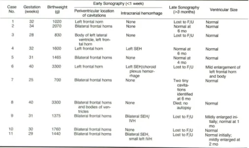
Clinical and sonographic findings of week-old infants with peri ventricular cavita -tions are summarized in Tables 1 and 2.
292 KELLER ET AL. AJNR:8, March/April1987
TABLE 1: Summary of Clinical Findings in Infants with Periventricular Cavitations
Case Gestation Birthweight Maternal Apgar Scores Respiratory Neurologic Findings
No, (weeks) (g) History at 1,5 min Status Neonatal Period Post neonatal Period
32 1020 Preeclampsia 2,8 Mild RDS Normal Lost to FlU
CS
2 34 2070 None 4,6 Mild RDS Abnormal EEG Normal at 7
mo; develop-mental delay
and seizures at 16 mo
3 28 830 None 8,9 RDS Normal Normal at 2 mo
4 32 1600 Urinary infection 9,9 Mild RDS Normal Normal at 4 mo
5 31 1465 None 7,9 Normal Normal Normal at 4 mo
6 40 3300 Intermittent leg 7, 9 Normal Hypotonia, abnormal Lost to FlU
and pedal EEG (right pari
e-edema tal)
7 25 700 Twins 1,6 RDS Normal Normal at 6 mo
8 40 3300 CS for fetal dis- 1,3 PFC Normal Died at 11
tress; nuchal days
cord
9 31 1375 Seizures; CS 1,4 RDS Normal Normal at 2 mo
for bleeding and fetal dis -tress
10 30 1760 None 7, 7 Mild RDS Normal Normal at 2 mo
11 29 1440 Meconium- 6, 7 RDS, PIE Normal Normal at 2 mo
tinged am-niotic fluid
[image:2.612.54.557.98.361.2]ection, RDS = respiratory distress syndrome, FlU = follow-up, PFC = persistent fetal circulation, PIE = pulmonary interstitial
TABLE 2: Sonographic Findings in Infants with Periventricular Cavitations
G tati n Birthweight Early Sonography «1 week) Late Sonography
s
Ventricular Size
N . (w ) (g) Periventricular location
Intracranial hemorrhage (>3 months)
of cavitations
2 1020 Left frontal horn None Lost to FlU Normal
4 2070 Bilateral frontal horns None Normal at Normal
6mo
2 0 Body of left lateral None Lost to FlU Normal
entricl ,left fron-tal hom
4 2 0 L ft frontal h m Left SEH Normal at Normal
6mo
14 5 Bil teral fr ntal hom None Normal at Normal
4 mo
4 Left front I h m Left SEH/choroid Lost to FlU Mild enlargement of
pie us hemor- left frontal hom
mage and body
7 5 Bilateral front I hom one Two tiny Normal
cavit
a-tions
identified at 6 mo
one Died; no Normal
autopsy
1 1 Bilateral SEHI Lo 1 to FlU Mildly enlarged
ini-IH tially; normal at 1
mo
1 B lfron Lost oFJU annal
11 1440 B Ifron Lost to FJU annal initially;
mildly enlarged al
2mo
[image:2.612.53.559.425.726.2]AJNR:8, March/April 1987 PERIVENTRICULAR CAVITATIONS IN INFANTS 293
A
B
c
Fig. 1.-Case 2. Coronal semiaxial (A), left (B) and right (C) para sagittal sonograms show bifrontal periventricular cavitations (arrows).
A
B
Fig. 2.-Case 6. Coronal semiaxial (A and B) and left parasagittal (C) sonograms show solitary right frontal cavitation (arrowhead) and linear cluster of polygonal cavitations along frontal hom and body of left lateral ventricle (arrows).
Occasionally, polygonal cavitations were grouped in linear
clusters.
While some of the infants had notable events in their
gestational histories, others did not. No prenatal or perinatal
clinical pattern emerged from the sonographic findings in this
small series.
Of the four infants who had follow-up studies, three (cases
2, 4, and 5) were seen at 6, 6, and 4 months of age,
respectively, with normal neurosonograms. One infant (case
2) was examined at 16 months and found to have
develop-mental delay and a seizure disorder but neither spasticity nor
a vision disorder. Two infants (cases 4 and 5) appeared
normal at 6 and 4 months of age. One infant with periventr
ic-ular cavitations (case 7) had a twin who did not have them.
At 6 months, both infants were developmentally normal, and
the follow-up neurosonogram of the smaller twin with initially
abnormal scans revealed two barely perceptible tiny residual
cavitations along the lateral aspect of each frontal horn (Fig. 3C).
Six of the infants were lost to follow-up. One infant died,
but permission for autopsy could not be obtained.
Discussion
[image:3.612.58.559.84.293.2] [image:3.612.54.560.324.539.2]294 KELLER ET AL. AJNR:8, March/April 1987
infants autopsied and described by Larroche [13]. Ten of
those infants were 2-3 days old, and 11 of the 22 cases had
subependymal pseudocysts located lateral to the external
angles of the lateral ventricles near the coronal plane of the foramina of Monro similar to those of most infants in our
series (Fig. 4). Four of the 17 cases in Larroche's series
involved the subependymal germinal matrix adjacent to the
caudate nuclei and had evidence of previous hemorrhage.
The subependymal portions of the occipital lobes were
in-volved in two cases.
In early gestation the germinal layer abundantly surrounds
the lateral ventricles until 30 weeks of gestation, when
invo-lution begins [14]. By 32-34 weeks, the germinal matrix is
limited to the area medial to the caudate heads. By 40 weeks,
the germinal matrix is largely involuted [13]. Because of the
occurrence of similarly sized cavitations in a periventricular
distribution, Larroche grouped all cases together and ascribed
small insults to the fetal germinal matrix as the common
pathogenic mechanism. Larroche admitted that some cavities
in her series were lined with macrophages, implying malacic
change, while in four other cases, positive tests for iron
pigment suggested resolved subependymal hemorrhages.
Thus, her case population appeared heterogeneous.
Sonographic findings of subependymal germinal matrix
hemorrhages evolving into subependymal cysts along the
medial aspect of the caudothalamic groove have been
de-scribed [8, 10]. Congenital neurotropic infection may result in
similarly located subependymal cysts [8-12] sonographically
indistinguishable from posthemorrhagic subependymal cysts
but without a documented prior hemorrhage [8]. Among
pathologists, the cause of cysts of the matrix zone has been
a subject of debate [15, 16]. While some of Larroche's cases
exhibited subependymal cysts medial to the caudate nuclei,
none were found in our patients who had no clinical or
serologic evidence for congenital infection and who were too
young to have cavitation within a neonatal subependymal
hemorrhage. Nevertheless, it is still possible that both the
Fig, 3.-Case 7. Coronal sonograms.
A, Bifrontal cavitations (arrows),
B, Follow-up coronal sonogram at age 6 months shows tiny residual cavitations (arrows),
Fig. 4.-Coronal brain section shows two subependymal pseudocysts as described by Larroche. (Reprinted from [13],)
cavitations and the high rate of prematurity in our series
represent sequelae of an unidentified intrauterine infection or
insult.
Periventricular cysts may be a manifestation of PVL, and
the significant neurodevelopmental sequelae of PVL have
been described [5, 7, 17]. Early recognition of PVL by
sonog-raphy helps to provide health care personnel with the ability to counsel parents about an infant's guarded neurologic
outlook.
The pathogenesis and evolution of PVL is thought to relate
to the circulation in the periventricular cerebral white matter
[5, 18]. In premature infants, the periventricular regions are
watersheds between ventriculopetal and ventriculofugal
cir-culations. These areas are therefore susceptible to infarct in
situations causing hypoxia and ischemia [5, 18]. The earliest
sonographic finding in PVL is irregular, coarse periventricular
hyperechogenicity [1-3]. Over 2-4 weeks, the infarcted brain
[image:4.613.55.393.87.294.2] [image:4.613.317.559.324.492.2]AJNR:8, March/April 1987 PERIVENTRICULAR CAVITATIONS IN INFANTS
295
and pathologically [1-7]. As PVL often involves the long motor tracts and optic radiations, affected infants commonly exhibit varying degrees of spasticity and cortical blindness [7, 17].
The infants in our series did not appear to have PVL as all had well-defined peri ventricular cavitations within the first week of life, and none of the four infants seen in follow-up had any neurologic abnormality associated with PVL. Even though several of our patients had cavitations in or near areas often involved by PVL, there was never any extension into the centrum semiovale. Moreover, the early sonographic de -tection of such well-defined cavities would preclude postna -tally acquired PVL.
Difficulty with interpretation of both the data of Larroche [13] and of our study is caused by sample bias. Larroche's autopsy study was performed before the advent of any high-resolution brain imaging, and all infants obviously had fatal illnesses. Our patients were identified because of clinical requests for neurosonography in infants with either prema-turity, low Apgar scores, or possible neurologic abnormality. Therefore, one cannot comment on the prevalence of these findings in infants but can state only that these findings are rare even in a group of selected high-risk infants. All but one of our 11 patients have survived, so a histologic comparison with Larroche's cases is not possible.
In our limited experience, the postnatal history of the cavi-tations appears to be resolution, as follow-up examinations in four of our patients have enabled us to see periventricular cavitations disappear in three infants and diminish in size and number in the other (Fig. 3C).
We call attention to small, lateral periventricular cavitations found in the first week of life. Whether they represent prenatal ischemia or infection with development of small areas of leukomalacia, or whether they represent degeneration cif peri -ventricular germinal matrix in early gestation, or whether they reflect a multiplicity of causes cannot be determined in this report. Neurosonologists and neonatologists should be aware that well-defined, small peri ventricular cavitations seen in infants in the first week of life is separate and distinguishable from postnatally acquired PVL. Identification of these infants
as a separate group will enable long-term neurodevelopmental follow-up that may ultimately give predictive clinical value to this finding.
REFERENCES
1. Bowerman RA, Donn SM, DiPietro MA, D'Amato CJ, Hicks SP. Periven
-tricular leukomalacia in the pre-term newborn infant: sonographic and clinical features. Radiology 1984;151 :383-388
2. Schellinger 0, Grant EG, Richardson JD. Cystic periventricular leukoma
-lacia: sonographic and CT findings. AJNR 1984;5:439-445
3. Schellinger 0, Grant EG, Richardson JD. Neonatal leukencephalopathy: common form of cerebral ischemia. Radiographics 1985;5:221-242
4. Banker BQ, Larroche JC. Peri ventricular leukomalacia of infancy. Arch NeuroI1962;7:386-410
5. DeReuck J, Chatta AS, Richardson EP Jr. Pathogenesis and evolution of
peri ventricular leukomalacia in infancy. Arch NeuroI1972;27:229-236
6. Leech RW, Alvord EC Jr. Morphologic variations in peri ventricular leuko-malacia. Am J PathoI1974;74:591-602
7. Shuman R M, Selednik LJ. Peri ventricular leukomalacia. A one-year autopsy
study. Arch NeuroI1980;37:231-235
8. Shackleford GO, Fulling KH, Glasier CM. Cysts of the subependymal
germinal matrix: sonographic demonstration with pathologic correlation.
Radiology 1983;149:117-121
9. Clair MR, Zalneraitis EL, Baim RS, Goodman K, Perkes EA. Neurosono-graphic recognition of subependymal cysts in high-risk neonates. AJNR 1984;5:761-764, AJR 1985;144:377-380
10. Butt W, Mackay RJ, deCrespigny LC, Murton LJ, Roy RND. Intracranial
lesions of congenital cytomegalovirus infection detected by ultrasound scanning. Pediatrics 1984;73:611-614
11. Shaw CM. Subependymal germinolysis. J Neuropathol Exp Neurol 1973;32:153
12. Shaw CM, Alvord EC. Subependymal germinolysis. Arch Neurol 1974;31 :374-381
13. Larroche JC. Subependymal pseudo-cysts in the newborn. Bioi Neonate 1972;21: 170-183
14. Friede RL. Developmental neuropathology. New York: Springer-Verlag,
1975:7
15. Rorke LB. Pathology of perinatal brain injury. New York: Raven, 1982:37
16. Gilles FH, Leviton A, Dooling EC. The developing human brain. Growth,
epidemiology, neuropathology. Boston: Wright, 1983:235-237
17. Armstrong 0, Norman MG. Peri ventricular leukomalacia in neonates.
Com-plications and sequelae. Arch Dis Child 1974;49:367-375
18. Takashima S, Tanaka K. Development of cerebrovascular architecture and its relationship to periventricular leukomalacia. Arch Neurol 1978;35:


