Supported by the Ministry of Agriculture of the Czech Republic (Project No. QJ1510138 and Project No. MZeRO0714), by the Ministry of Education, Youth and Sports of the Czech Republic (Project No. LO1503 under the NPU I program), by the Internal Grant Agency of the Czech University of Life Sciences Prague (CIGA) (Project No. 20132035), and by the Grant Agency of the Charles University in Prague (PRVOUK P36 program).
Bisphenol S instead of bisphenol A: a story
of reproductive disruption by regretable substitution
– a review
T. Žalmanová
1, K. Hošková
1, J. Nevoral
2,3, Š. Prokešová
1, K. Zámostná
1,
T. Kott
4, J. Petr
21
Department of Veterinary Sciences, Faculty of Agrobiology, Food and Natural Resources,
Czech University of Life Sciences Prague, Prague, Czech Republic
2
Laboratory of Reproductive Medicine, Biomedical Center, Faculty of Medicine in Pilsen,
Charles University in Prague, Pilsen, Czech Republic
3
Department of Histology and Embryology, Faculty of Medicine in Pilsen,
Charles University in Prague, Pilsen, Czech Republic
4
Institute of Animal Science, Prague-Uhříněves, Czech Republic
ABSTRACT
: A range of substances that are released into the environment, foodstuffs and drinking water as a
result of human activity were originally considered relatively harmless, and it was only later that their adverse effects
were discovered. In general the use of such substances is currently restricted, and they are often replaced by other
substances. This applies also in the case of a range of endocrine disruptors. These substances have the capacity to
disturb the balance of physiological functions of the organism on the level of hormonal regulation, and their
pleio-tropic spectrum of effects is very difficult to predict. Endocrine disruptors include the currently intensively studied
bisphenol A (BPA), a prevalent environmental pollutant and contaminant of both water and foodstuffs. BPA has
a significantly negative impact on human health, particularly on the regulation mechanisms of reproduction, and
influences fertility. The ever increasingly stringent restriction of the industrial production of BPA is leading to its
replacement with analogues, primarily with bisphenol S (BPS), which is not subject to these restrictions and whose
impacts on the regulation of reproduction have not yet been exhaustively studied. However, the limited number of
studies at disposal indicates that BPS may be at least as harmful as BPA. There is therefore a potential danger that
the replacement of BPA with BPS will become one of the cases of regrettable substitution, in which the newly used
substances manifest similar or even worse negative effects than the substances which they have replaced. The
objec-tive of this review is to draw attention to ill-advised replacements of endocrine disruptors with substances whose
effects are not yet tested, and which may represent the same risks for the environment, for the reproduction of males
and females, and for human health as have been demonstrated in the case of the originally used substances.
Keywords
:
human health; environment; endocrine disruptor; reproduction; oocyte; sperm
INTRODUCTION
Many substances have been introduced into use
with great hopes, only for it to be demonstrated
as a drug for pregnant women (McBride 1961), or
more recently neonicotinoid insecticides used for
the protection of fields against seed-destroying
insects (Blacquiere et al. 2012). Substances whose
negative effects on the environment or human health
were detected only after a long period of use also
include endocrine disruptors (Damstra et al. 2002).
The detection of the negative effects of
abun-dantly used substances leads to a dramatic
restric-tion of their use and their substiturestric-tion with other
substances. In a range of cases this brings about
a genuine improvement. For example, chromated
copper arsenate (CCA) used for wood
preserva-tion was demonstrated to be a substance with
carcinogenic effects, and as a result was replaced
with alkaline copper quaternary (ACQ). ACQ does
not contain arsenic or chrome, and although it is
just as effective as CCA against wood
destroy-ing arthropods, its impacts on the environment
and human health are fundamentally less serious
(Landrigan et al. 2004).
On the other hand, we have been witnesses to
substitutions of harmful substances which have later
been shown to be highly problematic. For example,
2,3-butanedione, which occurs naturally in butter,
has been produced synthetically and added to foods
in order to impart a buttery flavour. When it was
demonstrated that 2,3-butanedione damaged lung
tissue, it was replaced by 2,3-pentanedione, which
however was subsequently proven to have similar
negative effects on lung tissue as 2,3-butanedione
(Hubbs et al. 2012). There are far more similar
ex-amples of “regrettable substitutions” (Fahrenkamp-
Uppenbrink 2015; Zimmerman and Anastas 2015).
In these cases, negative impacts on reproduction are
often subsequently detected. For example, in the
case of pyrethroids, which replaced older insecticide
agents such as organocholorines, organophosphates
or carbamates, and which were considered harmless
to mammals, negative impacts were demonstrated on
the maturation of mammal oocytes (Petr et al. 2013).
From the perspective of reproductive risks, the
substitution of bisphenol A (BPA), a widely used
component of plastics and many other materials,
with its analogue bisphenol S (BPS) appears to be
potentially problematic. BPA has been proven to be
a strong endocrine disruptor, and its use has been
restricted. Many products are sold with a “BPA-free”
guarantee. Because BPA is substituted in a range
of cases by BPS, these products are not however
“bisphenol-free” (Glausiusz 2014), and their use
may be linked to significant reproductive risks. The
aim of this review is to point to the replacement of
BPA by BPS as a “regrettable substitution”.
Endocrine disruptors
A less harmful substitute is currently searched
for a number of substances that had previously
been considered safe from a toxicological
perspec-tive and finally appeared to exert various negaperspec-tive
effects on health. This category of compounds
includes substances referred to summarily as
en-docrine disruptors (Clayton 2011). According to
the US Environmental Protection Agency,
endo-crine disrupting chemicals (EDCs) are defined as
“exogenous agent(s) that interfere(s) in synthesis,
secretion, transport, metabolism, binding action,
or elimination of natural blood-borne hormones
that are present in the body and are responsible
for homeostasis, reproduction, and developmental
processes” (Diamanti-Kandarakis et al. 2009).
EDCs manifest a range of particular properties.
Their hormone-like effects may be suppressed or
may fade away entirely in the case that the
concen-tration of EDCs is higher than the physiological
level of their hormonal counterpart. This ability
of agents to attain paradoxically stronger effects
in low doses than in high ones (vom Saal and
Welshons 2005) is termed the “low dose effect”
(Grasselli et al. 2010; Vandenberg et al. 2012). The
low dose hypothesis posits that exogenous
che-micals that interact with hormone action can do
so in a quite specific manner. In accordance with
that, mentioned traditional toxicological endpoints
are not capable to preclude adverse outcome, as
EDCs act with dose responses, that are nonlinear
and potentially non-monotonic (Vandenberg et al.
2012). In the case the relationship between dose
and response is nonlinear, any prediction is even
more complex. Therefore, the low dose definition
was extended by the effects of non monotonic
response curves. The mechanisms responsible
for the non-linear effects are described in detail
(Vandenberg et al. 2012), usually in connection
with an interaction between a ligand (hormone or
EDC) and a hormone receptor (Vandenberg 2014).
(Calabrese and Baldwin 2001; Calabrese 2005).
Ho-wever, EDCs can also produce non monotonic dose
responses in which the slope of the curve changes
sign over the course of the dose-response (www.
who.int/ceh/publications/endocrine/en/index.html)
and low dose effects are described for the majority
of EDCs (Birnbaum 2012; Vandenberg et al. 2012,
2013; Zoeller et al. 2012; Bergman et al. 2013).
The concept of endocrine-disrupting chemicals
was proposed after these compounds had been
observed to affect various reproductive functions
in wildlife and humans (Colborn et al. 1993). The
influence of several EDCs was demonstrated on the
course of development of male gametes, sperm (Li
et al. 2011; Knez et al. 2014) and female gametes,
oocytes, as well as embryonic development of males
and females (Mok-Lin et al. 2010; Xiao et al. 2011).
Moreover, the effect of EDCs on the reproduction
of adult individuals, including transgenerational
inheritance, has been described (Susiarjo et al.
2015; Ziv-Gal et al. 2015). Therefore, reproductive
functions represent crucial targets of the EDCs’
negative effects. Recently intensively studied EDCs,
interfering with the regulation of physiological
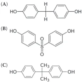
re-productive processes, include bisphenols, a family
of chemical compounds with two hydroxyphenyl
functional groups (Figure 1).
Bisphenol A
An example of a widely used substance, in which
endocrine-disrupting properties were detected
only later, is bisphenol A (BPA,
4,4'-(propane-2,2-diyl)diphenol) (Vandenberg et al. 2009). BPA was
first synthesized in 1891, and as early as in 1936
it was demonstrated that it imitates the activity of
the hormone estradiol (Dodds and Lawson 1936).
Despite a very strong estrogen activity, BPA has
been commercially used since 1957, and despite
the fact that its endocrine-disrupting activity was
discovered (Krishnan et al. 1993), BPA has become a
high production volume chemical (Wang et al. 2012).
Worldwide annual production, which in the case
of BPA reached 4.6 million t in 2012, is constantly
increasing. Its production was estimated at 5.4
mil-lion t in 2015 (Merchant Research & Consulting,
http://mcgroup.co.uk/researches/bisphenol-a-bpa).
BPA is present especially in polycarbonate
plas-tics, epoxide resins, and several paper products
(Ehrlich et al. 2014), and as a result it is used in a
variety of commonly used consumer products such
as thermal recipes, cosmetics, dental materials,
medicinal tubes, utensils, toys, baby feeding
bot-tles and dummies, etc. Heat, UV radiation, alkaline
treatment or intensive washing causes a release of
BPA monomer. It is estimated that the worldwide
release of BPA into the environment is almost half
million kg per year (Mileva et al. 2014).
BPA is released into the environment either
di-rectly from chemical, plastic coating, and staining
manufacturers, from paper or material recycling
companies, foundries which use BPA in casting
sand, or indirectly leaching from plastic, paper, and
waste in landfills (Yang et al. 2015). BPA passes into
foodstuffs or water directly from the lining of food
and beverage cans, where it is used as an
ingredi-ent in the plastic used to protect the food from
direct contact with the can (Goodson et al. 2002;
Vandenberg et al. 2009). The main path of human
exposure is the consumption of such contaminated
foodstuffs, drinking water or via dermal contact
with thermal paper and cosmetics or inhalation
(Miyamoto and Kotake 2005; Huang et al. 2012).
[image:3.595.115.259.93.238.2]It is therefore not surprising that a range of
stud-ies have now demonstrated the presence of BPA
in human tissue. Levels of BPA have been tested
in various populations worldwide, and the
pres-ence of BPA was demonstrated in 92.6% of
Ameri-cans (Wetherill et al. 2007) and 90% of Canadians
(Bushnik et al. 2010). Levels of BPA have been
demonstrated in various biological matrices, most
frequently in urine (Casas et al. 2013;
Salgueiro-Gonzalez et al. 2015), but also in blood serum.
Within the human reproductive system, levels of
BPA have been confirmed for example in testicle
tissue, seminal plasma (Manfo et al. 2014), in
ovar-ian follicular fluid (Ikezuki et al. 2002), mother’s
Figure 1. Chemical structure of bisphenol A (A),bisphe-nol S (B), bisphenol F (C) (A)
(B)
milk, fetal plasma (Shonfelder et al. 2002), amniotic
fluid (Yamada et al. 2002; Edlow et al. 2012), and the
placenta (Jimenez-Diaz et al. 2010; Cao et al. 2012)
(Table 1). Several studies have demonstrated a direct
correlation between exposure of the mother and the
BPA level of the fetus (Ikezuki et al. 2002;
Kuruto-Niwa et al. 2007). BPA may permeate the placenta
and thus influence the development of the fetus
(Edlow et al. 2012; Corbel et al. 2014). Newborns
may then be further exposed to the effect of BPA
during breastfeeding due to the presence of BPA in
mother’s milk (Mendonca et al. 2014).
The effects of BPA on humans are dependent not
only on the dose, but also on the window of exposure.
Exposure to BPA in the prenatal and neonatal period
probably affects the human organism in the most
receptive period (Fernandez et al. 2014).
Mechanism of BPA action
[image:4.595.62.532.111.311.2]A typical feature of endocrine disruptors is their
wide spectrum of outcomes (Figure 2).
Combi-nation of their action in various target systems
in the organism is one of causes of their non-
linear effects. In this respect, BPA acts as a typical
endocrine disruptor with multi-level impacts (Khan
and Ahmed 2015). Nongenomic effects of BPA have
been described, thus influencing cellular signalling
Table 1. Bisphenol A (BPA) levels in human fluidsSample Level of BPA References
Blood (ng/ml) 12.4–14.4 Bushnik et al. (2010)
Maternal blood (ng/ml) 0.63–14.36 Yamada et al. (2002) Fetal blood (ng/ml) 0.2–9.2 Schonfelder et al. (2002)
Urine (ng/ml) 0.02–21.0 Liao et al. (2012c)
Saliva (ng/ml) 0.3 Joskow et al. (2006)
Follicular fluid (ng/ml) 2.4 ± 0.8 Ikezuki et al. (2002) Amniotic fluid (ng/ml) 1.1–8.3 Ikezuki et al. (2002) Placental tissue (ng/g) 1.0–104.9 Schonfelder et al. (2002) Breast milk (ng/ml) 0.5–1.3 Mendonca et al. (2014)
Semen plasma (pg/ml) 132–179 (infertile men)66 (fertile men) Vitku et al. (2015)
[image:4.595.64.422.521.730.2](Nakagawa and Tayama 2000), as well as genomic,
which affect transcription regulation (Trapphoff et
al. 2013), and also epigenetic, responsible for the
methylation and acetylation of DNA and core
his-tones (Bromer et al. 2010). It is precisely pronounced
estrogen activity of BPA
in vitro
(vom Saal et al. 2007;
Wetherill et al. 2007) and
in vivo
that contributes to
its immense potential to afflict the hormonal system
and act as an endocrine disruptor.
BPA inhibits the activity of natural endogenous
estrogens and thus disrupts estrogen nuclear
hormone receptor action (Kitamura et al. 2005;
Wetherill et al. 2007; Grignard et al. 2012). BPA
affects hormonal homeostasis, for example through
bonding to the classic nuclear estrogen receptors
α, β, γ (ERα, ERβ, ERγ), where it manifests a
com-bination of agonistic and/or antagonistic actions
in dependence on the target tissue, cell types, ER
subtypes, and differential cofactors recruited by
ER-ligand complexes (Kurosawa et al. 2002). BPA
also bonds to non-classical membrane ERs and
causes activation of the nuclear receptor gamma
(Takayanagi et al. 2006; Matsushima et al. 2007).
BPA has been identified as an antagonist of
an-drogen receptors (Kitamura et al. 2005; Wetherill et
al. 2007; Vinggaard et al. 2008; Molina-Molina et al.
2013). Its anti-androgenic activity has been
docu-mented in several studies, but with changing values
of the maximum inhibition concentration (Xu et al.
2005; Bonefeld-Jorgensen et al. 2007). In contrast
with other known androgen receptor antagonists,
BPA inhibits the effective nuclear translocation of
the androgen receptors, and disrupts their function
by means of a number of mechanisms (Teng et al.
2013). The endocrine-related BPA action mechanism
also involves a reduction of aromatase expression
(Zhang et al. 2011; Chen et al. 2014) and a decrease
in aromatase activity
in vitro
(Bonefeld-Jorgensen
et al. 2007). Within this context, it is of interest
that a decline in the synthesis of testosterone and
estradiol
in vivo
has been documented following
exposure to BPA (Akingbemi et al. 2004).
The epigenetic mechanisms of the effect of BPA
include the alteration of certain DNA methylation
samples (Dolinoy et al. 2007; Susiarjo et al. 2013).
Prenatal exposure to BPA alters the expression of
genes coding individual subtypes of ERs in a sex-
and brain region-specific manner (Kundakovic et
al. 2013) and disrupts the normal development of
the placenta (Susiarjo et al. 2013). As a result, it is
possible that BPA predetermines the response to
steroid hormones in the very early phase of
devel-opment (Wilson and Sengoku 2013). It has been
documented that BPA also disrupts the gene
ex-pression of the regulating factors that control the
stability and flexibility of epigenetic regulation,
and as a result has an adverse influence on the
development of functions of the controlling organ
of hormonal regulation, the hypothalamus (Warita
et al. 2013). The impacts of these changes have
transgenerational effects (Manikkam et al. 2013).
Further demonstrated actions of BPA in the
organism include the bonding to the glucuronide
receptor, suppression of the transcription receptor
of the thyroid hormone, reduction of the transport
of cholesterol via the mitochondrial membrane,
increase of oxidation of fatty acids, stimulation of
prolactin release (Machtinger and Orvieto 2014)
or an agonistic effect on the human pregnane X
receptor (Sui et al. 2012).
BPA and human health
With such a wide spectrum of effects, it is
evi-dent that BPA has a negative influence on
hu-man health. Frequently discussed themes include
the possible association of BPA for example with
obesity (Trasande et al. 2012), diabetes (Lang et
al. 2008), neurobehavioural disorders (Jasarevic
et al. 2011), cancer (Jenkins et al. 2011), hepatic
(Peyre et al. 2014) and cardiovascular diseases,
hypertension, and disorders of the thyroid gland
function (Rochester 2013; Wang et al. 2013).
Especially in the area of reproduction in both
animal models and in humans, a wide range of
negative influences of BPA have been observed
(Kwintkiewicz et al. 2010; Trapphoff et al. 2013;
Zhang et al. 2014). BPA has varied and complex
mechanisms of action that may interfere with
normal reproductive development and functions.
In both males and females, BPA interferes with
hormonal regulation and influences the
hypotha-lamic–pituitary–gonadal axis on all levels (Navarro
et al. 2009; Patisaul et al. 2009; Xi et al. 2011).
im-mature spermatozoa was decreased and increased,
respectively (Sohoni et al. 2001) and also the sperm
motility and concentration were reduced
(Lahn-steiner et al. 2005). There is a large evidence that
BPA can induce sex reversal from male to female
in aquatic animals. Changes in sex ratio were
observed at zebrafish during embryonic
develop-ment (Drastichova et al. 2005) and
Xenopus
larvae
through metamorphosis (Kloas et al. 1999).
Experimental studies on the effects of BPA on
the reproduction of male rodents have revealed
an adverse influence on the development of testes
(Vrooman et al. 2015) and on the spermatogenesis
of adult individuals following prenatal
in utero
or early postnatal exposure. Exposure to BPA
during the period of development of the testes
is frequently linked to a range of negative effects
in adult testes, e.g. decreased levels of testicular
testosterone, decreased weights of the epididymis
and seminal vesicles, a decrease in daily sperm
production per gram testis, and increased weights
of the prostate and preputial (Richter et al. 2007).
Vrooman et al. (2015), with the help of
transplan-tation of spermatogonia from the testes of mice
exposed to the action of BPA into mice which were
not exposed, demonstrated permanent damage to
spermatogenesis. The influence of the exposure
of adult rodents to BPA on the quality of sperm
was also studied (Peretz et al. 2014).
Despite the differences in the experimental
de-signs used, certain findings appear repeatedly,
especially reduction in the number of sperm,
reduc-tion in the motility of sperm, increased amount of
apoptotic cells in the seminiferous tubules, changes
in the levels of hormones and steroid enzymes, and
damage to the DNA of sperm (Peretz et al. 2014).
Contemporary studies confirm that rodents are not
relevant for predicting the effect of low BPA
concen-trations on the endocrine function of human fetal
testis (N’Tumba-Byn et al. 2012). In a comparative
study by Maamar et al. (2015), the influence of BPA
was studied both on rats and on human fetal testes,
and it was determined that in both cases BPA had
dose-dependent anti-androgenic effects.
Neverthe-less, the authors urge caution in interpreting the
results obtained on rodents and their application
in human medicine (Maamar et al. 2015).
Unfortunately, there is only a limited number
of studies that have observed the influence of
exposure to BPA on the quality of sperm in adult
humans. In men exposed to BPA in the workplace
and patients in reproduction centres, a higher
level of BPA in urine was linked to a lower
num-ber, concentration, and motility of sperm (Knez
et al. 2014; Lassen et al. 2014). Nevertheless, in
a study conducted by Mendiola et al. (2010) on
fertile men, the concentration of BPA in urine did
not correlate with changes in semen parameters,
despite the fact that a significant correlation was
observed between the level of BPA in urine and
the volume of seminal plasma or markers of free
testosterone (Mendiola et al. 2010).
The following cohort study examined the
re-lationship between the concentration of BPA in
urine and the level of reproductive hormones
and semen in a group of 308 young healthy men.
It was determined that the concentration of BPA
strictly correlates with higher levels of selected
circulating reproductive hormones and reduced
motility of sperm. The results indicated that the
exposure to BPA on the level of environment has
an anti-androgenic and/or anti-estrogenic effect
due to the effect of BPA on the level of
recep-tors. The anti-estrogenic effect on the level of
the epididymis also explains the determined low
mobility of the sperm (Lassen et al. 2014).
Influences of BPA on reproduction of females
BPA markedly influences not only the
reproduc-tion of males, but also the reproducreproduc-tion of females.
In both
in vitro
and
in vivo
studies, the influence of
BPA has been demonstrated on fertility, function of
the womb i.e. formation of benign and malignant
lesions (Newbold et al. 2009), disruption apoptosis
of the uterine epithelium during estrus
(Mendoza-Rodriguez et al. 2011), function of ovaries and
quality of oocytes (Peretz et al. 2014), and
defec-tive folliculogenesis (Santamaria et al. 2016). In
females it is precisely the ovaries that are the key
organ responsible for reproductive and endocrine
functions, and BPA is frequently indicated as an
ovarian toxicant. BPA afflicts not only the overall
morphology and weight of the ovaries (Suzuki et al.
2002; Santamaria et al. 2016) but also demonstrably
reduces the quality of oocytes in both animal and
human models (Machtinger and
Orvieto
2014).
oocytes (Hunt et al. 2003). Similary, it was reported
that BPA exposure altered chromosome and spindle
organization which resulted in hyperploidy of mouse
oocytes during meiosis (Can et al. 2005) and it was
also demonstrated that low BPA doses are related
with aberration during meiotic prophase, including
increased incidence of recombination (Susiarjo et
al. 2007) and failure formation of primordial follicle
by inhibiting meiotic progression of oocytes (Zhang
et al. 2012). In contrast, Eichenlaub-Ritter and her
colleagues found no evidence that low BPA doses
increased hyperploidy at meiosis II. On the other
hand they observed cell cycle delay and meiotic
spindle abnormalities, changes in the distribution
of pericentriolar material and chromosome
align-ment (Eichenlaub-Ritter et al. 2008). Exposure of
mice, from mid-gestation to birth, causes synaptic
abnormalities in oocytes and an increased amount of
recombination between homologous chromosomes.
It is also of interest that identical effects have been
observed in homozygous mice with an intentionally
disrupted gene coding the ERβ. In mouse oocytes,
epigenetic changes have also been documented
fol-lowing cultivation of follicles in the presence of
BPA, in which a disruption of the configuration of
chromosomes took place, as well as disorders of
meiosis caused by faulty genomic imprinting and
altered posttranslational modification of histones
(Trapphoff et al. 2013). Chronic exposure of oocytes
was linked to an increased incidence of aberrant
metaphases II and prematurely segregated chromatids
(Pacchierotti et al. 2008).
Bovine oocytes cultivated in the presence of
BPA have also manifested disorders of the meiotic
spindle and the chromosomal configuration (Ferris
et al. 2015). In Barbary Macaques, negative effects
of BPA have been demonstrated in various stages
of the oogenesis of developing ovaries. Oocytes
in the prophase of meiosis and in fetal ovaries
exhibited an increased number of recombination,
and an increased number of abnormally formed
follicles containing multiple oocytes was recorded
in perinatal ovaries (Hunt et al. 2012).
Similary as in the aforementioned studies on
ro-dents, cattle, and primates, an increased number of
crossing over and degenerations in oocytes have been
determined also in human oocytes cultivated
in vitro
in the presence of BPA (Brieno-Enriquez et al. 2011).
In connected studies it has been demonstrated that
the exposure of human oocytes to BPA is linked to
up-regulation of genes involved in meiotic processes
connected to double strand breaks repair progression
(Brieno-Enriquez et al. 2012). A non-linear response
to BPA doses on the incidence of MII oocytes with
aligned chromosomes has also been determined
(Machtinger et al. 2013). The changes which have been
recorded in the development of oocytes exposed to
bisphenol may lead to disorders in the development
of embryos, fetal loss or genetic disorders (Rama
Raju et al. 2007; Ye et al. 2007; Tomari et al. 2011).
The result of maternal exposure to BPA may be the
disruption of the entire oogenesis in the developing
ovary (Susiarjo et al. 2007).
A number of cohort studies have been focused
on groups of persons who undergo treatment for
infertility through
in vitro
fertilization (IVF). The
measured levels of BPA in these persons were
ex-amined in connection with the ovarian response,
quality of embryos and implantation. A reduced
ovarian response was linked to a reduced success
rate of IVF (Mok-Lin et al. 2010). BPA also
dis-rupted embryonal development of fish via delay
hatching, yolk reabsorption, and larval growth of
trouts (Aluru et al. 2010), moreover lethality in
zebrafish larvae increased (Chan and
Chan 2012).
There is only a limited number of studies which
have observed the effects of BPA on the
develop-ment and quality of mammalian blastocysts. Failure
of embryonic development to mouse blastocyst
stage has been demonstrated after exposure of
females to BPA (Xiao et al. 2011). Disorder of
implantation of mouse blastocysts was also
dem-onstrated by Borman et al. (2015).
In human, Bloom et al. (2011) state a correlation
between the concentration of BPA in the urine
of men, though not in women, and a decline in
the quality of embryos generated by IVF. By
con-trast, in a study performed by Knez et al. (2014),
which confirms changes to the semen quality of
men with a determined environmental level of
BPA, undisrupted development of embryos into
blastocysts is described. As against this finding,
in women who have undergone IVF, a correlation
has been demonstrated between the concentration
of BPA in urine and a change to the formation of
blastocysts, though a reduced quality of embryos
was not recorded (Ehrlich et al. 2012).
The advent of BPS
range of cases its substitution with another
chemi-cal. On the basis of the effects on human health
and reproduction demonstrated with the help of
standardized toxicological testing procedures,
government agencies in the United States (the US
Environmental Protection Agency, USEPA), Canada
(Health Canada), and Europe (the European Food
Safety Authority, EFSA) have established tolerable
daily intake levels, ranging from 25 to 50 μg BPA/kg
of body weight (BW) per day (Rochester 2013).
With regard to the fact that several studies have
demonstrated BPA low dose effects (Vandenberg
et al. 2012), and that this possibility is
unfortu-nately not taken into account in the approach of
“traditional” toxicological studies, in which low
doses are not generally subjected to examination
(Vandenberg et al. 2012; Rochester 2013), scientists
have expressed concerns that the “safe” cut-off set
for BPA is too high (vom Saal and Hughes 2005).
In 2010 the Canadian government prohibited the
import, sale, and advertisement of baby feeding
bottles containing BPA. The European Union
re-sponded with a prohibition of the manufacture of
baby feeding bottles with BPA, which was passed
in 2011 (Commission Directive 2011). The Food
and Drug Administration (FDA) has indicated
BPA as a “chemical of concern”, and in July 2012 a
blanket prohibition of BPA in baby feeding bottles
and sippy cups was recommended (FDA 2011).
However, new data and refined methodologies
have led EFSA experts to considerably reduce
the safe level of BPA from 50 µg/kg of BW/day to
4 µg/kg of BW/day (EFSA 2014).
With regard to these restrictions and societal
pres-sures, manufacturers of plastics are now forced to
seek an alternative product which can replace BPA.
It is in the interest of chemical concerns that the
substitute which replaces BPA is inert or at least far
less toxic than BPA. Nevertheless, new chemicals
introduced onto the market are frequently
untest-ed, and may be equally or more harmful than the
originals, which are ultimately termed “regrettable
substitutions” (Rochester and Bolden 2015), as has
been the case of a number of perfluorinated
chemi-cals (Howard 2014), pesticides (Coggon 2002), and
self-extinguishing compounds (Bergman et al. 2012).
Manufacturers seeking BPA alternatives have turned
primarily to bisphenol S (BPS,
4,4'-sulfonyldiphe-nol) (see Figure 1), a structural analogue of BPA, to
produce “BPA-free” products (Grignard et al. 2012;
Barrett 2013). BPS is chemically more stable, worse
in terms of biodegradability than BPA, and shows
better dermal penetration than BPA (Ike et al. 2006;
Danzl et al. 2009; Liao et al. 2012a, b). It is
discon-certing that these properties may lead to a longer or
higher body burden or bioavailability of BPS versus
BPA (Helies-Toussaint et al. 2014). For these reasons,
too, at present the replacement of BPA with BPS is
considered a “regrettable substitution”
(Fahrenkamp-Uppenbrink 2015; Zimmerman and Anastas 2015).
With regard to the increase in production of BPS and
the indispensability of bisphenols in the production
of plastics, it is unfortunately possible to expect the
same widespread use of BPS as in the case of BPA
(Liao et al. 2012c). Now the presence of BPS can be
expected in almost all the consumer goods here in
which BPA was initially used (Mathew et al. 2014),
for example as a wash fastening agent in clearing
products, an electroplanting solvent, and a
constitu-ent of phenolic resins (Rochester and Bolden 2015).
One of the major industries that have replaced
BPA due its high occurrence (~3–22 g/kg) is that
of thermal paper (Mathew et al. 2014). In the
USA, Korea, Vietnam, Japan, and China (Liao et
al. 2012c), BPS has been detected in several
differ-ent “BPA free” paper products, including receipts
and paper money (Liao et al. 2012a). The presence
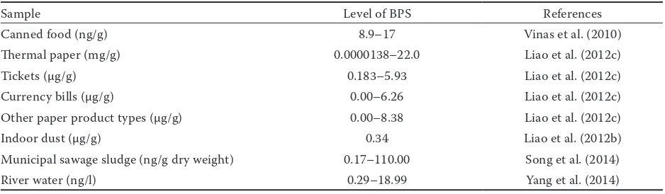
of BPS has been determined in tinned foodstuffs
(Vinas et al. 2010). The occurrence of BPS has
also been determined in indoor dust (Liao et al.
2012b), in fluvial water (Ike et al. 2006), surface
water, and waste waters (Song et al. 2014) (Table 2).
The main pathway to the human body is dermal,
dust ingestion, and dietary exposures (Liao et al.
2012b). Unfortunately, for example thermal paper
carries BPS into all recycled paper products,
mak-ing dermal exposure inevitable. Massive exposure
of the population to the effects of environmental
BPS has been demonstrated in a number of
differ-ent countries. Within the range of 0.02–21 ng/ml
(0.8–84nM) it has been detected in human urine
samples originating from seven Asian countries
and the USA (Liao et al. 2012a) in 81% of analyzed
samples. In the following study the presence of
BPS in urine was demonstrated in residents living
near a manufacturing plant in south China in a
concentration of 0.029 ng/ml (Yang et al. 2015).
Biological effects of BPS
endocrine-disrupting effects of BPS, the substitution of BPA
with BPS is raising concerns. The limited number
of studies available at the present time, dealing
with the biological interactions of BPS with the
organism, indicate that BPS is also capable of
imitating properties of hormones, interacting with
ER (Delfosse et al. 2012; Rosenmai et al. 2014;
Le Fol et al. 2015), and direct binding to nuclear
ERs (Yamasaki et al. 2004) and serum albumins
(Mathew et al. 2014) has been confirmed.
Some
in vitro
studies have demonstrated a weaker
estrogen activity of BPS than the activity manifested
by estradiol (Kuruto-Niwa et al. 2010; Grignard et
al. 2012; Molina-Molina et al. 2013; Rochester and
Bolden 2015). By contrast, a study conducted by
Vinas and Watson (2013a, b) demonstrated the same
or higher estrogen effectiveness than estradiol, BPS
was capable of stimulating the membrane
recep-tor pathways ordinarily up-regulated by estradiol.
After exposure to BPS there are also changes in
the expression of aromatase, the key enzyme in the
synthesis of estradiol (Kinch et al. 2015).
Like in the case of BPA, the androgenic activity
of BPS was confirmed (Kitamura et al. 2005), and
subsequently its anti-androgenic activity as well
(Molina-Molina et al. 2013). These observations
in
vitro
have also been confirmed by
in vivo
studies.
Chen et al. (2002) described acute toxicity of BPS in
Daphnia magna
and at the same time also
demon-strated estrogen activity of BPS
in vitro
. Yamasaki et
al. (2004) documented estrogen activity of BPS
in vivo
in rats with the assistance of postnatal exposure to
BPS, which in both low and high doses induced the
growth of the womb (Owens and Ashby 2002). An
in vivo
study on the effect of BPS in zebrafish
docu-mented not only changes in the mass of the gonads
and plasmatic levels of estrogen and testosterone,
but also a marked disruption of reproduction. The
study of Qiu and colleagues evaluated the impact of
BPA and BPS on the reproductive neuroendocrine
system during zebrafish embryonic development,
and explored potential mechanisms of action
as-sociated with ER, thyroid hormone receptor, and
enzyme aromatase pathways. All of these pathways
were necessary to observe the full effects of BPS on
the changes in gene expression in the reproductive
neuroendocrine axis (Qiu et al. 2016). These data
were substantiated by a decrease in egg production
and hatchability and an increasing number of embryo
malformations (Ji et al. 2013). These observations
were later extended upon by increased time to hatch,
reduced number of sperm, increasing number of
female to male ratio, and changes in the levels of
testosterone, estradiol, and vitellogenin (Naderi et
al. 2014). In further experiments provided in cell
cultures it has been demonstrated that BPS acts
cytotoxically, genotoxically (Lee et al. 2013), and
mutagenically (Fic et al. 2013).
The reason for these negative effects may be
for example binding to serum albumins or DNA
damage and subsequent influencing of several
signal cascades anywhere within the organism
(Lee et al. 2013; Mathew et al. 2014). Exposure to
BPS disrupts cellular signalling in the apoptotic
and survival pathways (Salvesen and Walsh 2014).
Evidently, it is possible to expect the interference
of BPS in signal pro-apoptotic pathways and signal
cascades described also in gametes, leading to an
altered cell cycle and cell death (Nevoral et al. 2013;
Sedmikova et al. 2013). Further studies focused on
the mechanism of BPS action are needed for a full
understanding its negative effect on reproduction
on the gamete level and cell cycle regulation.
[image:9.595.64.533.112.249.2]In respect to previous regrettable substitution,
another bisphenols, such as bisphenol F (BPF,
bis(4-hydroxyphenyl)methane; see Figure 1), do
Table 2. Bisphenol S (BPS) levels in the personal care products and environmentSample Level of BPS References
Canned food (ng/g) 8.9–17 Vinas et al. (2010)
Thermal paper (mg/g) 0.0000138–22.0 Liao et al. (2012c)
Tickets (µg/g) 0.183–5.93 Liao et al. (2012c)
Currency bills (µg/g) 0.00–6.26 Liao et al. (2012c) Other paper product types (µg/g) 0.00–8.38 Liao et al. (2012c)
Indoor dust (µg/g) 0.34 Liao et al. (2012b)
Municipal sawage sludge (ng/g dry weight) 0.17–110.00 Song et al. (2014)
not seem to be a suitable alternative. In
addi-tion to BPA and BPS, BPF has been described as
endocrine disruptor as well (Perez et al. 1998).
Surprisingly, natural presence of BPF has recently
been observed in mustard and, therefore, it is
a frequent compound of foodstuff (Zoller et al.
2016). Hence, BPF regulation is ambiguous for its
chronical intake by a major part of human
popula-tion (Dietrich and Hengstler 2016).
CONCLUSION
At present we are witnessing the substitution of
BPA with BPS in a whole range of materials, and
BPS is becoming a standard component of several
products. BPS is a substance which is structurally
very similar to BPA, it shows analogous
effective-ness and mechanism of
in vitro
action. Biological
changes occurring in the range of typical human
exposures were documented at doses below those
used in traditional toxicology. On the basis of the
described comparisons, it is possible to expect
that BPS, like BPA, is an endocrine disruptor,
and that it may have similar targets and manner
of action
in vivo
and may influence physiological
processes on several levels. With regard to its
slower degradation, BPS may act for a longer time
in the organism and thus interfere with the
regu-lation of reproduction of mammals in a yet more
dangerous manner than has been demonstrated
by a range of studies in the case of BPA.
The alarming results of the first reproduction
studies on BPS have generated an acute need for a
wider and at the same time more detailed
assess-ment of the impacts of BPS, with emphasis on the
area of reproduction of mammals, which is entirely
lacking at present. Should this not materialize,
due to the increasing industrial production of BPS
caused by the need to replace BPA, unfortunately
BPS may within the foreseeable future become
just as great an environmental health risk as BPA.
There is a need for very intensive research and
subsequently also legislative measures in order
to ensure that BPS will not become another
“re-grettable substitution” with pronounced negative
impacts on the environment and on human health,
including negative impacts on reproduction.
Acknowledgement
.
Professor František Jílek is
greatly acknowledged for his assistance in
manu-script writing.
REFERENCES
Akingbemi B.T., Sottas C.M., Koulova A.I., Klinefelter G.R., Hardy M.P. (2004): Inhibition of testicular steroidogen-esis by the xenoestrogen bisphenol A is associated with reduced pituitary luteinizing hormone secretion and decreased steroidogenic enzyme gene expression in rat Leydig cells. Endocrinology, 145, 592–603.
Aluru N., Leatherland J.F., Vijayan M.M. (2010): Bisphe-nol A in oocytes leads to growth suppression and altered stress performance in juvenile rainbow trout. PLoS ONE, 5, e10741.
Barrett J.R. (2013): Assessing the safety of a replacement chemical: nongenomic activity of bisphenol S. Environ-mental Health Perspectives, 121, 97.
Bergman A., Ryden A., Law R.J., de Boer J., Covaci A., Alaee M. (2012): A novel abbreviation standard for organo-bromine, organochlorine and organophosphorus flame retardants and some characteristics of the chemicals. Environment International, 49, 57–82.
Bergman A., Heindel J.J., Jobling S., Kidd K.A., Zoeller R.T. (eds) (2013): State of the Science of Endocrine Disrupting Chemicals – 2012. World Health Organization, Geneva, Switzerland/United Nations Environment Programme, Nairobi, Kenya.
Birnbaum L.S. (2012): Environmental chemicals: evaluating low-dose effects. Environmental Health Perspectives, 120, 143–144.
Blacquiere T., Smagghe G., van Gestel C.A., Mommaerts V. (2012): Neonicotinoids in bees: a review on concentra-tions, side-effects and risk assessment. Ecotoxicology, 21, 973–992.
Bloom M.S., vom Saal F.S., Kim D., Taylor J.A., Lamb J.D., Fujimoto V.Y. (2011): Serum unconjugated bisphenol A concentrations in men may influence embryo quality indicators during in vitro fertilization. Environmental Toxicology and Pharmacology, 32, 319–323.
Bonefeld-Jorgensen E.C., Long M., Hofmeister M.V., Ving-gaard A.M. (2007): Endocrine-disrupting potential of bis-phenol A, bisbis-phenol A dimethacrylate, 4-n-nonylbis-phenol, and 4-n-octylphenol in vitro: new data and a brief review. Environmental Health Perspectives, 1, 69–76.
Borman E.D., Foster W.G., Greenacre M.K., Muir C.C., de Catanzaro D. (2015): Stress lowers the threshold dose at which bisphenol A disrupts blastocyst implantation, in conjunction with decreased uterine closure and e-cadher-in. Chemico-Biological Interactions, 237, 87–95. Brieno-Enriquez M.A., Robles P., Camats-Tarruella N.,
human oocyte development. Human Reproduction, 26, 2807–2818.
Brieno-Enriquez M.A., Reig-Viader R., Cabero L., Toran N., Martinez F., Roig I., Garcia Caldes M. (2012): Gene expression is altered after bisphenol A exposure in human fetal oocytes in vitro. Molecular Human Reproduction, 18, 171–183.
Bromer J.G., Zhou Y., Taylor M.B., Doherty L., Taylor H.S. (2010): Bisphenol-A exposure in utero leads to epigenetic alterations in the developmental programming of uterine estrogen response. The FASEB Journal, 24, 2273–2280. Bushnik T., Haines D., Levallois P., Levesque J., van Oostdam
J., Viau C. (2010): Lead and bisphenol A concentrations in the Canadian population. Health Reports, 21, 7–18. Calabrese E.J. (2005): Toxicological awakenings: the rebirth
of hormesis as a central pillar of toxicology. Toxicology and Applied Pharmacology, 204, 1–8.
Calabrese E.J., Baldwin L.A. (2001): The frequency of U-shaped dose-responses in the toxicological literature. Toxicological Sciences, 62, 330–338.
Can A., Semiz O., Cinar O. (2005): Bisphenol-A induces cell cycle delay and alters centrosome and spindle microtu-bular organization in oocytes during meiosis. Molecular Human Reproduction, 11, 389–396.
Cao X., Zhang J., Goodyer C.G., Hayward S., Cooke G.M., Curran I.H.A. (2012): Bisphenol A in human placental and fetal liver tissues collected from Greater Montreal area (Quebec) during 1998–2008. Chemosphere, 89, 505–511. Casas M., Valvi D., Luque N., Ballesteros-Gomez A., Carsin
A.E., Fernandez M.F., Koch H.M., Mendez M.A., Sunyer J., Rubio S., Vrijheid M. (2013): Dietary and sociodemo-graphic determinants of bisphenol A urine concentra-tions in pregnant women and children. Environment International, 56, 10–18.
Chan W.K., Chan K.M. (2012): Disruption of the hypotha-lamic–pituitary–thyroid axis in zebrafish embryo-larvae following waterborne exposure to BDE-47, TBBPA and BPA. Aquatic Toxicology, 108, 106–111.
Chen M.Y., Ike M., Fujita M. (2002): Acute toxicity, mu-tagenicity, and estrogenicity of bisphenol A and other bisphenols. Environmental Toxicology, 17, 80–86. Chen S., Zhou D., Hsin L.Y., Kanaya N., Wong C., Yip R.,
Sakamuru S., Xia M., Yuan Y.C., Witt K., Teng C. (2014): AroER tri-screen is a biologically relevant assay for en-docrine disrupting chemicals modulating the activity of aromatase and/or the estrogen receptor. Toxicological Sciences, 139, 198–209.
Clayton R. (ed.) (2011): Endocrine Disrupters in the Environ-ment. Foundation for Water Research, Marlow, UK, 3–22. Coggon D. (2002): Work with pesticides and organophos-phate sheep dips. Occupational Medicine, 52, 467–470.
Colborn T., vom Saal F.S., Soto A.M. (1993): Developmen-tal effects of endocrine disrupting chemicals in wildlife and humans. Environmental Health Perspectives, 101, 378–384.
Corbel T., Gayrard V., Puel S., Lacroix M.Z., Berrebi A., Gil S., Viguie C., Toutain P.L., Picard-Hagen N. (2014): Bidirectional placental transfer of Bisphenol A and its main metabolite, Bisphenol A-Glucuronide, in the iso-lated perfused human placenta. Reproductive Toxicology, 47, 51–58.
Damstra T., Barlow S., Bergman A., Kavlock R., Van Der Kraak G. (eds) (2002): Global assessment of the state-of-the-science of endocrine disruptors. International Programme on Chemical Safety, World Health Organization. Available from http://www.who.int/ipcs/publications/new_issues/ endocrine_disruptors/en/ (accessed Aug 1, 2002). Danzl E., Sei K., Soda S., Ike M., Fujita M. (2009):
Biodeg-radation of bisphenol A, bisphenol F and bisphenol S in seawater. International Journal of Environmental Re-search and Public Health, 6, 1472–1484.
Delfosse V., Grimaldi M., Pons J.L., Boulahtouf A., le Maire A., Cavailles V., Labesse G., Bourguet W., Balaguer P. (2012): Structural and mechanistic insights into bisphe-nols action provide guidelines for risk assessment and discovery of bisphenol A substitutes. Proceedings of the National Academy of Sciences of the United States of America, 109, 14930–14935.
Diamanti-Kandarakis E., Bourguignon J.P., Giudice L.C. (2009): Endocrine-disrupting chemicals: an Endocrine So-ciety scientific statement. Endocrine Reviews, 30, 293–342. Dietrich D.R., Hengstler J.G. (2016): From bisphenol A to
bisphenol F and a ban of mustard due to chronic low-dose exposures? Archives of Toxicology, 90, 489–491. Dodds E.C., Lawson W. (1936): Synthetic estrogenic agents
without the phenanthrene nucleus. Nature, 137, 996. Dolinoy D.C., Huang D., Jirtle R.L. (2007): Maternal nutrient
supplementation counteracts bisphenol A-induced DNA hypomethylation in early development. Proceedings of the National Academy of Sciences of the United States of America, 104, 13056–13061.
Drastichova J., Svobodova Z., Groenland M., Dobsikova R., Zlabek V., Weissova D. (2005): Effect of exposure to bis-phenol A and 17b-estradiol on the sex differentiation in ze-brafish (Danio rerio). Acta Veterinaria Brno, 74, 287–291. Edlow A.G., Chen M., Smith N.A., Lu C., McElrath T.F.
(2012): Fetal bisphenol A exposure: concentration of conjugated and unconjugated bisphenol A in amniotic fluid in the second and third trimesters. Reproductive Toxicology, 34, 1–7.
Urinary bisphenol A concentrations and early reproduc-tive health outcomes among women undergoing IVF. Human Reproduction, 27, 3583–3592.
Ehrlich S., Calafat A.M., Humblet O., Smith T., Hauser R. (2014): Handling of thermal receipts as a source of ex-posure to bisphenol A. Journal of the American Medical Association, 311, 859–860.
Eichenlaub-Ritter U., Vogt E., Cukurcam S., Sun F., Pac-chierotti F., Parry J. (2008): Exposure of mouse oocytes to bisphenol A causes meiotic arrest but not aneuploidy. Mutation Research, 651, 82–92.
Commission Directive (2011): Commission Directive 2011/8/EU of 28 January 2011 amending Directive 2002/72/EC as regards the restriction of use of Bisphe-nol A in plastic infant feeding bottles. Official Journal of the European Union, L 26, 11–14.
EFSA (2014): Bisphenol A: EFSA consults on assessment of risks to human health. European Food Safety Authority. Available from www.efsa.europa.eu (accessed Jan 17, 2014). Fahrenkamp-Uppenbrink J. (2015): Using chemical design
to avoid regrets. Science, 347, 1213.
FDA (2011): Bisphenol A (BPA). US Food and Drug Ad-ministration. Available from http://www.fda.gov/Food/ FoodborneIllnessContaminants/ChemicalContaminants/ ucm166145.htm (accessed March 24, 2011).
Fernandez M.F., Roman M., Arrebola J.P., Olea N. (2014): Endocrine disruptors: time to act. Current Environmental Health Reports, 1, 325–332.
Ferris J., Favetta L.A., King W.A. (2015): Bisphenol A expo-sure during oocyte maturation in vitro results in spindle abnormalities and chromosome misalignment in Bos taurus. Cytogenetic and Genome Research, 145, 50–58. Fic A., Zegura B., Sollner Dolenc M., Filipic M., Peterlin
Masic L. (2013): Mutagenicity and DNA damage of bis-phenol A and its structural analogues in HepG2 cells. Ar-chives of Industrial Hygiene and Toxicology, 64, 189–200. Glausiusz J. (2014): Toxicology: the plastics puzzle. Nature,
508, 306–308.
Goodson A., Summerfield W., Cooper I. (2002): Survey of bisphenol A and bisphenol F in canned foods. Food Ad-ditives and Contaminants, 19, 796–802.
Grasselli F., Baratta L., Baioni L., Bussolati S., Ramoni R., Grolli S., Basini G. (2010): Bisphenol A disrupts granulosa cell function. Domestic Animal Endocrinology, 39, 34–39. Grignard E., Lapenna S., Bremer S. (2012): Weak estrogenic
transcriptional activities of Bisphenol A and Bisphenol S. Toxicology in Vitro, 26, 727–731.
Helies-Toussaint C., Peyre L., Costanzo C., Chagnon M.C., Rahmani R. (2014): Is bisphenol S a safe substitute for bis-phenol A in terms of metabolic function? An in vitro study. Toxicology and Applied Pharmacology, 280, 224–235.
Howard G.J. (2014): Chemical alternatives assessment: the case of flame retardants. Chemosphere, 116, 112–117. Huang Y.Q., Wong C.K.C., Zheng J.S., Bouwman H., Barra
R., Wahlstrom B., Wong M.H. (2012): Bisphenol A (BPA) in China: a review of sources, environmental levels, and potential human health impacts. Environment Interna-tional, 42, 91–99.
Hubbs A.F., Cumpston A.M., Goldsmith W.T., Battelli L.A., Kashon M.L., Jackson M.C., Frazer D.G., Fedan J.S., Gora-vanahally M.P., Castranova V., Kreiss K., Willard P.A., Friend S., Schwegler-Berry D., Fluharty K.L., Sriram K. (2012): Respiratory and olfactory cytotoxicity of inhaled 2,3-pentanedione in Sprague-Dawley rats. The American Journal of Pathology, 181, 829–844.
Hunt P.A., Koehler K.E., Susiarjo M., Hodges C.A., Ilagan A., Voigt R.C., Thomas S., Thomas B.F., Hassold T.J. (2003): Bisphenol A exposure causes meiotic aneuploidy in the female mouse. Current Biology, 13, 546–553.
Hunt P.A., Lawson C., Gieske M., Murdoch B., Smith H., Marre A., VandeVoort C.A. (2012): Bisphenol A alters early ooge-nesis and follicle formation in the fetal ovary of the rhesus monkey. Proceedings of the National Academy of Sciences of the United States of America, 109, 17525–17530. Ike M., Chen M.Y., Danzl E., Sei K., Fujita M. (2006):
Bio-degradation of a variety of bisphenols under aerobic and anaerobic conditions. Water Science and Technology, 53, 153–160.
Ikezuki Y., Tsutsumi O., Takai Y., Kamei Y., Taketani Y. (2002): Determination of bisphenol A concentrations in human biological fluids reveals significant early prenatal exposure. Human Reproduction, 17, 2839–2841. Jasarevic E., Sieli P.T., Twellman E.E., Welsh T.H., Schachtman
T.R., Roberts R.M. (2011): Disruption of adult expression of sexually selected traits by developmental exposure to bisphenol A. Proceedings of the National Academy of Sci-ences of the United States of America, 108, 11715–11720. Jenkins S., Wang J., Eltoum I., Desmond R., Lamartinie-re C.A. (2011): Chronic oral exposuLamartinie-re to bisphenol A results in a nonmonotonic dose response in mammary carcinogenesis and metastasis in MMTV-erbB2 mice. Environmental Health Perspectives, 119, 1604–1609. Ji K., Hong S., Kho Y., Choi K. (2013): Effects of bisphenol S
exposure on endocrine functions and reproduction of zebrafish. Environmental Science and Technology, 47, 8793–8800.
Joskow R., Barr D.B., Barr J.R., Calafat A.M., Needham L.L., Rubin C. (2006): Exposure to bisphenol A from bis-glyci-dyl dimethacrylate-based dental sealants. The Journal of the American Dental Association, 137, 353–362. Khan D., Ahmed S.A. (2015): Epigenetic regulation of
non-lymphoid cells by Bisphenol-A, a model endocrine disrupter: potential implications for immunoregulation. Frontiers in Endocrinology, 6, 91.
Kinch C.D., Ibhazehiebo K., Jeong J.H., Habibi H.R., Kur-rasch D.M. (2015): Low-dose exposure to bisphenol A and replacement bisphenol S induces precocious hypothala-mic neurogenesis in embryonic zebrafish. Proceedings of the National Academy of Sciences of the United States of America, 112, 1475–1480.
Kitamura S., Suzuki T., Sanoh S., Kohta R., Jinno N., Sugi-hara K., YoshiSugi-hara S., Fujimoto N., Watanabe H., Ohta S. (2005): Comparative study of the endocrine-disrupting activity of bisphenol A and 19 related compounds. The Journal of Toxicological Sciences, 84, 249–259.
Kloas W., Lutz I., Einspanier R. (1999): Amphibians as a mo-del to study endocrine disruptors: II. Estrogenic activity of environmental chemicals in vitro and in vivo. Science of the Total Environment, 225, 59–68.
Knez J., Kranvogl R., Breznik B.P., Voncina E., Vlaisavljevic V. (2014): Are urinary bisphenol A levels in men related to semen quality and embryo development after medi-cally assisted reproduction? Fertility and Sterility, 101, 215–221.
Krishnan A., Stathis P., Permuth S., Tokes L., Feldman D. (1993): Bisphenol-A – an estrogenic substance is released from polycarbonate flasks during autoclaving. Endocri-nology, 132, 2279–2286.
Kundakovic M., Gudsnuk K., Franks B., Madrid J., Miller R.L., Perera F.P., Champagne F.A. (2013): Sex-specific epigenetic disruption and behavioral changes following low-dose in utero bisphenol A exposure. Proceedings of the National Academy of Sciences of the United States of America, 110, 9956–9961.
Kurosawa T., Hiroi H., Tsutsumi O., Ishikawa T., Osuga Y., Fujiwara T., Inoue S., Muramatsu M., Momoeda M., Taketani Y. (2002): The activity of bisphenol A depends on both the estrogen receptor subtype and the cell type. Endocrine Journal, 49, 465–471.
Kuruto-Niwa R., Tateoka Y., Usuki Y., Nozawa R. (2007): Measurement of bisphenol A concentrations in human colostrum. Chemosphere, 66, 1160–1164.
Kuruto-Niwa R., Nozawa R., Miyakoshi T., Shiozawa T., Terao Y. (2010): Estrogenic activity of alkylphenols, bis-phenol S, and their chlorinated derivatives using a GFP expression system. Environmental Toxicology and Phar-macology, 19, 121–130.
Kwintkiewicz J., Nishi Y., Yanase T., Giudice L.C. (2010): Peroxisome proliferator-activated receptor-γ mediates bisphenol A inhibition of FSH-stimulated IGF-1, aro-matase, and estradiol in human granulosa cells. Environ-mental Health Perspectives, 118, 400–406.
Lahnsteiner F., Berger B., Kletz M., Weismann T. (2005): Effect of bisphenol A on maturation and quality of semen and eggs in the brown trout, Salmo trutta f. fario. Aquatic Toxicology, 75, 213–224.
Landrigan P.J., Kimmel C.A., Eskenazi B. (2004): Children’s health and the environment: public health issues and challenges for risk assessment. Environmental Health Perspectives, 112, 257–265.
Lang I.A., Galloway T.S., Scarlett A., Henley W.E., De-pledge M., Wallace R.B. (2008): Association of urinary bisphenol A concentration with medical disorders and laboratory abnormalities in adults. The Journal of the American Medical Association, 300, 1303–1310. Lassen T.H., Frederiksen H., Jensen T.K., Petersen J.H.,
Joensen U.N., Main K.M., Andersson A.M. (2014): Uri-nary bisphenol A levels in young men: association with reproductive hormones and semen quality. Environmen-tal Health Perspectives, 122, 478–484.
Lee S., Liu X., Takeda S., Choi K. (2013): Genotoxic poten-tials and related mechanisms of bisphenol A and other bisphenol compounds: a comparison study employing chicken DT40 cells. Chemosphere, 93, 434–440. Le Fol V., Ait-Aissa S., Cabaton N., Dolo L., Grimaldi M.,
Balaguer P., Perdu E., Debrauwer L., Brion F., Zalko D. (2015): Cell-specific biotransformation of benzophe-none-2 and bisphenol-S in zebrafish and human in vitro models used for toxicity and estrogenicity screening. Environmental Science and Technology, 49, 3860–3868. Li D.K., Zhou Z., Miao M., He Y., Wang J., Ferber J., Her-rinton L.J., Gao E., Yuan W. (2011): Urine bisphenol-A (BPA) level in relation to semen quality. Fertility and Sterility, 95, 625–630.
Liao C., Liu F., Alomirah H., Loi V.D., Mohd M.A., Moon H.B. (2012a): Bisphenol S in urine from the United States and seven Asian countries: occurrence and human ex-posures. Environmental Science and Technology, 46, 6860–6866.
Liao C., Liu F., Guo Y., Moon H.B., Nakata H., Wu Q. (2012b): Occurrence of eight bisphenol analogues in indoor dust from the United States and several Asian countries: im-plications for human exposure. Environmental Science and Technology, 46, 9138–9145.
Maamar M., Lesne L., Desdoits-Lethimonier C., Coiffec I., Lassurguere J., Lavoue V., Deceuninck Y., Antignac J.P., Le Bizec B., Perdu E., Zalko D., Pineau C., Chevrier C., Dejucq-Rainsford N., Mazaud-Guittot S., Jegou B. (2015): An investigation of the endocrine-disruptive effects of bisphenol A in human and rat fetal testes. PLoS ONE, 10, e0117226.
Machtinger R., Orvieto R. (2014): Bisphenol A, oocyte maturation, implantation, and IVF outcome: review of animal and human data. Reproductive BioMedicine On-line, 29, 404–410.
Machtinger R., Combelles C.M., Missmer S.A., Correia K.F., Williams P., Hauser R., Racowsky C. (2013): Bisphenol-A and human oocyte maturation in vitro. Human Reproduc-tion, 28, 2735–2745.
Manfo F.P., Jubendradass R., Nantia E.A., Moundipa P.F., Mathur P.P. (2014): Adverse effects of bisphenol A on male reproductive function. Reviews of Environmental Contamination and Toxicology, 228, 57–82.
Manikkam M., Tracey R., Guerrero-Bosagna C., Skinner M.K. (2013): Plastics derived endocrine disruptors (BPA, DEHP and DBP) induce epigenetic transgenerational inheritance of obesity, reproductive disease and sperm epimutations. PLoS ONE, 8, e55387.
Mathew M., Sreedhanya S., Manoj P., Aravindakumar C.T., Aravind U.K. (2014): Exploring the interaction of bisphe-nol-S with serum albumins: a better or worse alternative for bisphenol A? The Journal of Physical Chemistry B, 118, 3832–3843.
Matsushima A., Kakuta Y., Teramoto T., Koshiba T., Liu X., Okada H., Tokunaga T., Kawabata S., Kimura M., Shimohigashi Y. (2007): Structural evidence for endocrine disruptor bisphenol A binding to human nuclear receptor ERR gamma. The Journal of Biochemistry, 142, 517–524. McBride W.G. (1961): Thalidomide and congenital
abnor-malities. The Lancet, 278, 1358.
Mendiola J., Jorgensen N., Andersson A.M., Calafat A.M., Ye X., Redmon J.B. (2010): Are environmental levels of bisphenol A associated with reproductive function in fertile men? Environmental Health Perspectives, 118, 1286–1291.
Mendonca K., Hauser R., Calafat A.M., Arbuckle T.E., Duty S.M. (2014): Bisphenol A concentrations in maternal breast milk and infant urine. International Archives of Occupational and Environmental Health, 87, 13–20. Mendoza-Rodriguez C.A., Garcia-Guzman M.,
Baranda-Avila N., Morimoto S., Perrot-Applanat M., Cerbon M. (2011): Administration of bisphenol A to dams during perinatal period modifies molecular and morphological reproductive parameters of the offspring. Reproductive Toxicology, 31, 177–183.
Mileva G., Baker S.L., Konkle A.T., Bielajew C. (2014): Bisphenol-A: epigenetic reprogramming and effects on reproduction and behaviour. International Journal of En-vironmental Research and Public Health, 11, 7537–7561. Miyamoto K.I., Kotake M. (2005): Estimation of daily bis-phenol A intake of Japanese individuals with emphasis on uncertainty and variability. Environmental Sciences: an International Journal of Environmental Physiology and Toxicology, 13, 15–29.
Mok-Lin E., Ehrlich S., Williams P.L., Petrozza J., Wright D.L., Calafat A.M., Ye X., Hauser R. (2010): Urinary bis-phenol A concentrations and ovarian response among women undergoing IVF. International Journal of Androl-ogy, 33, 385–393.
Molina-Molina J.M., Amaya E., Grimaldi M., Saenz J.M., Real M., Fernandez M.F., Balaguer P., Olea N. (2013): In vitro study on the agonistic and antagonistic activities of bisphenol-S and other bisphenol-A congeners and derivatives via nuclear receptors. Toxicology and Applied Pharmacology, 272, 127–136.
Naderi M., Wong M.Y., Gholami F. (2014): Developmental exposure of zebrafish (Danio rerio) to bisphenol-S impairs subsequent reproduction potential and hormonal balance in adults. Aquatic Toxicology, 148, 195–203.
Nakagawa Y., Tayama S. (2000): Metabolism and cytotox-icity of bisphenol A and other bisphenols in isolated rat hepatocytes. Archives of Toxicology, 74, 99–105. Navarro V.M., Sanchez-Garrido M.A., Castellano J.M., Roa
J., Garcia-Galiano D., Pineda R., Tena-Sempere M. (2009): Persistent impairment of hypothalamic KiSS-1 system after exposures to estrogenic compounds at critical periods of brain sex differentiation. Endocrinology, 150, 2359–2367. Nevoral J., Krejcova T., Petr J., Melicharova P., Vyskocilova A., Dvorakova M., Sedmikova M. (2013): The role of nitric oxide synthase isoforms in aged porcine oocytes. Czech Journal of Animal Science, 58, 453–459.
Newbold R.R., Jefferson W.N., Padilla-Banks E. (2009): Pre-natal exposure to bisphenol A at environmentally relevant doses adversely affects the murine female reproductive tract later in life. Environmental Health Perspectives, 117, 879–885.
N’Tumba-Byn T., Moison D., Lacroix M., Lecureuil C., Lesage L. (2012): Differential effects of bisphenol A and diethylstilbestrol on human, rat and mouse fetal Leydig cell function. PLoS ONE, 7, e51579.
Pacchierotti F., Ranaldi R., Eichenlaub-Ritter U., Attia S., Adler I.D. (2008): Evaluation of aneugenic effects of bis-phenol A in static and germ cells of the mouse. Mutation Research, 651, 64–70.
Patisaul H.B., Todd K.L., Mickens J.A., Adewale H.B. (2009): Impact of neonatal exposure to the ERα agonist PPT, bis-phenol-A or phytoestrogens on hypothalamic kisspeptin fiber density in male and female rats. Neurotoxicology, 30, 350–357.
Peretz J., Vrooman L., Ricke W.A., Hunt P.A., Ehrlich S., Hauser R., Padmanabhan V., Taylor H.S., Swan S.H., Van-deVoort C.A., Flaws J.A. (2014): Bisphenol A and reproduc-tive health: update of experimental and human evidence. Environmental Health Perspectives, 122, 775–786. Perez P., Pulgar R., Olea-Serrano F., Villalobos M., Rivas A.,
Metzler M., Pedraza V., Olea N. (1998): The estrogenic-ity of bisphenol A-related diphenylalkanes with various substituents at the central carbon and the hydroxy groups. Environmental Health Perspectives, 106, 167–174. Petr J., Chmelikova E., Zalmanova T., Tumova L., Kheilova
K., Kucerova-Chrpova V., Jilek F. (2013): Pyrethroids cy-permethrin, deltamethrin and fenvalerate have different effects on in vitro maturation of pig oocytes at different stages of growth. Animal, 7, 134–142.
Peyre L., Rouimi P., de Sousa G., Helies-Toussaint C., Carre B., Barcellini S., Chagnon M.C., Rahmani R. (2014): Com-parative study of bisphenol A and its analogue bisphe-nol S on human hepatic cells: a focus on their potential involvement in nonalcoholic fatty liver disease. Food and Chemical Toxicology, 70, 9–18.
Qiu W., Zhao Y., Yang M., Farajzadeh M., Pan C., Wayne N.L. (2016): Actions of bisphenol A and bisphenol S on the reproductive neuroendocrine system during early development in zebrafish. Endocrinology, 157, 636–647. Rama Raju G.A., Prakash G.J., Krishna K.M., Madan K.
(2007): Meiotic spindle and zona pellucida characteristics as predictors of embryonic development: a preliminary study using PolScope imaging. Reproductive Biomedicine Online, 14, 166–174.
Richter C., Birnbaum L.S., Farabollini F., Newbold R.R., Ru-bin B.S., Talsness C.E., Vandenbergh J.G., Walser-Kuntz D.R., vom Saal F.S. (2007): In vivo effects of bisphenol A in laboratory rodent studies. Reproductive Toxicology, 24, 199–224.
Rochester J.R. (2013): Bisphenol A and human health: a review of the literature. Reproductive Toxicology, 42, 132–155.
Rochester J.R., Bolden A.L. (2015): Bisphenol S and F: a systematic review and comparison of the hormonal ac-tivity of bisphenol A substitutes. Environmental Health Perspectives, 123, 643–650.
Rosenmai A.K., Dybdahl M., Pedersen M., van Vugt-Lus-senburg B.M.A., Wedebye E.B., Taxvig C., Vinggaard A.M. (2014): Are structural analogues to bisphenol a safe alternatives? Toxicological Sciences, 139, 3–47.
Salgueiro-Gonzalez N., Turnes-Carou I., Vinas-Dieguez L., Muniategui-Lorenzo S., Lopez-Mahia P., Prada-Rod-riguez D. (2015): Occurrence of endocrine disrupting compounds in five estuaries of the northwest coast of Spain: ecological and human health impact. Chemosphe-re, 131, 241–247.
Salvesen G.S., Walsh C.M. (2014): Functions of caspase 8: the identified and the mysterious. Seminars in Immunol-ogy, 26, 246–252.
Santamaria C., Durando M., Munoz de Toro M., Luque E.H., Rodriguez H.A. (2016): Ovarian dysfunction in adult female rat offspring born to mothers perinatally exposed to low doses of bisphenol A. The Journal of Steroid Bio-chemistry and Molecular Biology, 158, 220–230. Schonfelder G., Wittfoht W., Hopp H., Talsness C.E., Paul
M., Chahoud I. (2002): Parent bisphenol A accumulation in the human maternal–fetal–placental unit. Environ-mental Health Perspectives, 110, 703–707.
Sedmikova M., Petr J., Dorflerova A., Nevoral J., Novotna B. (2013): Inhibition of c-Jun N-terminal kinase (JNK) suppresses porcine oocyte aging in vitro. Czech Journal of Animal Science, 58, 535–545.
Sohoni P.C.R.T., Hurd K., Caunter J., Hetheridge M., Wil-liams T., Woods C., Evans M., Toy R., Gargas M., Sumpter J.P. (2001): Reproductive effects of long-term exposure to bisphenol A in the fathead minnow (Pimephales promelas). Environmental Science and Technology, 35, 2917–2925. Song S., Song M., Zeng L., Wang T., Liu R., Ruan T. (2014):
Occurrence and profiles of bisphenol analogues in mu-nicipal sewage sludge in China. Environmental Pollution, 186, 14–19.
Sui Y., Ai N., Park S., Rios-Pilier J., Perkins J.T., Welsh W.J., Zhou C. (2012): Bisphenol A and its analogues activate human pregnane X receptor. Environmental Health Per-spectives, 120, 399–405.
Susiarjo M., Hassold T.J., Freeman E., Hunt P.A. (2007): Bisphenol A exposure in utero disrupts early oogenesis in the mouse. PLoS Genetics, 3, e5.
Susiarjo M., Sasson I., Mesaros C., Bartolomei M.S. (2013): Bisphenol A exposure disrupts genomic imprinting in the mouse. PLoS Genetics, 9, e1003401.
Susiarjo M., Xin F., Bansal A., Stefaniak M., Li C., Simmons R.A., Bartolomei M.S. (2015): Bisphenol A exposure dis-rupts metabolic health across multiple generations in the mouse. Endocrinology, 156, 2049–2058.
perinatal exposure to bisphenol-A and diethylstilbestrol on reproductive organs in female mice. Reproductive Toxicology, 16, 107–116.
Takayanagi S., Tokunaga T., Liu X., Okada H., Matsushima A., Shimohigashi Y. (2006): Endocrine disruptor bisphe-nol A strongly binds to human estrogen-related recep-tor gamma (ERRgamma) with high constitutive activity. Toxicology Letters, 167, 95–105.
Teng C., Goodwin B., Shockley K., Xia M., Huang R., Nor-ris J., Merrick B.A., Jetten A.M., Austin C.P., Tice R.R. (2013): Bisphenol A affects androgen receptor function via multiple mechanisms. Chemico-Biological Interac-tions, 203, 556–564.
Tomari H., Honjou K., Nagata Y., Horiuchi T. (2011): Re-lationship between meiotic spindle characteristics in human oocytes and the timing of the first zygotic cleav-age after intracytoplasmic sperm injection. Journal of Assisted Reproduction and Genetics, 28, 1099–1104. Trapphoff T., Heiligentag M., Hajj N.E., Haaf T.,
Eichenlaub-Ritter U. (2013): Chronic exposure to a low concentra-tion of bisphenol A during follicle culture affects the epigenetic status of germinal vesicles and metaphase II oocytes. Fertility and Sterility, 100, 1758–1767.
Trasande L., Attina T.M., Blustein J. (2012): Association between urinary bisphenol A concentration and obesity prevalence in children and adolescents. The Journal of the American Medical Association, 308, 1113–1121. Vandenberg L.N. (2014): Non-monotonic dose responses in
studies of endocrine disrupting chemicals: bisphenol A as a case study. Dose-Response, 12, 259–276.
Vandenberg L.N., Maffini M.V., Sonnenschein C., Rubin B.S., Soto A.M. (2009): Bisphenol-A and the great divide: a review of controversies in the field of endocrine disrup-tion. Endocrine Reviews, 30, 75–95.
Vanderberg L.N., Colborn T., Hazes T.B., Heindel J.J., Jacobs D.R., Lee D.H., Shioda T., Soto A.M., vom Saal F.S., Welshons W.V., Zoeller R.T., Myers J.P. (2012): Hor-mones and endocrine-disrupting chemicals: low-dose effects and nonmonotonic dose response. Endocrine Reviews, 33, 378–455.
Vandenberg L.N., Colborn T., Hayes T.B., Heindel J.J., Jacobs D.R., Lee D.H., Myers J.P., Shioda T., Soto A.M., Vom Saal F.S., Welshons W.V., Zoeller R.T. (2013). Regulatory decisions on endocrine disrupting chemicals should be based on the principles of endocrinology. Reproductive Toxicology, 38, 1–15.
Vinas P., Campillo N., Martinez-Castillo N., Hernandez-Cordoba M. (2010): Comparison of two derivatization-based methods for solid-phase microextraction–gas chromatography–mass spectrometric determination of bisphenol A, bisphenol S and biphenol migrated from
food cans. Analytical and Bioanalytical Chemistry, 397, 115–125.
Vinas R., Watson C.S. (2013a): Bisphenol S disrupts es-tradiol-induced nongenomic signaling in a rat pituitary cell line: effects on cell functions. Environmental Health Perspectives, 121, 352–358.
Vinas R., Watson C.S. (2013b): Mixtures of xenoestrogens disrupt estradiol-induced non-genomic signaling and downstream functions in pituitary cells. Environmental Health, 12, 26.
Vinggaard A.M., Niemela J., Wedebye E.B., Jensen G.E. (2008): Screening of 397 chemicals and development of a quantitative structure–activity relationship model for androgen receptor antagonism. Chemical Research in Toxicology, 21, 813–823.
Vitku J., Sosvor