Visualizing mRNA Translation in situ in Single Cells
Thesis by
Kelly Burke
In Partial Fulfillment of the Requirements for the degree of
Doctor of Philosophy in Chemistry
CALIFORNIA INSTITUTE OF TECHNOLOGY Pasadena, California
2018
ã 2017
Kelly Burke
ACKNOWLEDGEMENTS
First, thanks to my advisor, Prof. David Tirrell, for his infectious curiosity about science and genuine care for his students. I am especially grateful that Dave took a chance on me. As a third-year graduate student, I asked Dave if I could join his group and work on a project completely unrelated to the typical research in his lab. Without hesitation, Dave agreed. Dave’s openness to new ideas and belief in his students bring out the best creativity in people and are qualities I strive for in my own work.
Thanks to the members of my thesis committee, Prof. Jack Beauchamp, Prof. Bil Clemons, and Prof. Niles Pierce, for their support and advice. I thank Jack for serving as my committee chair and being available whenever I needed advice, whether related to my research or my personal goals. I thank Bil for always asking the ‘big picture’ questions about my research and encouraging me to think deeply about not just what I was doing but why. I thank Niles for sharing his expertise in nucleic acid hybridization technologies and meticulously developing methods in his lab that enabled me to carry out my research ideas. I also thank Dr. Harry Choi for his dedicated work building and perfecting these methods in Niles’ lab.
Thanks to the entire Tirrell group, both present and past, for fostering a supportive and fun working environment. It is rare to find a group of individuals who are so excited about the success of their peers and offer them help in any way possible.
independent researcher from day one. He challenged me to think critically about my work and take ownership of my project. He graciously allowed me to attend multiple conferences and helped me publish my first research publication. I could not have asked for a more supportive undergraduate advisor. Thanks in particular to Dr. Dzmitry Parul (Dima), who was a postdoc in Dr. Dyer’s lab and my first research mentor. Dima had the challenge of teaching me how to work in an academic research lab for the first time. I had so many questions and made so many mistakes, but Dima always responded with absolute patience and kindness. Thanks also to my best friends Diana Chen, Katie Slabach, Kaitlin Smith, and Jake Weiner for making my time at Emory an enjoyable one and for their continued friendship today.
ABSTRACT
Translation of mRNA is tightly regulated in cells to ensure that proteins are synthesized at the right time, in the right location, and at appropriate levels. Single-molecule fluorescence in situ hybridization (smFISH) is a simple and widely used method to measure mRNA transcription through determining the abundance and localization of mRNAs in single cells. A comparable single-molecule in situ method to measure mRNA translation would enable a more complete understanding of gene regulation. In this thesis, we describe the development and characterization of a fluorescence assay to detect ribosome interactions with mRNA (FLARIM). The method adapts smFISH to visualize and characterize translation of single molecules of mRNA in fixed cells. To visualize ribosome-mRNA interactions, we use pairs of oligonucleotide probes that bind separately to ribosomes (via ribosomal RNA) and to the mRNA of interest, and that produce strong fluorescence signals via the hybridization chain reaction (HCR) when the probes are in close proximity. FLARIM does not require genetic manipulation, is applicable to practically any endogenous mRNA transcript, and provides both spatial and temporal information.
PUBLISHED CONTENT AND CONTRIBUTIONS
Burke, K.S., Antilla, K.A., Tirrell, D.A. “A fluorescence in situ hybridization method to quantify mRNA translation by visualizing ribosome-mRNA interactions in single cells.” ACS Central Science, 2017, DOI: 10.1021/acscentsci.7b00048
TABLE OF CONTENTS
Acknowledgements………... .. iii
Abstract ……….v
Published Content and Contributions………...vii
Table of Contents………viii
List of Illustrations...………..x
List of Tables...………...……….xii
Nomenclature...………..xiii
Chapter I: Introduction ... 1
1.1 Regulation of mRNA Translation ... 1
1.2 Techniques to Measure mRNA Translation ... 3
1.3 References ... 8
Chapter II: Using Fluorescence in situ Hybridization to Quantify mRNA Translation in Single Cells ... 11
2.1 Summary ... 11
2.2 Introduction ... 12
2.3 Results and Discussion ... 14
2.4 Conclusions ... 21
2.5 Acknowledgements ... 23
2.6 Figures ... 24
2.7 Supplemental Data and Figures ... 30
2.8 Supplemental Tables ... 40
2.9 Methods ... 42
2.10 References ... 46
Chapter III: Monitoring local mRNA translation in situ in neurons ... 53
3.1 Contributions ... 53
3.2 Summary ... 54
3.3 Results and Discussion ... 58
3.4 Conclusions and Future Directions ... 63
3.5 Acknowledgments ... 65
3.6 Figures ... 66
3.7 Tables ... 72
3.8 Methods ... 73
LIST OF ILLUSTRATIONS
Number Page
2.1 Illustration of FLARIM ... 24 2.2 The translation inhibitor puromycin causes a significant decrease in ribosome-mRNA interaction as detected by FLARIM ... 26 2.3 Changes in FTH1 expression in response to added iron are detected by FLARIM. ... 28 2.S1 rRNA FISH in NIH 3T3 fibroblasts ... 30 2.S2 Schematic showing shared nucleotide sequences of ribosome probes and mRNA probes, as well as the sequence and binding location of the linker probe ... 31 2.S3 Effect of formamide concentration in the linker hybridization buffer and wash solution on FLARIM results ... 32 2.S4 Interaction of β-actin and FTH1 with ribosomes in cell nuclei ... 33 2.S5 Fraction of Arp3 transcripts spots colocalizing with ribosome-
mRNA interaction spots ... 34 2.S6 Colocalization between transcript probes and interaction probes for β-actin and FTH1 ... 35 2.S7 Levels of β-actin mRNA in control and puromycin-treated NIH 3T3 fibroblasts ... 36 2.S8 Effect of puromycin in combination with 4E1RCAT on interaction of
LIST OF TABLES
Number Page
2.S1 Sequences of all oligonucleotide probes used in Chapter 2 ... 40 2.S2 Probe sequences for human 18S rRNA ... 40 2.S3 Fraction of putative β-actin transcripts in the cytoplasm colocalized with Alexa 546 spots ... 40 2.S4 Fraction of putative FTH1 transcripts in the cytoplasm colocalized with Alexa 546 spots ... 40 2.S5 Fraction of ribosome-mRNA interaction spots colocalized with
β-actin transcript spots with or without puromycin treatment ... 41 2.S6 Fraction of ribosome-mRNA interaction spots colocalized with FTH1 transcript spots after different treatments with iron ... 41 3.1 Sequences of all oligonucleotide probes used in Chapter 3 ... 72 3.2 Fraction of putative β-actin transcripts in mouse hippocampal
NOMENCLATURE
CDS. Coding sequence.
FLARIM.Fluorescence assay to detect ribosome interactions with mRNA.
HCR.Hybridization chain reaction.
mRNA.Messenger RNA.
rRNA. Ribosomal RNA.
smFISH. Single-molecule fluorescence in situ hybridization.
smHCR. Single-molecule hybridization chain reaction.
UTR.Untranslated region.
β-actin. Beta Actin.
FTH1. Ferritin Heavy Chain.
Arc. Activity-regulated Cytoskeleton-associated Protein.
C h a p t e r 1
INTRODUCTION
1.1 Regulation of mRNA Translation
Translation, the process by which ribosomes synthesize protein from an mRNA transcript, is tightly regulated in cells to ensure that proteins are produced at the right time and in the right place. In eukaryotes, translation occurs in the cytoplasm and is carried out by the ribosome, which consists of two subunits composed of RNA and protein: the small subunit (40S) and the large subunit (60S). The small subunit of the mammalian ribosome contains one RNA species (the 18S ribosomal RNA (rRNA)) and 33 proteins. The large subunit of the mammalian ribosome contains three RNA species (28S, 5.8S, and 5S rRNA) and 47 proteins1.
2 synthesized. Termination of translation occurs when the 80S ribosome reaches the termination codon, which is not recognized by any tRNA. Instead, a release factor binds to the stop codon and promotes disassembly of the ribosome and mRNA complex to ultimately release the final protein product2.
3 neurons to respond to their local environment20. Multiple studies have reported that translation of specific mRNAs in dendrites or axons helps form memories7,21,22. One example mRNA is calcium/calmodulin-dependent protein kinase II alpha (CamKIIα). When the 3’UTR of CamKIIα is mutated such that the mRNA and protein are restricted to the soma instead of dendrites, mice demonstrate impaired spatial memory, fear conditioning, and object recognition memory23. Fully understanding the role of translational regulation in these and other biological phenomena requires techniques to detect and quantify mRNA translation in a cellular context.
1.2 Techniques to measure mRNA Translation
Transcription and translation of mRNA can be measured with both ensemble and single-cell techniques. Global mRNA transcription is most commonly measured using RNA sequencing (RNA-seq). With RNA-seq, RNA is isolated from homogenized cells or tissues and is converted into cDNA via reverse transcription. The cDNA fragments are then sequenced to determine the mRNA species that were expressed and their relative abundances24. Another common tool to measure global transcription is a DNA microarray, which consists of a solid surface containing discrete spots of single-stranded DNA probes. The sequence of the probes in each spot corresponds to a specific gene in the sample of interest25. The microarray can be designed to cover an entire genome26. To measure transcription, purified mRNA from cell or tissue samples is reverse transcribed into cDNA, fluorescently labeled, and added to the microarray. The cDNAs hybridize to their complementary DNA probes on the microarray, thereby producing a fluorescence readout at the microarray spots containing those probes. The fluorescence intensity of each spot provides a relative measure of mRNA expression level25.
4 profiling determines which mRNAs in a sample are being actively translated. In polysome fractionation, ribosomes are separated into polysome and non-polysome fractions using sucrose density gradient centrifugation. The relative abundances of an mRNA in the polysome and non-polysome fractions is then measured via Northern blot or reverse transcription polymerase chain reaction (RT-PCR)27. In ribosome profiling, mRNA fragments bound by ribosomes are isolated and sequenced to identify which mRNAs are translated and the relative level of their translation, as well as to which mRNA regions ribosomes bind28–30.
In addition to techniques that detect ribosome association with mRNA, methods to identify and quantify proteins in a sample can be used to assess translation levels. Traditional quantitative proteomics is performed with two-dimensional gel electrophoresis (2-DE) and or mass spectrometry (MS). 2-DE separates proteins in a sample by both their isoelectric points and molecular weights31. Once proteins have been separated, the resulting gel bands can be compared between samples to identify proteins that are more or less abundant. These proteins can be identified from gel databases, such as the Swiss 2D-PAGE database. More commonly, the proteins are excised from the gel, digested, and analyzed by MS. With MS, proteins from a sample are ionized and their mass-to-charge ratio is measured to identify individual proteins and quantify their abundance32. Stable isotope labeling by amino acids in cell culture (SILAC) and bioorthogonal noncanonical amino acid tagging (BONCAT) are additional MS-based proteomic methods that can detect differences in protein abundance between samples and identify newly synthesized proteins, respectively33,34.
5 Fluorescence in situ hybridization (FISH) is a widely used method to study mRNA expression in fixed cells and tissue samples. FISH utilizes fluorescently labeled oligonucleotides (either DNA or RNA) that hybridize a target mRNA sequence and thereby produce a fluorescent signal at the site of the mRNA. Traditional FISH probes are 200-600 nucleotides long. They are generated by vector expression or PCR followed by nick translation, in vitro transcription, or random-primed DNA synthesis to incorporate nucleotides bearing fluorescent tags into the probes35. Because the probes are relatively long and therefore have low target specificity, traditional FISH is often limited to longer, higher copy transcripts and results in a broad staining pattern in cells36,37. More recently, single molecule FISH (smFISH) has been developed to detect individual mRNAs inside single cells38,39. smFISH utilizes multiple short, single-stranded and singly-labeled DNA probes that hybridize to an mRNA of interest. Probes are generally 20 nt long, and up to 48 probes are designed per mRNA transcript. Single molecules of a target mRNA are then visible as diffraction-limited fluorescent spots under a fluorescence microscope39. Various methods such as hybridization chain reaction (HCR) and RNAscope can be used to enhance the brightness and specificity of smFISH signals40,41. Most recently, single-molecule hybridization chain reaction (smHCR) has been used to generate bright, robust signals for detection of mRNA in cultured cells as well as in thick tissue samples42. Compared to smFISH, which uses probes carrying a single fluorophore, smHCR uses probes containing a single-stranded initiator sequence that binds and opens fluorescently labeled DNA hairpins. These hairpins assemble into a fluorescent polymer, resulting in a bright fluorescent signal at the site of amplification.
6 concentration of the tRNA analog puromycin, which is recognized by the ribosome and incorporated into nascent peptide chains. Cells are fixed, and the puromyclated peptides are labeled with a fluorescent anti-puromycin antibody45,46. Alternatively, puromycin conjugated with fluorophores is used47. FUNCAT follows a similar procedure but uses amino acid analogues instead of puromycin. Cells are treated with chemically modified amino acids, which are incorporated into newly synthesized proteins by the cells’ endogenous protein synthesis machinery. Cells are fixed and the amino acid analogues are coupled to fluorophores to highlight the cellular locations and relative level of protein synthesis48. Puromycylation and FUNCAT can also be combined with the proximity ligation assay (PLA) to detect translation of specific proteins in situ49,50.
8
1.3 References
(1) Wilson, D. N.; Doudna Cate, J. H. The Structure and Function of the Eukaryotic Ribosome. Cold Spring Harb. Perspect. Biol. 2012, 4, 1–17 DOI: 10.1101/cshperspect.a011536.
(2) Hershey, J. W. B. Translational Control in Mammalian Cells. Annu. Rev. Biochem. 1991, 60, 717–755 DOI: 10.1146/annurev.bi.60.070191.003441.
(3) Martínez-Salas, E.; Piñeiro, D.; Fernández, N. Alternative Mechanisms to Initiate Translation in Eukaryotic mRNAs. Comp. Funct. Genomics 2012, 2012 DOI: 10.1155/2012/391546.
(4) Curtis, D.; Lehmann, R.; Zamore, P. D. Translational Regulation in Development. Cell 1995, 81, 171–178 DOI: 10.1016/0092-8674(95)90325-9.
(5) Liao, G.; Mingle, L.; Van De Water, L.; Liu, G. Control of Cell Migration through mRNA Localization and Local Translation. Wiley Interdiscip. Rev. RNA 2015, 6, 1–15 DOI: 10.1002/wrna.1265.
(6) Holcik, M.; Sonenberg, N. Translational Control in Stress and Apoptosis. Nat. Rev. Mol. Cell Biol. 2005, 6, 318–327 DOI: 10.1038/nrm1618.
(7) Costa-mattioli, M.; Sossin, W. S.; Klann, E.; Sonenberg, N. Translational Control of Long-Lasting Synaptic Plasticity and Memory. Neuron 2009, 61, 10–26 DOI: 10.1016/j.neuron.2008.10.055.
(8) Johnstone, O.; Lasko, P. Translational Regulation and RNA Localization in Drosophila Oocytes and Embryos. Annu. Rev. Genet. 2001, 35, 365–406.
(9) King, M. Lou; Messitt, T. J.; Mowry, K. L. Putting RNAs in the Right Place at the Right Time: RNA Localization in the Frog Oocyte. Biol. Cell 2005, 97, 19–33 DOI: 10.1042/BC20040067.
(10) Kaufman, R. Regulation of mRNA Translation by Protein Folding in the Endoplasmic Reticulum. Trends Biochem. Sci. 2004, 29, 152–158 DOI: 10.1016/j.tibs.2004.01.004.
(11) Ventoso, I.; Kochetov, A.; Montaner, D.; Dopazo, J.; Santoyo, J. Extensive Translatome Remodeling during ER Stress Response in Mammalian Cells. PLoS One 2012, 7, 1–12 DOI: 10.1371/journal.pone.0035915.
(12) Vattem, K. M.; Wek, R. C. Reinitiation Involving Upstream ORFs Regulates ATF4 mRNA Translation in Mammalian Cells. Proc. Natl. Acad. Sci. U. S. A.
9 (13) Lawrence, J. B.; Singer, R. H. Intracellular Localization of Messenger RNAs
for Cytoskeletal Proteins. Cell 1986, 45, 407–415 DOI: 10.1016/0092-8674(86)90326-0.
(14) Kislauskis, E. H.; Zhu, X.; Singer, R. H. Beta-Actin Messenger RNA Localization and Protein Synthesis Augment Cell Motility. J. Cell Biol. 1997, 136, 1263–1270 DOI: 10.1083/jcb.136.6.1263.
(15) Shestakova, E. A.; Singer, R. H.; Condeelis, J. The Physiological Significance of β-Actin mRNA Localization in Determining Cell Polarity and Directional Motility. Proc. Natl. Acad. Sci. U. S. A. 2001, 98, 7045–7050 DOI: 10.1073/pnas.121146098.
(16) Katz, Z. B.; Wells, A. L.; Park, H. Y.; Wu, B.; Shenoy, S. M.; Singer, R. H.
β-Actin mRNA Compartmentalization Enhances Focal Adhesion Stability and Directs Cell Migration. Genes Dev. 2012, 26, 1885–1890 DOI: 10.1101/gad.190413.112.
(17) Eom, T.; Antar, L. N.; Singer, R. H.; Bassell, G. J. Localization of a Beta-Actin Messenger Ribonucleoprotein Complex with Zipcode-Binding Protein Modulates the Density of Dendritic Filopodia and Filopodial Synapses. J. Neurosci. 2003, 23, 10433–10444 DOI: 0270-6474/03/2310433-12.
(18) Hüttelmaier, S.; Zenklusen, D.; Lederer, M.; Dictenberg, J.; Lorenz, M.; Meng, X.; Bassell, G. J.; Condeelis, J.; Singer, R. H. Spatial Regulation of β -Actin Translation by Src-Dependent Phosphorylation of ZBP1. Nature 2005, 438, 512–515 DOI: 10.1038/nature04115.
(19) Cajigas, J.; Tushev, G.; Will, T. J.; Dieck, S.; Fuerst, N.; Schuman, E. M. The Local Transcriptome in the Synaptic Neuropil Revealed by Deep Sequencing and High-Resolution Imaging. Neuron 2012, 74, 453–466 DOI: 10.1016/j.neuron.2012.02.036.
(20) Lin, A. C.; Holt, C. E. Local Translation and Directional Steering in Axons. EMBO J. 2007, 26, 3729–3736 DOI: 10.1038/sj.emboj.7601808.
(21) Davis, H. P.; Squire, L. R. Protein Synthesis and Memory: A Review. Psychol. Bull. 1984, 96, 518–559 DOI: 10.1037/0033-2909.96.3.518.
(22) Sutton, M. A.; Schuman, E. M. Dendritic Protein Synthesis, Synaptic Plasticity, and Memory. Cell 2006, 127, 49–58 DOI: 10.1016/j.cell.2006.09.014.
10 Disruption of Dendritic Translation of CaMKIIalpha Impairs Stabilization of Synaptic Plasticity and Memory Consolidation. Neuron 2002, 36, 507–519 DOI: 10.1016/S0896-6273(02)00978-9.
(24) Mortazavi, A.; Williams, B. A.; McCue, K.; Schaeffer, L.; Wold, B. Mapping and Quantifying Mammalian Transcriptomes by RNA-Seq. Nat. Methods
2008, 5, 621–628 DOI: 10.1038/nmeth.1226.
(25) Schena, M.; Shalon, D.; Davis, R. W.; Brown, P. O. Quantitative Monitoring of Gene Expression Patterns with a Complementary DNA Microarray. Science 1995, 270, 467–470 DOI: 10.1126/science.270.5235.467.
(26) Bertone, P.; Stolc, V.; Royce, T. E.; Rozowsky, J. S.; Urban, A. E.; Zhu, X.; Rinn, J. L.; Tongprasit, W.; Samanta, M.; Weissman, S.; Gerstein, M.; Snyder, M. Global Identification of Human Transcribed Sequences with Genome Tiling Arrays. Science 2004, 306, 2242–2246 DOI: 10.1126/science.1103388.
(27) Gandin, V.; Sikström, K.; Alain, T.; Morita, M.; McLaughlan, S.; Larsson, O.; Topisirovic, I. Polysome Fractionation and Analysis of Mammalian Translatomes on a Genome-Wide Scale. J. Vis. Exp. 2014, 87, 1–10 DOI: 10.3791/51455.
(28) Ingolia, N. T.; Ghaemmaghami, S.; Newman, J. R. S.; Weissman, J. S. Genome-Wide Analysis in Vivo of Translation with Nucleotide Resolution Using Ribosome Profiling. Science 2009, 324, 218–223 DOI: 10.1126/science.1168978.
(29) Jan, C. H.; Williams, C. C.; Weissman, J. S. Principles of ER Cotranslational Translocation Revealed by Proximity-Specific Ribosome Profiling. Science
2014, 346, 1–8 DOI: 10.1126/science.1257521.
(30) Williams, C. C.; Jan, C. H.; Weissman, J. S. Targeting and Plasticity of Mitochondrial Proteins Revealed by Proximity-Specific Ribosome Profiling. Science 2014, 346, 748–751 DOI: 10.1126/science.1257522.
(31) Rabilloud, T.; Lelong, C. Two-Dimensional Gel Electrophoresis in Proteomics: A Tutorial. J. Proteomics 2011, 74, 1829–1841 DOI: 10.1016/j.jprot.2011.05.040.
(32) Yates, J. R.; Ruse, C. I.; Nakorchevsky, A. Proteomics by Mass Spectrometry: Approaches, Advances, and Applications. Annu. Rev. Biomed. Eng. 2009, 11, 49–79 DOI: 10.1146/annurev-bioeng-061008-124934.
11 a Simple and Accurate Approach to Expression Proteomics. Mol. Cell. Proteomics 2002, 1, 376–386 DOI: 10.1074/mcp.M200025-MCP200.
(34) Dieterich, D. C.; Link, A. J.; Graumann, J.; Tirrell, D. A.; Schuman, E. M. Selective Identification of Newly Synthesized Proteins in Mammalian Cells Using Bioorthogonal Noncanonical Amino Acid Tagging (BONCAT). Proc. Natl. Acad. Sci. U. S. A. 2006, 103, 9482–9487 DOI: 10.1073/pnas.0601637103.
(35) Morrison, L. E.; Ramakrishnan, R.; Ruffalo, T. M.; Wilber, K. A. Labeling Fluorescence In Situ Hybridization Probes for Genomic Targets. In Methods in Molecular Biology, Vol 204: Molecular Cytogenetics: Protocols and Applications; 2003; Vol. 204, pp 21–40.
(36) Levsky, J. M.; Singer, R. H. Fluorescence in Situ Hybridization: Past, Present and Future. J. Cell Sci. 2003, 116, 2833–2838 DOI: 10.1242/jcs.00633.
(37) Singer, R. H.; Ward, D. C. Actin Gene Expression Visualized in Chicken Muscle Tissue Culture by Using in Situ Hybridization with a Biotinated Nucleotide Analog. Proc. Natl. Acad. Sci. U. S. A. 1982, 79, 7331–7335 DOI: 10.1073/pnas.79.23.7331.
(38) Raj, A.; Peskin, C. S.; Tranchina, D.; Vargas, D. Y.; Tyagi, S. Stochastic mRNA Synthesis in Mammalian Cells. PLoS Biol. 2006, 4, 1707–1719 DOI: 10.1371/journal.pbio.0040309.
(39) Raj, A.; Bogaard, P. Van Den; Rifkin, S. A.; Oudenaarden, A. Van; Tyagi, S. Imaging Individual mRNA Molecules Using Multiple Singly Labeled Probes. Nat. Methods 2008, 5, 877–879 DOI: 10.1038/NMETH.1253.
(40) Choi, H. M. T.; Beck, V. A.; Pierce, N. A. Next-Generation in Situ Hybridization Chain Reaction : Higher Gain , Lower Cost , Greater Durability. ACS Nano 2014, 8, 4284–4294 DOI: 10.1021/nn405717p C2014.
(41) Wang, F.; Flanagan, J.; Su, N.; Wang, L. C.; Bui, S.; Nielson, A.; Wu, X.; Vo, H. T.; Ma, X. J.; Luo, Y. RNAscope: A Novel in Situ RNA Analysis Platform for Formalin-Fixed, Paraffin-Embedded Tissues. J. Mol. Diagnostics 2012, 14, 22–29 DOI: 10.1016/j.jmoldx.2011.08.002.
12 (43) Beutner, E. H. Immunofluorescent Staining: The Fluorescent Antibody
Method. Bacteriol. Rev. 1961, 25, 49–76.
(44) Ramos-Vara, J. A. Technical Aspects of Immunohistochemistry. Vet. Pathol.
2005, 42, 405–426 DOI: 10.1354/vp.42-4-405.
(45) Schmidt, E. K.; Clavarino, G.; Ceppi, M.; Pierre, P. SUnSET, a Nonradioactive Method to Monitor Protein Synthesis. Nat. Methods 2009, 6, 275–277 DOI: 10.1038/nmeth.1314.
(46) David, A.; Dolan, B. P.; Hickman, H. D.; Knowlton, J. J.; Clavarino, G.; Pierre, P.; Bennink, J. R.; Yewdell, J. W. Nuclear Translation Visualized by Ribosome-Bound Nascent Chain Puromycylation. J. Cell Biol. 2012, 197, 45– 57 DOI: 10.1083/jcb.201112145.
(47) Starck, S. R.; Green, H. M.; Alberola-Ila, J.; Roberts, R. W. A General Approach to Detect Protein Expression In Vivo Using Fluorescent Puromycin Conjugates. Chem. Biol. 2004, 11, 999–1008 DOI: 10.1016/j.chembiol .2004.05.011.
(48) Dieterich, D. C.; Hodas, J. J. L.; Gouzer, G.; Shadrin, I. Y.; Ngo, J. T.; Triller, A.; Tirrell, D. A.; Schuman, E. M. In Situ Visualization and Dynamics of Newly Synthesized Proteins in Rat Hippocampal Neurons. Nat. Neurosci.
2010, 13, 897–905 DOI: 10.1038/nn.2580.
(49) Söderberg, O.; Gullberg, M.; Jarvius, M.; Ridderstråle, K.; Leuchowius, K.-J.; Jarvius, K.-J.; Wester, K.; Hydbring, P.; Bahram, F.; Larsson, L.-G.; Landegren, U. Direct Observation of Individual Endogenous Protein Complexes in Situ by Proximity Ligation. Nat. Methods 2006, 3, 995–1000 DOI: 10.1038/nmeth947.
(50) tom Dieck, S.; Kochen, L.; Hanus, C.; Heumüller, M.; Bartnik, I.; Nassim-Assir, B.; Merk, K.; Mosler, T.; Garg, S.; Bunse, S.; Tirrell, D. A.; Schuman, E. M. Direct Visualization of Newly Synthesized Target Proteins in Situ. Nat. Methods 2015, 12, 411–414 DOI: 10.1038/nmeth.3319.
(51) Griffin, B. A.; Adams, S. R.; Tsien, R. Y. Specific Covalent Labeling of Recombinant Protein Molecules Inside Live Cells. Science 1998, 281, 269– 272 DOI: 10.1126/science.281.5374.269.
13 (53) Halstead, J. M.; Lionnet, T.; Wilbertz, J. H.; Wippich, F.; Ephrussi, A.;
Singer, R. H.; Chao, J. A. An RNA Biosensor for Imaging the First Round of Translation from Single Cells to Living Animals. Science 2015, 347, 1367– 1370 DOI: 10.1126/science.aaa3380.
(54) Wu, B.; Buxbaum, A. R.; Katz, Z. B.; Yoon, Y. J.; Singer, R. H. Quantifying Protein-mRNA Interactions in Single Live Cells. Cell 2015, 162, 211–220 DOI: 10.1016/j.cell.2015.05.054.
(55) Katz, Z. B.; English, B. P.; Lionnet, T.; Yoon, Y. J.; Monnier, N.; Ovryn, B.; Bathe, M.; Singer, R. H. Mapping Translation “Hot-Spots” in Live Cells by Tracking Single Molecules of mRNA and Ribosomes. eLife 2016, 5, e10415 DOI: 10.7554/eLife.10415.
(56) Morisaki, T.; Lyon, K.; Deluca, K. F.; Deluca, J. G.; English, B. P.; Lavis, L. D.; Grimm, J. B.; Viswanathan, S.; Looger, L. L.; Lionnet, T.; Stasevich, T. J. Real-Time Quantification of Single RNA Translation Dynamics in Living Cells. Science 2016, 899, 1–10 DOI: 10.1126/science.aaf0899.
(57) Wang, C.; Han, B.; Zhou, R.; Zhuang, X. Real-Time Imaging of Translation on Single mRNA Transcripts in Live Cells. Cell 2016, 165, 990–1001 DOI: 10.1016/j.cell.2016.04.040.
(58) Wu, B.; Eliscovich, C.; Yoon, Y. J.; Singer, R. H. Translation Dynamics of Single mRNAs in Live Cells and Neurons. Science 2016, 1084, 1–10 DOI: 10.1126/science.aaf1084.
(59) Yan, X.; Hoek, T. A.; Vale, R. D.; Tanenbaum, M. E. Dynamics of Translation of Single mRNA Molecules In Vivo. Cell 2016, 165, 976–989 DOI: 10.1016/j.cell.2016.04.034.
C h a p t e r 2
USING FLUORESCENCE
IN SITU
HYBRIDIZATION TO
QUANTIFY MRNA TRANSLATION IN SINGLE CELLS
Published as:
Burke, K.S., Antilla, K.A., Tirrell, D.A. “A fluorescence in situ hybridization method to quantify mRNA translation by visualizing ribosome-mRNA interactions
in single cells.” ACS Central Science, 2017, DOI: 10.1021/acscentsci.7b00048
2.1 Summary
Precise spatiotemporal control of mRNA translation is critical for regulating
cellular processes. Single-molecule detection of mRNA via fluorescence in situ
hybridization (FISH) has revealed when specific mRNAs are produced and where they are located in cells. However, current FISH methods cannot determine when and where these single molecules of mRNA are translated into protein. While live-cell imaging techniques have been developed to track when an mRNA associates with ribosomes or produces its peptide product, these techniques require genetic manipulation that may be difficult and time-consuming to accomplish. In addition, the methods are not applicable to fixed samples. We sought to develop a simple,
inexpensive method to visualizeand quantify changes in mRNA translation in fixed
cells. Our method – termed FLARIM, for fluorescence assay to detect ribosome
interactions with mRNA – utilizes RNA in situ hybridization and the hybridization
2.2 Introduction
Gene expression is regulated at both the transcriptional and translational
levels. For many genes, changes in mRNA and protein levels are not correlated1–4,
and protein abundance is often dominated by translation rather than transcription5.
Translational regulation is necessary to coordinate the timing, amount, and location of protein synthesis, and is essential to biological processes including cell
morphogenesis and migration6,7, organismal development8, responses to cell stress9,
and memory formation10.
Ensemble biochemical methods are widely used to measure global changes in mRNA transcription and translation in cells. The most common method for
transcriptomic analysis is RNA-seq11. Ribosome profiling is an extension of
RNA-seq in which mRNA fragments bound by ribosomes are isolated and RNA-sequenced to
assess mRNA translation3,12,13. The translation efficiency of a particular mRNA can
also be determined by fractionating ribosomes using sucrose density gradient centrifugation and measuring the relative abundances of the mRNA in the polysome
and non-polysome fractions14. These methods measure genome-wide expression
levels in populations of cells (although RNA-seq has been modified to measure
mRNA expression in single cells as well15). Because these methods require cell
disruption, they provide no information about subcellular localization of transcription
and translation for particular genes. In order to study local gene regulation, in situ
methods that detect mRNA and protein within cells are required.
Various in situ fluorescence imaging techniques have been developed to
study transcription and translation in single cells with spatial and temporal
resolution16. Transcription is commonly measured with single-molecule fluorescence
in situ hybridization (smFISH), in which mRNAs are probed with fluorophore-labeled DNA oligonucleotides so that their numbers and locations in single cells can be quantified. Several methods have been developed to enhance the brightness and
specificity of smFISH17,18. Recently, the single-molecule hybridization chain
reaction (smHCR) has been developed to achieve bright, robust signals for detection
translation can be measured via 35S-methionine labeling20, bioorthogonal
non-canonical amino acid tagging (BONCAT)21,22 or puromycylation23–25. The latter
methods can be combined with the proximity ligation assay (PLA)26 to detect
translation of specific proteins in situ27, but none of these approaches provides
information about mRNA abundance or location.
Live-cell fluorescence imaging techniques have utilized dual labeling systems in which an mRNA is genetically tagged with stem-loop recognition elements (MS2 or PP7 stem loops) to which a fluorescently-labeled coat protein binds, and the interaction of the mRNA with ribosomes is detected by colocalization
of a second label28–30. Recently, multiple live-cell imaging methods have been
developed to simultaneously detect an mRNA and its nascent polypeptide product. Here, a reporter mRNA contains MS2 or PP7 stem loops and the nascent polypeptide encodes an array of epitope tags to which genetically-encoded fluorescent antibodies bind31–35. Colocalization of fluorescence signals from an mRNA and its nascent polypeptide indicates active translation of that mRNA. These methods are the first to provide single-molecule resolution of mRNA translation events in single living cells. However, they are limited to monitoring reporter mRNAs rather than endogenous transcripts. The cloning required to implement these methods may affect cellular behavior and may not be feasible for all cell types and mRNAs of interest.
We have developed an imaging method that uses RNA in situ hybridization
perturbations. Because FLARIM yields images of fixed cells, the subcellular locations where mRNAs interact with ribosomes can be determined.
Here we introduce and characterize FLARIM in NIH 3T3 mouse fibroblasts.
We first demonstrate the method by detecting the interaction of β-actin mRNA with
ribosomes, and by probing how this interaction changes upon treatment with the translation inhibitor puromycin. Both the fraction of mRNAs bound to ribosomes and the intensities of individual interaction signals decrease. We then examine translational regulation of ferritin heavy chain (FTH1) mRNA in response to added iron. We observe an increase in ribosome-mRNA interaction over a 24-h iron incubation period. We also note a small increase in FTH1 mRNA copy number for cells treated with iron, in contrast to previous reports that FTH1 mRNA levels are
unchanged by addition of iron36,37. FLARIM thus provides both spatial and temporal
information about two of the key steps in regulation of gene expression.
2.3 Results and Discussion
To visualize ribosome-bound mRNAs, we use pairs of oligonucleotide probes that bind separately to ribosomes (via ribosomal RNA) and to the mRNA of interest, and that produce strong fluorescence signals via the hybridization chain reaction (HCR) when in close proximity (Figure 2.1A). Ribosomes are hybridized with multiple oligonucleotide probes that bind to 18S ribosomal RNA (rRNA). We
used the mouse rRNA secondary structure of Holmberg et al.38 to identify regions on
the 18S rRNA that are relatively unstructured and that contain bases shown to be accessible for chemical modification. We preferentially targeted our ribosome probes to these regions and designed 24 unique probes in total. We verified that the corresponding sense probes for 18S rRNA do not produce fluorescence via FISH (Figure 2.S1). An mRNA of interest is hybridized with two different sets of oligonucleotide probes: one (designated “mRNA interaction probes”) that pairs with
the ribosome probes to form binding sites for a linker probe that carries an HCR
sequence (CDS), the region of the mRNA that is translated by ribosomes. Transcript probes are primarily targeted to untranslated regions (5’UTR and 3’UTR) of the mRNA. We targeted each mRNA region with 15 - 36 probes. Multiple probes are used per target in order to increase the signal to noise ratio and to discriminate signal arising from true mRNAs from signal resulting from nonspecific binding of
individual probes39,40. Sequences of all oligonucleotide probes used in this study are
listed in Supplemental Table 1.
Each mRNA transcript probe contains an HCR initiator sequence and a 25-nucleotide (nt) region complementary to the mRNA. In HCR, a single-stranded initiator sequence is used to bind and open fluorescently labeled DNA hairpins that then assemble into a fluorescent polymer, resulting in a bright fluorescent signal at the site of amplification. Choi and coworkers have introduced five unique HCR initiator sequences (designated B1-B5), each with a corresponding pair of HCR
hairpins for fluorescence amplification17. We used the B2 HCR initiator and its
corresponding hairpins coupled to Alexa Fluor 488 for all mRNA transcript probes (Figure S2, Table S1). Each mRNA interaction probe and each ribosome probe contains a 25-nt region complementary to its target RNA, a 13-nt polyA spacer, and a 22-nt extension sequence. A common extension sequence is used for all mRNA interaction probes, and a different extension sequence is used for all ribosome probes (Figure 2.S2, Table 2.S1).
Extension sequences on the mRNA interaction and ribosome probes are hybridized with an oligonucleotide linker probe bearing a 26-nt binding sequence that spans both extensions when they are in close proximity (15 nt hybridize to the mRNA extensions; 11 nt hybridize to the ribosome extensions) (Figure 2.S2). The binding strength of the linker is tuned with formamide, which lowers the melting
temperature of DNA41. The amount of formamide in solution during the linker
hybridization step. We found that 35% formamide removed the most background signal without comprising hybridization of mRNA transcript probes (Figure 2.S3).
The linker probe also contains an HCR initiator sequence. We used the B3 HCR initiator and its corresponding HCR hairpins coupled to Alexa Fluor 546 to amplify fluorescence signals associated with the linker probe. Signals from the linker probes and from mRNA transcript probes appear as single, diffraction-limited spots when visualized by confocal microscopy (Figure 2.1C). In the ideal FLARIM scheme, spots that colocalize in the Alexa 488 (shown in red throughout this study) and Alexa 546 (shown in green throughout this study) channels indicate mRNAs bound to ribosomes; spots that appear only in the Alexa 488 channel indicate mRNAs that do not interact with ribosomes. On the basis of the assumption that three
nucleotides add ~1 nm to the length of a DNA probe42, we estimated that ribosome
probes and mRNA interaction probes must be separated by no more than ~18 nm if they are to produce interaction signals.
We first tested FLARIM in situ in NIH 3T3 fibroblasts, using probes designed
for β-actin mRNA. Control experiments in which either the mRNA interaction probes
or the ribosome probes were omitted showed little or no labeling from the linker probe, consistent with our expectation that the linker can be effectively washed out of cells when it binds only to an mRNA interaction probe or to a ribosome probe. However, when both probe sets were present, we saw a significant increase in signal from the linker probe (Figure 2.1B) and substantial colocalization of these signals
with those derived from the β-actin transcript probes (Figure 2.1C). We found on
average that 61 ± 4% (n = 21 cells) of β-actin transcripts in the cytoplasm were
colocalized with ribosomes.
We examined β-actin transcript probes in control experiments with either
ribosome probes or mRNA interaction probes to ensure that signals from the combination of the latter two probe sets showed significantly higher colocalization
to β-actin transcripts than to background signals from either probe set alone. Both
which only 8 ± 1% (n = 10 cells) and 7 ± 2% (n = 11 cells) of Alexa 488 spots colocalized, respectively (Figure 2.1D, Table 2.S3). We also checked potential background colocalization from the linker probe. Cells treated with the linker probe alone produced spots of Alexa 546 emission that colocalized to fewer than 1% (n = 11 cells) of Alexa 488 spots (Table 2.S3). We conclude that the levels of false positive signals arising from nonspecific binding of the linker, or from the HCR amplification step, are low. As a further check on the method, we analyzed ribosome
interaction with β-actin mRNA in cell nuclei, where translation is not expected to
occur.We found that only 12 ± 12% (n = 10 cells) of β-actin transcripts in cell nuclei
colocalized with ribosome signal, consistent with the results of the control experiments described above (Figure 2.S4). The uncertainty in the measurement of nuclear colocalization arises from the small number (average of 11 ± 4 spots per
nucleus for 10 cells) of β-actin mRNA spots in the nucleus. To determine the utility
of FLARIM for the study of transcripts characterized by lower copy numbers, we examined Actin-related protein 3 (Arp3) mRNA, which is ~10X less abundant than
β-actin mRNA (we measured an average of 190 Arp3 mRNAs per cell, n = 31 cells).
As with β-actin, we found that the fraction Arp3 transcripts that colocalized to
background signal from ribosome or mRNA interaction probes alone was at least 8X lower than the fraction that colocalized to signal from the combination of both probe sets (Figure 2.S5).
The percentage of cytoplasmic β-actin transcripts observed to bind ribosomes
(61 ± 4%) is almost certainly an underestimate. Fluorescence signals generated by smFISH and smHCR invariably yield less than 100% colocalization for probes
targeted to the same message19,39,43. For example, Shah and coworkers found that
when using three sets of smHCR probes per transcript, approximately 85% of spots
from a single channel colocalized with spots from at least one other channel19. To set
an upper bound on the extent of ribosome-mRNA interaction to be expected in our
β-actin experiments, we performed another control experiment in which we replaced
This experiment showed 74 ± 3% of Alexa 488 spots to colocalize with spots in the Alexa 546 channel (Figure 2.S6). This result indicates that the measured value of 61
± 4% is indeed an underestimate of the percent of cytoplasmic β-actin transcripts
bound to ribosomes. Our results are consistent with the polysome profiling data of
Ventoso and coworkers, who found the fraction of β-actin mRNAs associated with
polysomes in NIH 3T3 cells to be 0.7244.
We tested the sensitivity of FLARIM to changes in ribosome-mRNA binding by treating cells with puromycin, a translation inhibitor that causes dissociation of
ribosomal subunits from mRNA45. The effect of puromycin is apparent in a
comparison of side-by-side images of treated and untreated cells (Figure 2.2A). Cells treated with puromycin show less colocalization (yellow) between transcript spots
(red) and ribosome-mRNA interaction spots (green). The percentage of β-actin
transcripts interacting with ribosomes in the cytoplasm decreased from 61 ± 4% in control cells to 38 ± 6% in puromycin-treated cells (Figure 2.2B). Furthermore, the intensities of the fluorescence signals associated with single ribosome-mRNA interaction spots shifted to lower values (Figure 2.2C), indicating a reduction in the
number of ribosomes bound per β-actin transcript.
The observed changes in signal colocalization and signal intensity with puromycin treatment demonstrate the sensitivity of the FLARIM method to perturbations in ribosome association with mRNA. We found no change in the
average number of β-actin mRNAs per cell after puromycin treatment (Figure 2.S7).
We measured an average of ~2000 β-actin transcripts per cell, in agreement
Schwanhäusser’s estimate of ~2200 β-actin transcripts per NIH 3T3 cell, as
determined by mRNA sequencing5. We also found no change in the fraction of
ribosome-mRNA interaction spots colocalizing with β-actin transcript spots after
puromycin treatment (Table 2.S5). To estimate the total change in ribosome
interaction withβ-actin mRNA after puromycin treatment, we multiplied the fraction
of β-actin mRNAs colocalized with ribosomes by the average intensity of the
per mRNA forming a successful interaction with ribosomes. A ribosome-mRNA interaction signal could still be produced, but it would have a lower fluorescence intensity. We observed a 2.6-fold decrease in ribosome interaction based on this metric (average of two independent experiments, n = 7-16 cells per condition per experiment). As an additional check on the sensitivity of FLARIM measurements to changes in ribosome association with mRNA, we treated cells with both puromycin and 4E1RCat, which inhibits formation of the translation initiation complex and
hence prevents recruitment of the small ribosomal subunit to mRNA46. The
percentage of β-actin transcripts interacting with ribosomes in this experiment
dropped to 23 ± 5% (Figure 2.S8), indicating that the signal observed with puromycin treatment alone may have reflected binding of the small ribosomal subunit to the 5’
UTR of β-actin mRNA.
We subjected the FLARIM method to a second test by examining the
translational regulation of ferritin synthesis in response to iron treatment47. Under
standard conditions in cell culture, ferritin heavy chain (FTH1) mRNA is translationally repressed by binding of an iron regulatory protein (IRP) to an iron response element (IRE) in the 5’UTR. Upon addition of iron, the IRP is released from
the IRE, ribosomes bind to the mRNA, and FTH1 is efficiently translated5,48,49
(Figure 2.3A). Increases in FTH1 protein levels in mammalian cells in response to elevated iron are attributed to increased translation (not transcription), as the levels
of FTH1 mRNA have been shown to remain constant36,37.
We used hemin, an iron porphyrin, as the source of iron. When added to cells in culture, hemin rapidly releases iron intracellularly, and has been shown to induce
ferritin synthesis36. We added hemin at a final concentration of 50 µM to cell culture
extent of increased interaction was essentially constant over 24 h (Figure 2.3C). The intensities of the fluorescence signals associated with single ribosome-mRNA interaction spots also shifted to higher values (Figure 2.3D, Figure 2.S10). We found no significant colocalization of FTH1 transcript signals to background signals in control experiments containing only ribosome probes or only mRNA interaction probes (Table 2.S4). We also found no difference in the fraction of ribosome-mRNA interaction spots colocalizing with FTH1 transcript spots between vehicle-treated and iron-treated cells (Table 2.S6).
As discussed previously with respect to β-actin, FLARIM almost certainly
provides underestimates of the fractions of ribosome-bound transcripts, owing to imperfect colocalization of the mRNA transcript and interaction probes (Figure 2.S6). Nevertheless, the method reveals distinct increases in ribosome association with FTH1 when translation of the mRNA is upregulated. Our finding that FTH1
transcripts are translated at a lower rate than β-actin transcripts is consistent with the
polysome profiling data reported by Ventoso et al., who found that only about 6% of
FTH1 mRNAs are associated with polysomes in NIH 3T3 cells in the absence of iron
treatment44.
As before, we estimated the change in ribosome interaction with FTH1
mRNA after addition of iron by multiplying the fraction of FTH1 mRNAs
colocalized with ribosomes by the average intensity of the associated Alexa 546 spots for each treatment condition. After 4 h, 12 h, and 24 h of iron treatment, we observed 2.7-, 2.2-, and 2.3-fold increases, respectively, in ribosome interaction with FTH1 mRNA compared to the vehicle control (average of three independent experiments, n = 10-17 cells per condition per experiment).
We detected an average of ~1000 FTH1 transcripts per NIH 3T3 cell in our
vehicle control condition. In comparison, Schwanhäusser et al. estimated ~2200
FTH1 transcripts per NIH 3T3 cell in media with no added iron5. Although several
previous studies report that FTH1 mRNA levels in mammalian cells are unchanged
upon iron treatment36,37,we observed a slight but statistically significant (P<0.0002
after iron treatment (Figure 2.3E). On average, cells treated with iron for 4 – 24 h contained roughly 40% more copies of FTH1 mRNA than untreated cells. This modest increase in mRNA may not have been detectable with previous studies, which
used northern blotting for quantification of FTH1 mRNA abundance36,37. The
increase may also be a unique response in NIH 3T3 cells under our experimental conditions. Studies that suggest unchanged levels of FTH1 mRNA upon treatment
with iron have focused on rat liver cells and transgenic mouse fibroblasts36,37,50.
However, an investigation of Friend erythroleukemia cells (FLCs) found that FTH1
mRNA expression increased by up to 10-fold upon treatment with hemin51. The fact
that FLARIM reveals changes in both mRNA interaction with ribosomes and mRNA copy number illustrates the utility of the method in assessing both translational and transcriptional control of gene expression in single cells.
2.4 Conclusions
This study shows that changes in ribosome association with endogenous,
unmodified mRNAs can be imaged and quantified in situ using standard DNA
oligonucleotide probes and HCR. We characterized this method, which we termed FLARIM, in NIH 3T3 mouse fibroblasts. We first measured ribosome-mRNA
interactions for β-actin in single cells and detected a decrease in these interactions
when cells were treated with the translation inhibitor puromycin. We observed no significant ribosome-mRNA interactions in cell nuclei, where translation is not
expected to occur, although a few studies report conflicting evidence24,52. We also
FLARIM is simple and inexpensive, uses commercially available reagents and common laboratory equipment, and requires no genetic manipulation of the cells of interest. Experiments can be completed in 2-3 days from cell fixation to image
collection and analysis. Compared to various proximity ligation assays53,54, which
could conceivably be adapted to analyze interactions between ribosomes and mRNA, FLARIM requires fewer steps and is enzyme-free, making it cheaper and easier to modify for different sample types. The method is also amenable to the study of fixed clinical samples, which are inaccessible to techniques that require cloning. We anticipate that FLARIM will be especially useful in studies of local mRNA translation. For example, neurons contain thousands of different mRNA transcripts
in their dendrites and or axons55, and FLARIM is well suited to the monitoring of
changes in ribosomal association of these transcripts in response to external stimuli.
In similar fashion, studies of local translation during embryonic development56
should prove fruitful.
FLARIM should be applicable to essentially any mRNA of interest; however, it does require that the mRNA be efficiently hybridized with oligonucleotide probes. It is conceivable that short mRNAs may not bind a sufficient number of probes to produce reliable signals. Using a higher number of probes is known to improve the
robustness of mRNA detection39 and to increase the ratio of signal to
autofluorescence40, although the number of probes needed for reliable mRNA
detection may depend on the target transcript. It is also important to note that FLARIM does not yield a numerically accurate measure of the number of mRNAs being translated in the cell. Rather, it provides an approximate measure of translation, useful for comparisons among samples, along with spatial information and a measure of mRNA copy number. We use ribosome interaction as a proxy for translation, but it is known that mRNAs can be bound by ribosomes without being translated, e.g. in
the case of ribosome stalling57.
conjugated to DNA oligonucleotides or by using aptamer probes58. Interactions with DNA might be measured by combining the method with DNA FISH techniques. The method is designed in a manner that makes it highly tunable. In adapting the method to detect interactions between different molecular species and in different cell types, the probe sequences and the stringency of the wash buffer can easily be adjusted to lower background and ensure that HCR amplification occurs essentially only from interacting probe pairs. Probe sequences can also be engineered to increase or decrease the maximum distance between probes that allows for signal generation.
2.5 Acknowledgements
Figure 2.1 (previous page): Illustration of FLARIM. (A) Schematic of method to
detect ribosome-mRNA interactions in situ. Multiple DNA oligonucleotide probes
are hybridized to ribosomes via rRNA and two different mRNA regions. For illustration purposes, only a single probe per ribosome and mRNA region is shown. Top: When an mRNA is bound by ribosomes, the linker probe can hybridize across the extension sequences of both the ribosome probes and the mRNA interaction probes, and thereby produce a fluorescence signal via HCR. Bottom: When an mRNA is not bound by ribosomes, the linker probe hybridizes weakly to extensions on the ribosome probes and the mRNA interaction probes and can be washed out of cells. (B) NIH 3T3 fibroblasts hybridized with either ribosome probes, mRNA
interaction probes to β-actin, or both ribosome and β-actin mRNA interaction probes
(top, green, Alexa 546 fluorescence). Cells are simultaneously hybridized with β
-actin transcript probes (middle, red, Alexa 488 fluorescence). Nuclei are stained with DAPI (blue). Merge of ribosome-mRNA interaction signals and transcript signals
(bottom). Scale bar = 10 µm. (C) Zoom of single mRNA molecules. Red spot: mRNA
transcript without ribosome interaction. Yellow spots: mRNA transcripts with
ribosome interaction. Scale bar = 1 µm. (D) Fraction of β-actin transcript spots
Figure 2.2 (previous page): The translation inhibitor puromycin causes a significant decrease in ribosome-mRNA interaction as detected by FLARIM. (A)
Ribosome-mRNA interaction images for β-actin in NIH 3T3 cells that were either untreated
(left) or treated (right) with puromycin at 200 µg/mL for 1 h. In the puromycin sample, there is a noticeable decrease in detectable colocalization (yellow) between
β-actin mRNA transcript signals (red, Alexa 488 fluorescence) and ribosome-mRNA
interaction signals (green, Alexa 546 fluorescence). Top, scale bar = 20 µm. Bottom,
scale bar = 2 µm. (B) Fraction of β-actin mRNA transcript spots per cell colocalized
with a ribosome-mRNA interaction spot, with and without puromycin treatment. Dots represent single cells. Data represent two independent experiments. n = 7 – 16 cells per condition per experiment. Error bars, standard deviation. ****P<0.0001, Student’s t-test. (C) Distribution of fluorescence intensities of ribosome-mRNA
interaction spots for β-actin, with and without puromycin treatment. Representative
Figure 2.3 (previous page): Changes in FTH1 expression in response to added iron are detected by FLARIM. (A) Schematic of translational regulation of FTH1 mRNA by iron. (B) Images illustrating increase in ribosome-mRNA interaction for FTH1 after iron treatment for 4 h (right) compared to a vehicle control (left). In the iron-treated sample, there is a noticeable increase in detectable colocalization (yellow) between FTH1 mRNA transcript signals (red, Alexa 488 fluorescence) and ribosome-mRNA interaction signals (green, Alexa 546 fluorescence). Representative
results from three independent experiments are shown. Scale bar = 20 µm. (C)
Fraction of FTH1 mRNA transcript spots per cell colocalized with a ribosome-mRNA interaction spot, with and without iron treatment over time. Dots represent
single cells. Data represent three independent experiments. n = 10-17 cells per
condition per experiment. Error bars, standard deviation. ****P<0.0001 (one-way ANOVA with Dunnett’s test). (D) Distribution of fluorescence intensities of ribosome-mRNA interaction spots for FTH1 in cells treated with iron for 4 h compared to a vehicle control. Representative results from one experiment. Vehicle,
n = 17 cells and 3,503 spots; 4 h, n = 15 cells and 9,106 spots.(E) Changes in FTH1
2.7 Supplemental Data and Figures
Figure 2.S3: Effect of formamide concentration in the linker hybridization buffer
and wash solution on FLARIM results. Effect on (A) colocalization of β-actin mRNA
transcript signals to background signal relative to ribosome-mRNA interaction signal
and (B) hybridization of the β-actin mRNA transcript probes. In solutions with 25%
and 30% formamide, the colocalization of β-actin transcript signals to ribosome or
Figure 2.S4: Interaction of β-actin and FTH1 with ribosomes in cell nuclei. Confocal
images of ribosome-mRNA interactions for β-actin (left) and FTH1 (right). Images
show merged signals from Alexa 488 and Alexa 546 channels. Red = Alexa 488 fluorescence from mRNA transcript probes. Green = Alexa 546 fluorescence from mRNA interaction probes. Yellow = colocalization of red and green indicating an
mRNA with bound ribosomes. Circled red spots in the nucleus illustrate lack of
ribosome interaction with these transcripts. Scale bar = 10 µm. The fraction of β-actin
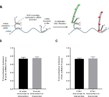
Figure 2.S6: Colocalization between transcript probes and interaction probes for β -actin and FTH1. (A) Schematic of experimental design to test extent of colocalization between mRNA interaction and transcript probes. Multiple interaction and transcript probes are hybridized to a single mRNA, but for illustration purposes, only a single
probe per type is shown. (B) Average colocalization between β-actin mRNA probes
in the cytoplasm. The fraction of β-actin transcript probes colocalized with
interaction probes is 0.74 ± 0.03. The fraction of interaction probes colocalized with transcript probes is 0.75 ± 0.03. Data represent two independent experiments, n = 8 cells. (C) Average colocalization between FTH1 mRNA probes in the cytoplasm. The fraction of FTH1 transcript probes colocalized with interaction probes is 0.73 ± 0.02. The fraction of interaction probes colocalized with transcript probes is 0.74 ± 0.04. Data represent three independent experiments, n = 20 cells.
B -a c tin tr a n s c r ip t to
in te r a c tio n
B -a c tin in te r a c tio n to
tr a n s c r ip t 0 .0 0 .2 0 .4 0 .6 0 .8 1 .0 C o lo c a li z a ti o n b e tw e e n B -a c ti n m R N A P ro b e s
F T H 1 tr a n s c r ip t to
in te r a c tio n
F T H 1 in te r a c tio n to
Figure 2.S7: Levels of β-actin mRNA in control and puromycin-treated NIH 3T3
fibroblasts. The average numbers (± standard deviation) of β-actin mRNAs per cell
are 1999 ± 645 (n = 21 cells) and 1995 ± 608 (n = 23 cells) for control and puromycin-treated cells, respectively. Differences in the values are not statistically significant, P = 0.9843.
C o n t r o l P u r o m yc in
0 1 0 0 0 2 0 0 0 3 0 0 0 4 0 0 0
A
v
e
ra
g
e
#
o
f
B
-a
c
ti
n
m
R
N
A
p
e
r
c
e
Figure 2.S8: Effect of puromycin in combination with 4E1RCAT on interaction of
β-actin mRNA with ribosomes. Fraction of β-actin mRNA transcript spots per cell
colocalized with a ribosome-mRNA interaction spot after no treatment (Control),
treatment with 200 ug/mL of puromycin for 1 h, or treatment 200 µg/mL of
puromycin and 5 µM 4E1RCat for 1 h. n = 3-14 cells per measurement. Error bars,
Figure 2.S9: (A) Western blot and (B) quantification of increased FTH1 protein levels in NIH 3T3 cells treated with iron in the form of hemin at 50 µM for 4, 12, and
24 h. Lysates were first blotted against primary antibodies for FTH1 and β-actin and
then blotted against a goat anti-rabbit IgG secondary antibody coupled to Alexa 488.
β-actin was used as a loading control. L = ladder, 1 = No treatment, 2 = 0.2% DMSO
for 24 h (vehicle), 3 = 4 h hemin, 4 = 12 h hemin, 5 = 24 h hemin. We used ImageQuant TL software to quantify the fold change in FTH1 protein level per treatment condition compared to the control. We first performed a background subtraction of the Western blot with the rolling ball method to remove the baseline intensity. We then measured the integrated intensity of each band and determined the
ratio of the FTH1 band intensity to the β-actin band intensity per lane. The fold
[image:54.612.118.498.100.286.2]Figure 2.S10: Distribution of fluorescence intensities of ribosome-mRNA interaction spots for FTH1 in cells treated with iron for 4 h, 12 h, or 24 h compared to a vehicle control. Representative results from one experiment. Vehicle, n = 10 cells and 2770 spots; 4 h, n = 16 cells and 13360 spots; 12 h, n = 16 cells and 11392 spots; 24 h, n = 15 cells and 11890 spots.
0 0.05 0.1 0.15 0.2 0.25 0.3 0.35 0.4 0.
05 0.1
0.
15 0.2
0.
25 0.3
0.
35 0.4
0.
45 0.5
0.
55 0.6
0.
65 0.7
0.
75 0.8
0.
85 0.9
0. 95 1 Fr a c ti on of S pot s
[image:55.612.113.494.113.335.2]2.8 Supplemental Tables
Table 2.S1: Sequences of all oligonucleotide probes used in Chapter 2. Provided as a separate Excel file.
Table 2.S2: Probe sequences for human 18S rRNA. Provided as a separate Excel file.
Table 2.S3: Fraction (average ± standard deviation) of putative β-actin transcripts (Alexa 488 spots) in the cytoplasm colocalized with Alexa 546 spots, which may
correspond to linker probes or dye-labeled HCR hairpins, when only β-actin
interaction probes, only ribosomes probes, or neither are added. Data represent two independent experiments. n = 4-6 cells per measurement per experiment.
Ribosome probes only mRNA interaction probes only Neither ribosome nor mRNA interaction probes
(Linker + HCR background)
Control 0.08 ± 0.01 0.07 ± 0.02 0.007 ± 0.003
Puromycin 0.09 ± 0.03 0.09 ± 0.03 0.004 ± 0.002
Table 2.S4: Fraction (average ± standard deviation) of putative FTH1 transcripts (Alexa 488 spots) in the cytoplasm colocalized with Alexa 546 spots, which may correspond to linker probes or dye-labeled HCR hairpins, when FTH1 interaction probes, ribosomes probes, or both are omitted. Data represent three independent
experiments. n = 5-9cells per measurement per experiment.
Ribosome probes only mRNA interaction probes only Neither ribosome nor mRNA interaction probes
(Linker + HCR background)
Vehicle 0.06 ± 0.02 0.02 ± 0.02 0.006 ± 0.004
4h 0.06 ± 0.02 0.04 ± 0.01 0.005 ± 0.003
12h 0.05 ± 0.02 0.04 ± 0.02 0.005 ± 0.003
[image:56.612.108.504.278.428.2] [image:56.612.109.506.506.700.2]Table 2.S5: Fraction of ribosome-mRNA interaction spots colocalized with β-actin transcript spots with or without puromycin treatment. Data represent two independent experiments. n = 4-6 cells per measurement per experiment.
Fraction of spots
Control 0.68 ± 0.04
[image:57.612.105.505.263.369.2]Puromycin 0.68 ± 0.03
Table 2.S6: Fraction of ribosome-mRNA interaction spots colocalized with FTH1 transcript spots after different treatments with iron. Data represent three independent
experiments. n = 5-9cells per measurement per experiment.
Fraction of spots
Vehicle 0.54 ± 0.05
4h 0.57 ± 0.04
12h 0.55 ± 0.05
2.9 Methods
Dulbecco’s Modified Eagle Medium (DMEM) (#12491015), Fetal Bovine Serum (FBS) (#10438026), Penicillin-Streptomycin (5,000 U/mL) (#15070063), Trypsin-EDTA (0.05%) (#25300054), RIPA buffer (#89900), and Human Plasma Fibronectin (#33016015) were purchased from ThermoFisher (Tustin, CA). SecureSeal hybridization chambers (8 well, 7mm x 7mm x 0.8mm, SKU 621503) were
purchased from Grace BioLabs (Bend, OR), and 22mm x 50mm No. 1 glass
coverslips were purchased from VWR (Brisbane, CA). All DNA oligonucleotide probes were designed using Stellaris Probe Designer version 4.2 (LGC Biosearch Technologies) and purchased from Integrated DNA Technologies (San Diego, CA). HCR hairpins were purchased pre-coupled to fluorophores from Molecular Instruments (Pasadena, CA). Formamide (SKU F9037), dextran sulfate (SKU D8906), puromycin (SKU P8833), hemin (SKU 51280), Benzonase Nuclease (SKU E1014) and cOmplete Protease Inhibitor Cocktail (SKU 4693116001) were purchased from Sigma-Aldrich (St. Louis, MO). Primary antibodies for FTH1 (ab183781) and ACTB (ab8227) were purchased from Abcam (Burlingame, CA). A goat anti-rabbit IgG secondary antibody coupled to Alexa 488 (A-11034) was purchased from Life Technologies (Carlsbad, CA).
Images were acquired on a Zeiss LSM 800 laser scanning confocal microscope operated by the Biological Imaging Facility of the Beckman Institute at Caltech. Imaging data were analyzed with the FISH-quant program written by Florian Mueller, the Cell Profiler program developed by the Broad Institute Imaging Platform, and the XPIWIT software tool developed by Johannes Stegmaier and the Center for Advanced Methods in Biological Image Analysis at the Beckman Institute (CAMBIA).
Cell Culture. NIH 3T3 fibroblasts were grown in DMEM supplemented with 10%
fetal bovine serum (FBS), 50 units/mL penicillin, and 50 µg/mLstreptomycin at 37˚C
in 5% CO2. Cells were passaged 2-3 times per week. In preparation for each imaging