ISSN Online: 2325-7083 ISSN Print: 2325-7075
DOI: 10.4236/crcm.2017.610029 Oct. 23, 2017 255 Case Reports in Clinical Medicine
“End-Stage” Constrictive Pericarditis—A Case
Report
Ramachandran Muthiah
Thoothukudi Medical College Hospital, Thoothukudi, India
Abstract
Aim: To report a case of “end-stage” constrictive pericarditis with clinical manifestations such as ascites mimicking as cirrhosis of liver. Introduction: In “End-stage” constrictive pericarditis, the etiology remains unknown in majority of cases and inflammation plays a central role in its development. It has been readily confused with cirrhosis of liver in which there may be ascites, but venous pressure is normal, the neck veins are not engorged and cardiac enlargement is frequent in other causes of heart failure. Case Report: A 67 years old male presented with sudden onset of tachycardia. Clinical examina-tion revealed right-sided heart failure, “Egg-shell” calcificaexamina-tion in Chest X-ray and echocardiographic features of pericardial constriction such as septal bounce and dynamic respiratory changes in mitral inflow velocity. The patient was advised medical measures. Conclusion: When clinical signs of right heart failure become unresponsive to increased doses of diuretics, constrictive peri-carditis is more likely the underlying disease since severe, right-sided failure develops in very advanced, the “end-stage” of the disease.
Keywords
“End-Stage” Constrictive Pericarditis, Engorged Neck Vein, Septal Bounce, Waffle Procedure, Amniotic Stem Cell Therapy
1. Introduction
The normal pericardium is a fibroelastic sac enveloping the heart and consists of two layers. The visceral pericardium (serous pericardium) is a single layer of mesothelial cells contiguous with epicardium and a tough, fibrous layer as a pa-rietal pericardium. When the pericardium limits the heart’s ability to function normally either due to accumulation of fluid (pericardial effusion) or scarred
How to cite this paper: Muthiah R. (2017)
“End-Stage” Constrictive Pericarditis—A Case Report. Case Reports in Clinical Medi-cine, 6, 255-273.
https://doi.org/10.4236/crcm.2017.610029 Received: August 28, 2017
Accepted: October 20, 2017 Published: October 23, 2017 Copyright © 2017 by author and Scientific Research Publishing Inc. This work is licensed under the Creative Commons Attribution International License (CC BY 4.0).
http://creativecommons.org/licenses/by/4.0/
DOI: 10.4236/crcm.2017.610029 256 Case Reports in Clinical Medicine and inelastic (constriction), the pericardial compression syndromes may occur.
The constrictive pericarditis is typically chronic, but variants including acute, subacute, transient, occult and end-stage may occur. Historically, the eponym “Pick’s disease” was given to constrictive pericarditis with ascites and hepato-megaly [1] and it was diagnosed as having chronic liver disease and so this case had been reported.
2. Case Report
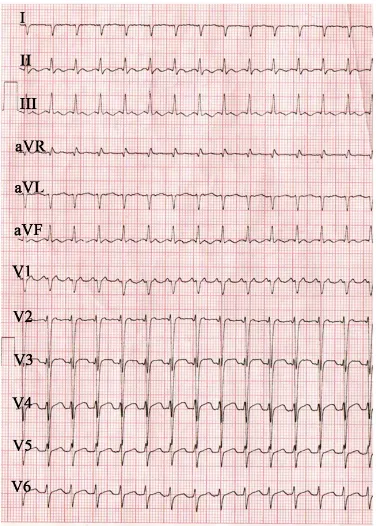
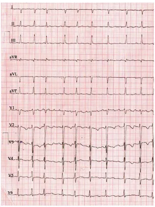
[image:2.595.280.468.431.694.2]A 67 years old male was admitted with sudden onset of palpitations in the emergency room. ECG revealed tachycardia with a heart rate of 150 bpm as in
Figure 1 and blood pressure 110/70 mmHg. Blood chemistry revealed normal.
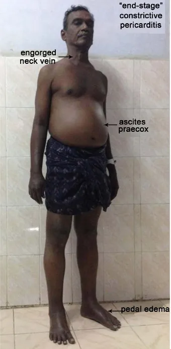
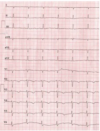
Physical examination showed an engorged neck vein as in Figure 2 which fails to decrease with inspiration (Kussmaul’s sign) with a deep Y descent (Frei-dreich’s sign) reflecting the predominant ventricular filling during early diastole, ascites and pedal edema as shown in Figure 3 suggesting a right-sided heart failure. Auscultation revealed pericardial knock, an early diastolic sound occurs due to cessation in diastolic filling and retraction of apical impulse in systole. X-ray chest revealed “egg-shell’ calcification as shown in Figure 4. Transthoracic echocardiography revealed the features of constrictive pericarditis as in Figures 5-10. Since the patient was in “end-stage” disease, he was given conservative med-ical measures such as diuretics, antibiotics, anti-inflammatory drugs and the rhythm was controlled with calcium channel antagonist, verapamil 40 mg three times daily as shown in Figure 11 and Figure 12.
Figure 1. ECG showing tachycardia (rate 150 bpm) in a 67 year old male with “end-stage”
DOI: 10.4236/crcm.2017.610029 257 Case Reports in Clinical Medicine
Figure 2. Showing the “engorged neck vein” as a feature of elevated venous pressure in
“end-stage” constrictive pericarditis (Photo image with consent).
Figure 3. Showing the clinical features of “end-stage” Constrictive pericarditis (Photo
[image:3.595.288.458.346.693.2]DOI: 10.4236/crcm.2017.610029 258 Case Reports in Clinical Medicine
Figure 4. X-ray chest PA (postero-anterior) view showing the “egg-shell” calcification–
[image:4.595.289.458.380.508.2]“tortoise-shell” like and flattening of right heart border in “end-stage” constrictive peri-carditis.
Figure 5. Showing the “acute angle” between the LA (left atrium) and LV (left ventricle)
posterior walls and a pericardial thickness of 8 mm.
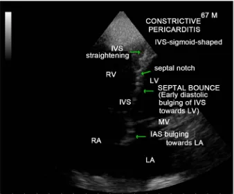
Figure 6. Apical view showing the “septal bounce” as a sign of ventricular
[image:4.595.289.458.554.694.2]DOI: 10.4236/crcm.2017.610029 259 Case Reports in Clinical Medicine
Figure 7. M-mode LV study showing the “septal notch” and “dip and flattening” of LV
[image:5.595.260.489.299.471.2]posterior wall.
Figure 8. Pulsed Doppler imaging showing “the dynamic respiratory change” of the
mi-tral inflow velocity of constrictive pattern.
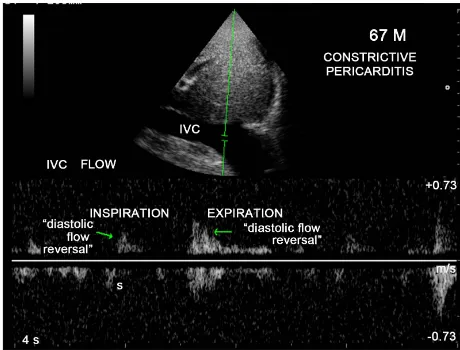
Figure 9. Pulsed Doppler imaging showing the increase in diastolic IVC flow reversal in
[image:5.595.257.487.517.692.2]DOI: 10.4236/crcm.2017.610029 260 Case Reports in Clinical Medicine
Figure 10. Subcostal view showing the dilated IVC (inferior vena cava-plethoric) with no
respiratory variation.
[image:6.595.221.525.299.704.2]DOI: 10.4236/crcm.2017.610029 261 Case Reports in Clinical Medicine
Figure 12. ECG—Rhythn is normalizing on continuation of verapamil.
3. Discussion
Review of literatureDOI: 10.4236/crcm.2017.610029 262 Case Reports in Clinical Medicine Concato described the effusion in serous cavities (polyserositis) in patients with constrictive pericarditis is due to the result of cardiac compression and inflam-mation of serous membranes is absent or occur secondarily.
Etiopathogenesis
Constrictive pericarditis is most commonly caused by conditions or events that cause inflammation to develop around the heart. Inflammatory process of the pericardium typically causes pain and fluid accumulation and more chroni-cally results in fibrosis and calcification of pericardium with pericardial constric-tion, the process that inhibit diastolic filling of the heart. The most common an-tecedents are idiopathic and tuberculosis. The tuberculosis accounted for 49% of cases of constrictive pericarditis in a series reported in 1962 [4] and it was found to be the most common cause in third-world countries such as India [5] [6]. Viral pericarditis is more common in the west and in Europe and North, it is of-ten a sequelae of cardiac surgery and mediastinal irradiation.
Constrictive pericarditis can occur after many pericardial disease processes. All causes of pericarditis can lead to subsequent constriction [7] and in acute pericar-ditis, only 9% of cases may go for constriction. Rheumatic fever, although fre-quently accompanied by pancarditis, does not result in chronic constrictive peri-carditis and may have pericardial adhesion which are not maximally constricting. The pericarditis associated with uremia and myocardial infarction is not of the constricting type and most cases of effusive-constrictive pericarditis are often idi-opathic, can occur in malignancy of breast and lung, tuberculosis, endomyocardial fibrosis and hypothyroidism (cholesterol pericarditis or “gold paint” pericarditis).
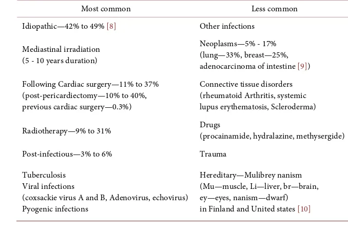
The causes of constrictive pericarditis are shown in Table 1. Hemodynamic changes
[image:8.595.193.532.500.725.2]The normal pericardium can stretch to accommodate the physiological changes in cardiac volume. In constrictive pericarditis, the visceral and parietal pericar-
Table 1. Showing the causes of constrictive pericarditis.
Most common Less common Idiopathic—42% to 49% [8] Other infections
Mediastinal irradiation (5 - 10 years duration)
Neoplasms—5% - 17% (lung—33%, breast—25%, adenocarcinoma of intestine [9]) Following Cardiac surgery—11% to 37%
(post-pericardiectomy—10% to 40%, previous cardiac surgery—0.3%)
Connective tissue disorders (rheumatoid Arthritis, systemic lupus erythematosis, Scleroderma) Radiotherapy—9% to 31% Drugs (procainamide, hydralazine, methysergide) Post-infectious—3% to 6% Trauma
Tuberculosis Viral infections
(coxsackie virus A and B, Adenovirus, echovirus) Pyogenic infections
DOI: 10.4236/crcm.2017.610029 263 Case Reports in Clinical Medicine dium are fibrosed and fused together [11], although not necessarily always thickened [12], prevent the heart from expansion and resulting in minimal abil-ity to adapt to volume changes and significant dynamic respiratory variation in blood flow in the chambers of the heart attributed to isolation of the cardiac chambers from intrathoracic respiratory pressure changes, i.e., dissociation be-tween intrathoracic and intracardiac pressures with enhanced ventricular inte-raction as reported by Hatle et al. in 1989 [13].
In the heart with a normal pericardium, inspiration causes a decrease in in-trathoracic pressure, which is reflected in the cardiac chambers as decrease in intracardiac pressures simultaneously and there is no change in the driving pressure from the lungs across the pulmonary veins into the left atrium and across the mitral valve into the left ventricle. There is some increase in the filling of the right ventricle because of enhanced venous return, but filling of the left ventricle is unaffected throughout the cardiac cycle. In patients with constrictive pericarditis, the rigid pericardium does not allow the decrease in intrathoracic pressure to be transmitted to the left -sided chambers and there is a lower driv-ing force from the lungs into the left side of the heart and the left ventricle be-comes underfilled with a reciprocal increase in the filling of right ventricle and therefore a septal shift occurs [14]. Conversely, during expiration, there is de-creased filling of the right ventricle and inde-creased filling of the left ventricle. As both ventricles are sharing the same limited space, the chamber size and func-tion of one ventricle affect the other ventricle and this interacfunc-tion is known as “ventricular interdependence” since the amount of blood flow into one ventricle is dependent on the amount of blood flow into the other ventricle and it is en-hanced in constrictive pericarditis with a discordance in right and left heart fil-lings.
DOI: 10.4236/crcm.2017.610029 264 Case Reports in Clinical Medicine the edema appears first and ascites much later.
In isolated constrictive pericarditis, the myocardium is unaffected and therefore the systolic function and early diastolic filling are normal. In the mixed form (con-strictive—restrictive—mainly due to radiation—induced, post cardiac surgery), atrophy of myocardial cells and fibrosis may develop during long-term compres-sion by the pericardium. Both the irritation of heart by the actual process involving the myocardium and the constricting effect of left heart chambers on the right ventricle and right atrium result in atrial arrhythmias such as atrial fibrillation as shown in Figure 11 and less commonly atrial flutter as complications in chronic constrictive pericarditis. With diminution in the output of heart, the blood pres-sure, especially the pulse pressure tends to be low and the blood pressure decreas-ing even to the point of disappearance durdecreas-ing inspiration, manifested as absence of pulse, an important sign called as “paradoxical pulse” in some of the more ad-vanced cases. The venous pressure, on the other hand, is very much elevated and frequently exceeding 200 mm of H2O, even exceeding 300 mm of H2O. The salient
[image:10.595.208.540.345.479.2]features of end-stage constrictive pericarditis are shown in Table 2.
Table 2. Showing the salient features of “end-stage” constrictive pericarditis) [17].
High venous pressure Diminution of blood pressure
Paradoxical pulse Atrial fibrillation
Ascites
Low cardiac output (cardiac index ≤ 1.2 L/m2/minute)
Pseudocirrhosis Pedal edema
Diagnostic Methods Radiological
The plain radiograph is frequently abnormal in patients with hemodynami-cally significant constrictive pericarditis [18]. A typical X-ray chest of a patient with constrictive pericarditis shows a normal sized heart (47%) or only mildly en-larged (16%) and moderate to marked enlargement (37%) in effusive-constrictive pericarditis [19]. Cardiac contour abnormalities, particularly the flattening of right cardiac border is a characteristic feature of constrictive pericarditis, but in-frequently present. The left atrium, which is covered only partly by the pericar-dium may be enlarged.
DOI: 10.4236/crcm.2017.610029 265 Case Reports in Clinical Medicine ing a “napkin” ring shape is termed as “napkin-ring” constrictive pericarditis [20]. Once calcification has developed, it represents chronic pericarditis and roughly visible on plain films in 50% of cases [21] [22] which, if present excludes restrictive cardiomyopathy with an overall incidence of 5% to 27% [23], but it may be as high as 44% in patients with tuberculous pericarditis [24]. Two classic patterns of pericardial calcification have been found.
1) The calcium can be thin and linear, and appears as “egg-shell calcification” around the margins of the heart (tortoise shell like) as shown in Figure 4.
2) The calcification also can appear as thick, shaggy, amorphous and histori-cally believed to be specific for tuberculous pericarditis.
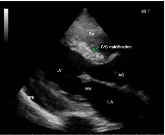
Occasionally, extensive calcification involving the interventricular septum [25] may occur in constrictive pericarditis which indicates an associated disease such as endomyocardial fibrosis as shown in Figure 13. Patients with pericardial calcification are more likely to be idiopathic in origin with features of pericardial knock, atrial arrhythmias as in this patient and have high perioperative mortali-ty.
[image:11.595.210.537.409.678.2]Thickening of pericardium occurs heterogeneously with some areas thicker than other (thinnest over the left ventricle (0.7 - 1.2 mm). A thickened pericar-dium (>4 mm) on its own does not indicate constrictive pericarditis [26]. In 20% of cases of constrictive pericarditis, imaging methods show a pericardium of normal thickness and in such cases, the constrictive process may be caused by epicardial rather than pericardial constriction [27]. Tuberculous constrictive peri-
Figure 13. Showing the IVS (interventricular septum) calcification in effusive-constrictive
DOI: 10.4236/crcm.2017.610029 266 Case Reports in Clinical Medicine carditis is almost always associated with pericardial thickening and mostly, the pericardial thickening is >3 mm by the time the patient becomes symptomatic and >6 mm when the patient is clinically in heart failure.
Two-dimensional Echocardiography
The thickened, constricting pericardium affects the posterior left ventricle (pericardial thickness is 8 mm) more than the posterior left atrium, which then expands at a more acute angle respected to the LV wall [28] as shown in Figure 5. IVS bounce (septal shudder or shivering septum) is the most consistent sign of CP as shown in Figure 6 with a sensitivity of 62% and specificity of 93% [29]. It is an abnormal ventricular coupling manifest by pronounced septal displace-ment during deep inspiration as a “paradoxical bouncing motion” of IVS initial-ly directed towards and then away from the LV during earinitial-ly diastole. The infe-rior vena cava is plethoric (dilated without any respiratory variation in its di-ameter) in constrictive pericarditis as shown in Figure 10 as a manifestation of elevated venous pressures, but right ventricle may show normal contour with tubular morphology (tubularization).
M-mode findings
In constrictive pericarditis, the LV posterior wall rapidly expands posteriorly during early diastole, followed by abrupt cessation of such movement during mid and late diastole, which corresponds to abrupt termination of rapid ventri-cular filling [30] [31] and this lack of motion, termed “flattening” can be best observed with M-mode echocardiography [32] [33] as in Figure 7 [34] and the septum is “sigmoid-shaped” with a bulging towards left ventricle with a septal notch and distal straightening as in Figure 6.
Doppler Echocardiography
Doppler echocardiographic findings include:
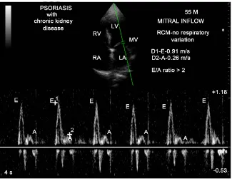
1) Prominent, usually >25% increase in initial E velocity during expiration and decrease during inspiration as shown in Figure 8, the E wave is greater than A wave in both phases of respiration.
Patients with restrictive cardiomyopathy (an infiltrative process that leads to myocardial stiffening) may not show any respiratory variation in mitral inflow velocity as in Figure 14 (restrictive physiology).
Atrial fibrillation may complicate the interpretation of respiratory variation of Doppler velocities, but respiratory variation can still be appreciated regardless of cardiac cycle length.
2) The Figure 9 shows the prominant expiratory diastolic flow reversal in IVC (inferior vena cava) in constrictive pericarditis whereas in restrictive cardi-omyopathy, it is more prominent during inspiration.
Cardiac catheterization
DOI: 10.4236/crcm.2017.610029 267 Case Reports in Clinical Medicine
Figure 14. Pulsed Doppler imaging showing the mitral inflow velocity with no
respirato-ry variation of restrictive pattern.
proximately equal on both sides in simultaneous recordings, a fluid bolus should theoretically increase LVEDP above RVEDP in restrictive cardiomyopathy [36]. Normally, the systolic pressure in the right ventricle and pulmonary artery does not exceed 25 mmHg and if there is predominant constriction of left heart chambers, pulmonary pressure may be twice or thrice the normal. Tachycardia, which abbreviates diastasis, may abolish the plateau in mid and late diastole, but “dip” persists in ventricular pressure tracing of constrictive pericarditis and there is such overlap that these criterias are difficult to apply in an individual case [37] [38].
Management
The treatment of chronic constrictive pericarditis is very much discouraging, but both medical and surgical treatments have been improved greatly.
Medical therapy
The constriction may be transient or reversible. In these conditions, the con-striction is due to inflammation (acute inflammatory pericarditis) and non-ste- roidal anti-inflammatory drugs (NSAIDs) are the most frequent treatment. Fol-low-up studies showing resolution within 2 months to 2 years (usually respond-ing in an average of 8 weeks) and typically effective in idiopathic cases.
tubercul-DOI: 10.4236/crcm.2017.610029 268 Case Reports in Clinical Medicine ous etiology and the use of adjunctive corticosteroids remain controversial since the published trials showed a reduction in mortality and no significant decrease in pericardial fluid reaccumulation or progression to constriction [40] [41]. Pa-tients with tachycardia due to auricular flutter or auricular fibrillation may be-come better with the control of heart rate by digitalis [42] or it may prove of li-mited value [43] and ineffective in heart failure of constrictive pericarditis.
Surgical therapy
The surgical treatment of chronic constrictive pericarditis was first recom-mended by Delorme in 1898 and some years later, the first pericardiectomy for constrictive pericarditis was performed by Franz Volhard collaborates with Vik-tor Schmieden in 1923 and in United States by Churchill in 1929. Decreased cardiac output resulting from a chronic constrictive process may require surgical intervention.
Pericardial resection (or pericardial stripping) is a surgical procedure where the entire pericardium is pealed away from the heart, is a delicate time-consuming procedure, somewhat hazardous and it is different from Brauer’s operation (freeing the heart from thoracic cage). It was the custom to approach the heart anteriorly and to free the right heart chambers and if the whole heart is affected or elevated pulmonary pressure, it would be wise to decorticate the left heart chambers posteriorly first. In patients with heavy calcification penetrating the myocardium, the pericardium could be resected in patches and some islands of epicardium and pericardium were left intact with multiple turtle shell incisions. It is difficult to predict preoperatively which patients are likely to respond to to-tal pericardiectomy. Therefore, recommendations of surgery should be done cau-tiously in patients with mild, very advanced disease, mixed constrictive-restrictive disease, myocardial dysfunction, significant renal dysfunction and radiation in-duced constriction since the prognosis after pericardiectomy was reported to be poor in these disorders [44] [45].
Waffle procedure
Constrictive pericarditis is somewhat associated with constrictive epicarditis especially in Japanese people. Constrictive epicardial thickness might leads to repeat surgery in some cases. Attempts are made to decorticate the white, fibr-ous, thickened layer of epicardium over the ventricles. The waffle procedure performed by incising the tight, fibrotic epicardium in a crosshatched manner releases the epicardial constriction [46] [47] [48].
Amniotic membrane patches (amniotic stem cell therapy)
Current treatment focused on targeting inflammation cental to this disease process and has shown overall positive outcomes [49]. Amniotic stem cell ther-apy consisting of either stem cells with extracellular matrix or extracellular ma-trix alone in the form of human amniotic membrane allograft, an emerging an-ti-inflammatory and antifibrotic treatment [50] [51], applied intraoperatively [52] prior to closure.
DOI: 10.4236/crcm.2017.610029 269 Case Reports in Clinical Medicine The prognosis varies after pericardiectomy according to the etiology of con-strictive pericarditis [53]. Idiopathic and tuberculous patients showed good prognosis at 5 years after pericardiectomy [54].
Preoperative clinical status such as older age, pulmonary hypertension with concomitant myocardial dysfunction, multi-organ failure, atrial fibrillation and high mitral inflow E velocity in Doppler study [55] are related to poor prognosis and 16% of cases may not show any postoperative improvement. “Low-output syndrome” during early postoperative period occurs [56] in patients with long standing symptomatic pericardial constriction due to remodeling of ventricles and weakening of myocardium and it may gradually improves in most of the pa-tients [57].
Case analysis
This case was presented with sudden onset of tachycardia with features of right heart failure. Imaging studies showed an “egg-shell” calcification of peri-cardium, dynamic respiratory changes of mitral inflow velocity and a plethoric inferior vena cava suggesting a constrictive physiology. High venous pressure as “engorged neck vein”, atrial fibrillation, ascites and pedal edema is the manife-stations of this “end-stage” disease.
4. Conclusion
Cardiac catheterization is no longer performed to diagnose constrictive pericar-ditis [58]. Two-dimensional echocardiography and Doppler echocardiography may provide additional diagnostic information and confirm the presence of constrictive physiology. Medical therapy may be used as a palliative measure to control symptoms and to optimize hemodynamics in this “end-stage” disease, who are not candidates for surgery [59].
References
[1] Fowler, N.O. (1995) Constrictive Pericarditis: Its History and Current Status. Clini-cal Cardiology, 18, 341-350. https://doi.org/10.1002/clc.4960180610
[2] Chevers, N. (1842) Observations on the Diseases of the Orifice and Valves of the Aorta. Guy’s Hospital Reports, 7, 387-392.
[3] Wilks, S. (1870-1871) Adherent Pericardium as a Cause of Cardiac Disease. Guy’s Hospital Reports, 16, 196.
[4] Robertson, R. and Arnold, C.R. (1962) Constrictive Pericarditis with Particular Ref-erence to Etiology. Circulation, 26, 525-529.
https://doi.org/10.1161/01.CIR.26.4.525
[5] Bawa, Y.S., Wahi, P.L. and Mehta, M.C. (1960) Pericarditis. A Clinical Survey of 35 Cases. Indian Journal of Medical Society, 14, 111-121.
[6] Bashi, V.V., John, S., Ravikumar, E., Jairaj,J., Shyamsunder, K. and Krishnaswami, S. (1988) Early and Late Results of Pericardiectomy in 118 Cases of Constrictive Pe-ricarditis. Thorax, 43, 637-641. https://doi.org/10.1136/thx.43.8.637
[7] Hancock, E.W. (1990) Neoplastic Pericardial Disease, Cardiology Clinics, 8, 673-682.
DOI: 10.4236/crcm.2017.610029 270 Case Reports in Clinical Medicine
Etiology and Cause-Specific Survival after Pericardiectomy, Journal of American College of Cardiology, 43, 1445-1452. https://doi.org/10.1016/j.jacc.2003.11.048
[9] Flood, S.P., Ayah, O., Furukawa, S. and Norris, R.B. (2017) A Rare Cause of Con-strictive Pericarditis. BMJ (British Medical Journal) Case Reports.
[10] Voorhes, M.L., Husson, G.S. and Blackman, M.S. (1976) Growth Failure with Peri-cardial Constriction, the Syndrome of Mulibrey Nanism. American Journal of Dis-eases of Children, 130, 1146-1148.
https://doi.org/10.1001/archpedi.1976.02120110108017
[11] White, P.D. (1951) Chronic Constrictive Pericarditis. Circulation, 4, 288-294.
https://doi.org/10.1161/01.CIR.4.2.288
[12] Talreja, D.R., Edwards, W.D., Danielson, G.K., Schaff, H.V., Tajik, A.J., Tazelaar, H.D., et al. (2003) Constrictive Pericarditis in 26 Patients with Histologically Nor-mal Pericardial Thickness. Circulation, 108, 1852-1857.
https://doi.org/10.1161/01.CIR.0000087606.18453.FD
[13] Hatle, L.K., Appleton, C.P. and Popp, R.L. (1989) Differentiation of Constrictive Pericarditis and Restrictive Cardiomyopathy by Doppler Echocardiography. Circu-lation, 79, 357-370. https://doi.org/10.1161/01.CIR.79.2.357
[14] Camm, D., Katritsis, G., Gersh, B.J. and John, A. (2013) Constrictive Pericarditis.
Clinical Cardiology, Current Practice Guidelines, 388.
[15] Shabetai, R., Fowler, N.O. and Guntheroth, W.G. (1970) The Hemodynamics of Cardiac Tamponade and Constrictive Pericarditis. American Journal of Cardiology, 26, 480-489. https://doi.org/10.1016/0002-9149(70)90706-X
[16] Bilchick, K.C. and Wise, R.A. (2002) Paradoxical Physical Findings Described by Kussmaul: Pulsus Paradoxus and Kussmaul’s Sign. Lancet, 359, 1940-1942.
https://doi.org/10.1016/S0140-6736(02)08763-9
[17] Hoit, B.D. (2011) Constrictive Pericarditis. UpToDate.
[18] Webb, W.R. and Higgins, C.B. (2005) Thoracic Imaging: Pulmonary and Cardi-ovascular Radiology. Lippincott Williams and Wilkins, Philadelphia.
[19] Hancock, E.W. (1971) Subacute Effusive, Constrictive Pericarditis. Circulation, 43, 183-192. https://doi.org/10.1161/01.CIR.43.2.183
[20] Milkas, A., Van Mieghem, C., Van Hoe, L., Barbato, E. and De Bruyne, B. (2016) The “Napkin-Ring” Constrictive Pericarditis. European Heart Journal—Cardiovascular Imaging, 17, 1436. https://doi.org/10.1093/ehjci/jew203
[21] Miller, S.W. (2005) Cardiac Imaging: The Requisites. 2nd Edition, Elsevier Mosby, Philadelphia.
[22] Masui, T., Finck, S. and Higgins, C.B. (1992) Constrictive Pericarditis and Restric-tive Cardiomyopathy: Evaluation with MR Imaging. Radiology, 182, 369-373.
https://doi.org/10.1148/radiology.182.2.1732952
[23] Ling, L.H., Oh, J.K., Breen, J.F., Schaff, H.V., Danielson, G.K., Mahoney, D.W., et al.
(2000) Calcific Constrictive Pericarditis. Annals of Internal Medicine, 132, 444-450.
https://doi.org/10.7326/0003-4819-132-6-200003210-00004
[24] Bozbuga, N., Erentug, V., Eren, E., Erdogan, H.B., Kirali, K., Antal, A., et al. (2003) Pericardiectomy for Chronic Constrictive Tuberculous Pericarditis: Risks and Pre-dictors of Survival. Texas Heart Institute Journal, 30, 180-185.
DOI: 10.4236/crcm.2017.610029 271 Case Reports in Clinical Medicine
[26] O’ Leary, S.M., Williams, P.L., Williams, M.P., et al. (2010) Imaging the Pericar-dium: Appearances on ECG-Gated 64-Detector Row Cardiac Computed Tomogra-phy. British Journal of Radiology, 83, 194-205. https://doi.org/10.1259/bjr/55699491
[27] Nishimura, R.A. (2001) Constrictive Pericarditis in the Modern Era: A Diagnostic Dilemma. Heart, 86, 619-623. https://doi.org/10.1136/heart.86.6.619
[28] D’Cruz, I.A., Dick, A., Gross, C.M., Hand, C.R. and Lalmalani, G.G. (1989) Abnor-mal Left Ventricular-Left Atrial Posterior Wall Contour: A New Two-Dimensional Echocardiographic Sign in Constrictive Pericarditis. American Heart Journal, 118, 128-132. https://doi.org/10.1016/0002-8703(89)90082-3
[29] Himelman, R.B., Lee, E. and Schiller, N.B. (1988) Septal Bounce, Vena Cava Ple-thora, and Pericardial Adhesion: Informative Two-Dimensional Echocardiographic Signs in the Diagnosis of Pericardial Constriction. Journal of American Society of Echocardiography, 1, 333- 340. https://doi.org/10.1016/S0894-7317(88)80007-5
[30] Feigenbaum, H. (1972) Echocardiography. Lee & Febiger, Philadelphia.
[31] Popp, R.L. (1976) Echocardiographic Assessment of Cardiac Disease. Circulation,
54, 538-552. https://doi.org/10.1161/01.CIR.54.4.538
[32] Schnittger, I., Bowden, R.E., Abrams, I. and Popp, R.L. (1978) Echocardiography: Pericardial Thickening and Constrictive Pericarditis. American Journal of Cardiol-ogy, 42, 388-395. https://doi.org/10.1016/0002-9149(78)90933-5
[33] Voelkel, A.G., Pietro, D.A., Folland, E.D., Fisher, M.L. and Parisi, A.F. (1978) Echocardiographic Features of Constrictive Pericarditis. Circulation, 58, 871-875.
https://doi.org/10.1161/01.CIR.58.5.871
[34] Dai-Biano, J.P., et al. (2009) Role of Echocardiography in the Diagnosis of Constric-tive Pericarditis. Journal of the American Society of Echocardiography, 22, 24-33.
https://doi.org/10.1016/j.echo.2008.11.004
[35] Hansen, A.T., Eskildsen, P. and Gotzsche, H. (1961) Pressure Curves from the Right Auricle and the Right Ventricle in Chronic Constrictive Pericarditis. Circulation, 3, 881-888. https://doi.org/10.1161/01.CIR.3.6.881
[36] Tyberg, T.I., Goodyer, A.V., Hurst 3rd, V.W., et al. (1981) Left Ventricular Filling in
Differentiating Restrictive Amyloid Cardiomyopathy and Constrictive Pericarditis,
American Journal of Cardiology, 47, 791.
https://doi.org/10.1016/0002-9149(81)90175-2
[37] Shabetai, R. (1992) Controversial Issues in Restrictive Cardiomyopathy. Postgra-duate Medical Journal, 68, 547-551.
[38] Vaitkus, P.T. and Kussmaul, W.G. (1991) Constrictive Pericarditis versus Restric-tive Cardiomyopathy: A Reappraisal and Update of Diagnostic Criteria. American Heart Journal, 122, 1431-1441. https://doi.org/10.1016/0002-8703(91)90587-8
[39] Marshall, A., Ring, N. and Lewis, T. (2006) Constrictive Pericarditis: Lessons from the Past Five Years Experience in the South West Cardiothoracic Centre. Clinical Medicine, 6, 592-597. https://doi.org/10.7861/clinmedicine.6-6-592
[40] Mayosi, B.M., Burgess, L.J. and Doubell, A.F. (2005) Tuberculous Pericarditis. Cir-culation, 112, 3608-3616. https://doi.org/10.1161/CIRCULATIONAHA.105.543066
[41] Ntsekhe, M., Wiysonge, C., Volmink, J.A., Commerford, P.J. and Mayosi, B.M. (2003) Adjuvant Corticosteroids for Tuberculous Pericarditis: Promising, But Not Proven. Quarterly Journal of Medicine, 96, 593-599.
https://doi.org/10.1093/qjmed/hcg100
[42] White, P.D. (1951) Chronic Constrictive Pericarditis. Circulation, 4, 288-294.
DOI: 10.4236/crcm.2017.610029 272 Case Reports in Clinical Medicine
[43] Mehta, A., Mehta, M. and Jain, A.C. (1999) Constrictive Pericarditis. Clinical Car-diology, 22, 334-344. https://doi.org/10.1002/clc.4960220509
[44] Ling, L.H., Oh, J.K., Schaff, H.V., et al. (1999) Constrictive Pericarditis in the Mod-ern Era: Evolving Clinical Spectrum and Impact on Outcome after Pericardiectomy,
Circulation, 100, 1380-1386. https://doi.org/10.1161/01.CIR.100.13.1380
[45] Seifert, F.C., Miller, D.C., Oesterle, S.N., Oyer, P.E., Stinson, E.B. and Shumway, N.E. (1985) Surgical Treatment of Constrictive Pericarditis: Analysis of Outcome and Diagnostic Error. Circulation, 72, 264-273.
[46] Heimbecker, R.O., Smith, D., Shimizu, S. and Kestle, J. (1983) Surgical Technique for the Management of Constrictive Epicarditis Complicating Constrictive Pericar-ditis (The Waffle Procedure). Annals of Thoracic Surgery, 36, 605-606.
https://doi.org/10.1016/S0003-4975(10)60693-5
[47] Shiraishi, M., Yamaguchi, A., Muramatsu, K., Kimura, N., Yuri, K., Matsumoto, H.,
et al. (2015) Validation of Waffle Procedure for Constrictive Pericarditis with Epi-cardial Thickening. General Thoracic and Cardiovascular Surgery, 63, 30-37.
https://doi.org/10.1007/s11748-014-0434-6
[48] Matsuura, K., Mogi, K. and Takahara, Y. (2015) Off-Pump Waffle Procedure Using an Ultrasonic Scalpal for Constrictive Pericarditis. European Journal of Cardiotho-racic Surgery, 47, e220-222. https://doi.org/10.1093/ejcts/ezu554
[49] Syed, F.F., Schaff, H.V. and Oh, J.K. (2014) Constrictive Pericarditis—A Curable Diastolic Heart Failure. Nature Reviews Cardiology, 11, 530-544.
https://doi.org/10.1038/nrcardio.2014.100
[50] Hemphil, C., Stavoe, K. and Khalpey, Z. (2014) First in Man: Amniotic Stem Cell Injection Promotes Scar Remodeling and Healing Processes in Late-Stage Fibrosis,
International Journal of Cardiology, 174, 442-443.
https://doi.org/10.1016/j.ijcard.2014.04.023
[51] Khalpey, Z., Marsh, K.M., Ferng, A., Riaz, I.B., Friedman, M., Indik, J., Avery, R., Jokerst, C. and Oliva, I. (2015) First in Man: Amniotic Patch Reduces Post-Operative Inflammation. American Journal of Medicine, 128, e5-e6.
https://doi.org/10.1016/j.amjmed.2014.08.028
[52] Marsh, K.M., et al. (2017) Anti-Inflammatory Properties of Amniotic Membrane Patch Following Pericardiectomy for Constrictive Pericarditis. Journal of Cardio-thoracic Surgery, 12, 6. https://doi.org/10.1186/s13019-017-0567-7
[53] Avgerinos, D., Rabitnokov, Y., Worku, B., Neragi-Miandoab, S. and Girardi, L.N. (2014) Fifteen-Year Experience and Outcome of Pericardiectomy for Constrictive Pericarditis. Journal of Cardiac Surgery, 29, 434-438.
https://doi.org/10.1111/jocs.12344
[54] Szabo, G., Schmack, B., Bulut, C., Soos, P., Weymann, A., Stadtfeld, K., et al. (2013) Constrictive Pericarditis: Risks, Aetiologies and Outcomes after Total Pericardiec-tomy: 24 Years of Experience. European Journal of Cardiothoracic Surgery, 44, 1023-1028. https://doi.org/10.1093/ejcts/ezt138
[55] Kang, S.H., Song, J.M., Kim, M., Choo, S.J., Chung, C.H., Kang, D.H., et al. (2014) Prognostic Predictors in Pericardiectomy for Chronic Constrictive Pericarditis.
Journal of Thoracic and Cardiovascular Surgery, 147, 598-605.
https://doi.org/10.1016/j.jtcvs.2013.01.022
DOI: 10.4236/crcm.2017.610029 273 Case Reports in Clinical Medicine
[57] Omoto, T., Minami, K., Varvaras, D., Bothig, D. and Korfer, R. (2001) Radical Pe-ricardiectomy for Chronic Constrictive Pericarditis. Asian Cardiovascular and Thoracic Annals, 9, 286-290. https://doi.org/10.1177/021849230100900409
[58] Sohn, D.-W. (2012) Constrictive Pericarditis as a Never Ending Story: What’s New?
Korean Circulation Journal, 42, 143-150. https://doi.org/10.4070/kcj.2012.42.3.143
[59] Adler, Y., Charron, P., Imazio, M., Badano, L., Baron-Esquivias, G., Bogaert, J., et al.
(2015) ESC Guidelines for the Diagnosis and Management of Pericardial Disease,
![Table 2. Showing the salient features of “end-stage” constrictive pericarditis) [17].](https://thumb-us.123doks.com/thumbv2/123dok_us/83116.508625/10.595.208.540.345.479/table-showing-salient-features-end-stage-constrictive-pericarditis.webp)