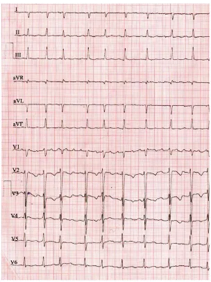
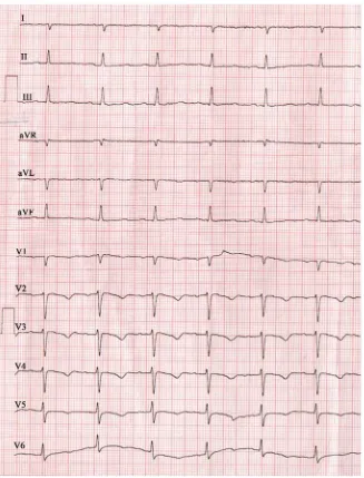
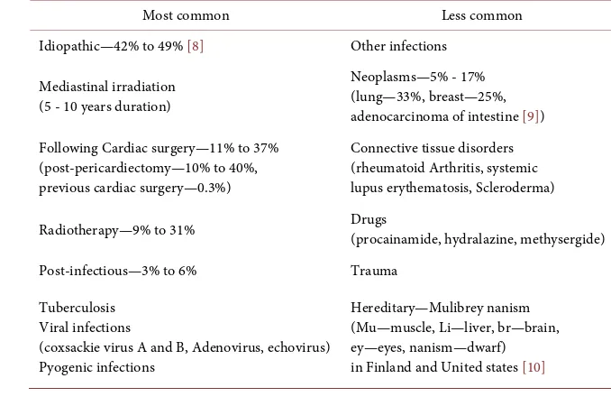
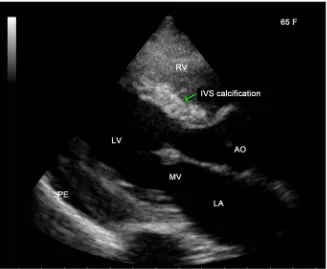
“End Stage” Constrictive Pericarditis—A Case Report
Full text
Figure
![Table 2. Showing the salient features of “end-stage” constrictive pericarditis) [17].](https://thumb-us.123doks.com/thumbv2/123dok_us/83116.508625/10.595.208.540.345.479/table-showing-salient-features-end-stage-constrictive-pericarditis.webp)
Related documents
The proportional and integral gains of the controller are calculated so that the controller guarantees stability, minimization of the closed loop response and robustness
At the very least, the Health Department should have sent someone out to the hospital to assess Patient X and create a plan for after care – standard protocol, according to
By using the concept of a fundamental theorem of neutro-homomorphism and neutro-isomorphism theorems, the relation between neutrosophic algebraic structures (neutrosophic triplet
Conclusion- In this paper the finding of optic disk is made by means of skin locustechniques, blood vessel segmentation and exudates detection by means of
Subgroup analyses on the type of PAM, diagnoses, feed- back frequency, risk of bias judgement and type of PA measure on the effect of the intervention on PA (Add- itional file 1
line. Key Words: Computer Vision; Concealed Firearm Detection; Human Gait Analysis; Video Surveillance.. Background of the Study.. The increase in crime and risk of terrorist
Figure 4 Schematic representation of filament dynamics in presence of nucleation promoting factors (NPFs). a) Filaments treadmill by attachment of monomers at one end and detachment
This study has shown that treatment delay of up to several days plays no significant role, optimal repositioning of root fractures with dislocation of the coronal fragment of up to 1
