Original Article
Loss of Mac-1 causes lung respiratory failure
through affecting type I alveolar epithelial cells
Yanbin Ci1*, Cuili Liu2*, Jun Wang2, Hongmei Sun3, Huahua Liu2
Departments of 1ICU, 2Respiratory Medicine, 3Nursing, Laizhou People’s Hospital, Laizhou, Shandong, China. *Equal contributors.
Received March 14, 2017; Accepted April 25, 2017; Epub December 15, 2017; Published December 30, 2017
Abstract: Type I alveolar cell damage associates with a variety of lung diseases, and severe damages can lead to respiratory failure. Mac-1 as a member of integrin family has been studied for a long time. This study investigated
the relationship between Mac-1 deficiency and respiratory failure in Mac-1 Knockout (Mac-1-/-) mice. C
57 mice were used as a control. The newborn survival rate of Mac-1-/- mice was calculated; HE staining of mice lung tissue was performed for histological tests; Western Blotting, Q-PCR detection were used to detect the expression of type I and type II alveolar epithelial cells, as well as alveolar surfactant secreted by type II alveolar epithelial cells. Birth survival rate of Mac-1-/- mice was significantly lower than that of C
57 mice; in lung floating experiment, the lung of C57 mice
were floating upwards, but for Mac-1-/- mice, the lungs sank downwards to the bottom of the EP tube. Compared with C57 mice, the ProSP-C, as the specific protein of type II alveolar epithelial cells, and the alveolar surfactants in
Mac-1-/- mice had no significant differences, and the structure and function were basically complete. However, western
blotting showed that expression of T1α, Aqp5 and Snx5 in Mac-1-/- mice, as the specific proteins of type I alveolar
epithelial cells, was decreased significantly than those in C57 mice (P<0.05). Mac-1 may play an important role in
the development of respiratory failure. Lack of Mac-1 leading to respiratory failure is not affected by type II alveolar
epithelial cells or their secretive surfactant, but rather by reducing type I alveolar cells.
Keywords: Mac-1, type I alveolar epithelial cells, respiratory failure, type II alveolar epithelial cells
Introduction
Respiratory failure is a common disease in clin-ic, and severe cases, if not get the timely and effective treatments, can lead to a series of serious complications, such as multiple organ dysfunction syndrome (MODS), and eventually death [1, 2]. Alveolar epithelial cells are com-posed of type I and type II alveolar cells, of which, type II alveolar epithelial cells are syn-thesized by the original stem cells, having the potential to differentiate into type I alveolar epi-thelial cells, which can synthesize and secrete
alveolar surfactant, and are of great signifi
-cance to maintain the stability of alveolar [3, 4]. There are many in-depth studies on the type II alveolar cells in the pathogenesis of lung injury.
Type I alveolar epithelial cell, a large and flat
cell, covers more than 95% of the alveolar sur-face area, which is the target cell of various types of damage. Type I alveolar epithelial cells
may have a variety of biological functions, besides being involved in the formation of air-blood barriers, and they can also transport water and ions with certain immune regulating functions [5, 6]. However, the role of type I alve-olar cells in the pathogenesis of lung injury remain unclear.
Mac-1 is a member of β2 integrin family. As an
important adhesion molecule, it is involved in the body defense and immune response, and expressed widely in partial white blood cells, such as neutrophils, monocytes, eosinophilic
cells and NK cells, with few expressions in T cells, macrophages, and B cells [7-9]. Lack of
Mac-1 affects the migration and tissue infiltra
-tion of white blood cells, which play a biological
function in the inflammatory response, and inflammation has a close relationship with the
Mac-1 in the occurrence and development of lung injury, especially of its role in the type I alveolar epithelial cells. This article focuses on the role of Mac-1 in respiratory failure and its mechanism.
Materials and methods
Materials
Experimental animals and breeding: Mac-1
Knockout mice (B6.129S6-Itgam tm1Myd/J),
homozygous (Mac-1-/-), were purchased from
the Jackson Laboratory (US), coded as 003991.
C57 BL/6J mice (hereinafter referred to as C57)
were used as the background control mice, pur
-chased from in the Medical Laboratory Animal Center of Shandong Province, the production
license was SCXK 2008-0002. These mice
were raised in VMC64S7 independent delivery
and return air purification baskets (Suhang Experimental Animals Equipment Factory) in SPF environment, room temperature was 22 to
28°C, relative humidity was 50% to 70%, auto
-matic light control (12 h light/12 h dark). Feed
were purchased from Shandong Animal Center, which underwent 60Co irradiation sterilization.
Drinking water was sterilized urban water.
Mice were used for all experiments, and all pro-cedures were approved by the Animal Ethics Committee of People’s Hospital of Laizhou City.
Main instruments and reagents
PCR (Biometra Company); Stereoscopic
micro-scope (Motic Company); Proteinase K, dNTP (Shanghai Univ-bio Company); PCR primers (Invitrogen Trading (Shanghai) Co., Ltd); Taq
polymerase, Tris saturated phenol, chloroform (sigma Co., Ltd).
Methods
Mac-1-/- gene identification: Identification
se-quence of Mac-1 knockout mice: mutations
stripe: primer 1: 5’-TAG GCT ATC CAG AGG TAG AC-3’; Primer 2: 5’-ATC GCC TTC TTG ACG AGT
TCA-3’, amplification bands were 700 bp. Wild
type stripe: primer 1: 5’-TAG GCT ATC CAG AGG TAG AC-3’; Primer 2: 5’-CAT ACC TGT GAC CAG
AAG AGC-3’, amplification bands were 325 bp.
The PCR reaction conditions: denaturations
were at 94°C for 3 minutes, 94°C for 30 sec
-onds, 58°C for 1 minute, 72°C for 2 minutes,
with 35 cycles; Extensions were at 72°C for 2 minutes, annealing was at 10°C.
Study group and the control
The male and female C57 mice, in the propor-tion of 1:2, mated in the full siblings mating way, and the pregnant female mice, as the con-trol group, were placed separately in a cage. The male and female Mac-1-/- mice, in the pro-portion of 1:2, mated in the full siblings mating way, and the pregnant female mice, as the experimental group, were placed separately in another cage. The feeding conditions for the two mice were the same. All operations were conducted within the scope of ethics.
Lung floating experiment
The lung tissues, after Mac-1-/- mice were
sacri-ficed, were immediately removed to a centri
-fuge tube with 0.5 ml of PBS.
HE staining
The lung tissues were placed in formalin to be
fixed for one night, and then dehydrated.
Dehydration was performed as follows: 70%
ethanol for 3 h, 80% ethane for 3 h, 95% etha
-nol for 2 h, 100% etha-nol I for 1.5 h, 100% ethanol II for 1.5 h, Xylene I/II each for 0.5 h,
paraffin I for 1 h, paraffin II for 2 h (60°C); Conventional slice and the thickness was 3 μm.
Dewaxing: per the conventional dewaxing
method: 10 minutes were taken in xylene,
anhydrous ethanol, 95% ethanol, 90% ethanol,
85% ethanol and 80% ethanol respectively.
Conduct hematoxylin stain for 1 min, and wash it with water to blue, then conduct eosin stain for 10 sec, wash it with water, dry it and mount followed by observation under the microscope,
take 20× of the middle part in the visual field,
and then three different pathology technicians read and diagnosed it.
Immunofluorescence staining
After the tissue was fixed, use sucrose for gra
-dient dehydration, freeze and then slice by 6
mm of thickness, repair at high temperature for
5 min. After it cools, PBS washed it for three
times, five minutes per time followed by addi
three times, 5 min for each time, then conduct
second antibody fluorescence staining. Primary
antibody was proSP-C (anti-rabbit), purchased from Santa, 1:100; second antibody was
anti-rabbit (488 nm), purchased from Santa, 1:100.
Dry mounting. Calculate the ratio of positive
cell number to the area of vision field by J image
software. Compare the difference of positive rates that was the positive expression differ-ence. Get rid of bubble from the plate before sampling.
Western blotting
Tissue proteins were extracted from protein kit, and use BCA for quantitative analysis. Prepare BCA solution by the ratio of A:B to 50:1. Take 2 μl of collected cell supernatant, add 18 μl of PBS and 200 μl of AB mixed solution. All protein
samples were adjusted to the same
concentra-tion, and add 1× bromphenol blue, which
accounted for 1/5 of the total volume. All pro-tein samples were transferred to the same
con-centration, and then sample, while adding 6 μl of protein marker. run in concentration gel at the initial voltage of 80 V, and then raise to 120 V to run in the separation gel. finish the process
when the target protein swam to 1 cm above
the edge of the gel. immerse PVDF membrane
in methyl for 5 min. Constant current was at
250 mA for 90 minutes. Take the membrane
out of electronic transfection pool; rinse slightly
with TBST, swing slowly in 5% skim milk sealing
solution for 1 h. Rinse slightly with TBST. Incubate with primary antibody overnight, and warm it at room temperature for 40 minutes on the second day. Rinse the membrane with TBST three times, 5 minutes for each time. Choose the secondary antibody per the primary anti-body, and swing slightly at room temperature for 1 hour. After incubation with the secondary antibody, wash the membrane with TBST three times, 5 minutes for each time. The washed
PVDF film was developed with ECL with the con
-figuration of ECL liquid being A:B as 1:1.
Statistical analysis
Western-Blot results were processed by the Image-J software which were repeated at least three times and showed as mean ± standard deviation (SD). SPSS11.0 was used for statisti-cal analysis. Comparison of difference was assessed by student t test. P<0.05 indicated
the difference was statistically significant.
Results
The survival rates of C57 and Mac-1-/- newborn
mice
The survival rate of C57 newborn mice was 100% at 10 hours after birth (Figure 1A), but the survival rate of Mac-1-/- newborn mice reduced to 60% at 1 hour after birth, which was decreased more with time increasing, and at 70 hours after birth, it was reduced to less than Figure 1. A. The Survival Rates of C57 and Mac-1-/- Newborn Mice, *indicates P<0.05; B. The Obvious Cyanosis of Mac-1-/- Newborn Mice Body, Compared with C
40%. The difference of survival rates between
these two groups was statistically significant
(P<0.05); Compared with newborn C57 mice, the body of Mac-1-/- newborn mice showed
obvi-ous cyanosis. The specimens were taken from
mice at 24 hours after birth (Figure 1B).
Alveolar layer thickening of Mac-1-/- mice
The lung tissue of C57 mice was floating in PBS
buffer fluid, but for Mac-1-/- mice, some of the
failure in newborn Mac-1-/- mice, expression of
the specific protein proSP-C of type II alveolar
epithelial cells was detected by immunofluores
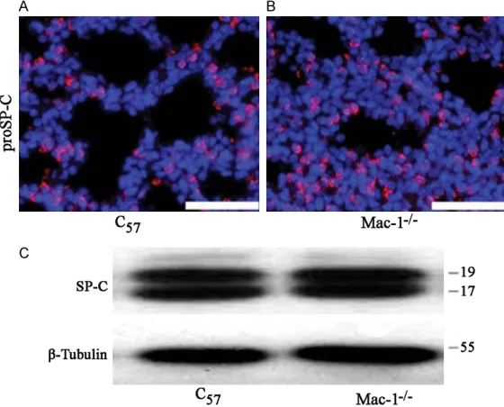
-cence assay. Figure 3A and 3B showed no
sig-nificant difference of the expressions of
proSP-C between proSP-C57 mice and in Mac-1-/- mice. Consistent with this, the results of Western blot
showed that there was no significant difference
[image:4.612.94.526.72.222.2]in the expression of SP-C secreted protein between C57 and Mac-1-/- mice (Figure 3C). These results indicate that the structure and Figure 2. Alveolar Layer Thickening of Mac-1-/- Mice. A. Indicated that compared with C
57 mice, Mac-1-/- mice may have respiratory failure at birth. B. Showed that compared with that of C57 mice, the alveolar layer of mac-1-/- mice
became thickening.
Figure 3. The difference between the alveolar surfactant proteins of type II alveolar epithelial cells in C57 and Mac-1-/- mice was not significant. A and B.
Showed the results of proSP-C detected by immunofluorescence assay. C.
Showed the western blotting results of SP-C.
lung tissue was floating, most
was falling down to the bot-tom of the tube due to in- creased density caused by
insufficient lung expansion
(Figure 2A). These
phenome-na indicated that Mac-1-/- mice may have respiratory failure at birth. And the results of lung tissue HE staining sh- owed that the alveolar layer
became thickening, monolay
-er alveolar cells became dou-ble and even multi-layer cells in Mac-1-/- mice. The lung tis-sue was drawn from the new-born mice at 24 hours after birth (Figure 2B).
The structure and function of type II alveolar cells in
Mac-1-/- mice were relatively
complete
[image:4.612.95.375.288.514.2]function of the alveolar surface of Mac-1-/- mice
were not significantly different from that of C57
mice, and there was no significant difference
between the expressions of alveolar surfactant
proteins in these two kinds of mice.
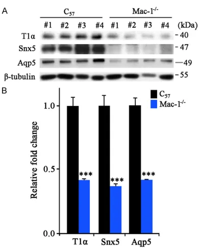
The specific proteins of type I alveolar cells in
Mac-1-/- mice decreased
The expression of specific proteins of type I alveolar cells, T1α, Aqp5 and Snx5, were
detected. Western blotting results showed that compared with those in C57 mice, the expres-sion of these three proteins in Mac-1-/- mice
was decreased with statistical significance
(P<0.001), indicating that the occurrence of respiratory failure may be related to the decrease of type I alveolar epithelial cells, as shown in Figure 4.
Discussion
Mac-1 is a member of β2 integrin family, which,
as an important adhesion molecule, is involved in body defense and immune response, as well
as an important participant in inflammatory reaction, and inflammatory injury is an exact
important factor in the lung injury [12]. It has
been reported that Mac-1 plays an role in the occurrence of the lung disease [13], however, the study of Mac-1 in vivo experiment is very rare. This study investigated the effect of Mac-1 on respiratory failure. Compared with that of C57 mice, the perinatal mortality of Mac-1-/- mice
was increased significantly, and the body of
died mice showed obvious cyanosis, indicating that respiratory failure may be developed
before mice died. In lung floating experiments,
the lung tissue of Mac-1-/- mice was falling down to the bottom of the tube, indicating excessive lung tissue hyperplasia leading to density
increase. It was not hard to find by pathological
examination that the cause of mice death was
very likely to be respiratory failure. Some stud
-ies have indicated that the respiratory failure is caused by the incomplete differentiation of type I or II alveolar epithelial cells, so we have a further study on alveolar epithelial cells. To further explore the mechanism of lung injury,
the specific markers of alveolar epithelial cells
were detected, and results found that Mac-1 gene absence caused the impaired differentia-tion of type I alveolar epithelial cells, which resulted in the reduction of type I alveolar epi-thelial cells, eventually leading to the occur-rence of respiratory failure. Other study has indicated that Mac-1 plays an important role in the maturation and differentiation of type I alveolar epithelial cells, so our study further investigated the role of Mac-1 in type I alveolar epithelial cells [14-17].
Type I alveolar epithelial cells cover more than 95% of the alveolar surface area, which are the target cells of various injuries. Besides partici-pating the formation of air-blood barrier, they also transport water and ions, and have certain
immune regulation functions [18, 19]. Type I
alveolar cells are differentiated from type II alveolar cells, the mechanism of which in lung injury has been studied deeply [20]. Our study found that Mac-1 did not directly affect type II alveolar epithelial cells and the secretion of SP-C.
In conclusion, loss of Mac-1 affects the differ-entiation of type 1 alveolar cells and
subse-quent resulting in the incomplete of the lung
[image:5.612.91.286.67.311.2]structure and function, eventually leading to respiratory failure, suggesting the role of Mac-1 in the development and pathogenesis of respi-ratory failure.
Figure 4. The specific proteins of type I alveolar cells, T1α, Aqp5 and Snx5 decreased significantly in
Acknowledgements
This project supported by the National Natural
Science Foundation of China (NO. 8242010-8089).
Disclosure of conflict of interest
None.
Address correspondence to: Dr. Huahua Liu, De partment of Respiratory Medicine, Laizhou People’s
Hospital, No. 1718, Wuli Street, Laizhou, Shandong, China. Tel: 2276081; Fax:
+86-0535-2212735; E-mail: [email protected]
References
[1] Morrisey EE, Hogan BL. Preparing for the first
breath: genetic and cellular mechanisms in
lung development. Dev Cell 2010; 18: 8-23.
[2] Pei L, Leblanc M, Barish G, Atkins A, Nofsinger
R. Thyroid hormone receptor repression is
linked to type I pneumocyte-associated respi -ratory distress syndrome. Nat Med 2011; 17: 1466-1472.
[3] McGough IJ. CP. Recent advances in retromer
biology. Traffic 2011; 12: 963-971.
[4] Weinmaster G, Fischer JA. Notch ligand ubiqui -tylation: what is it good for? Dev Cell 2011; 21: 134-144.
[5] Geng Y, Dong Y, Yu M, Zhang L, Yan X. Fol
-listatin-Like 1 (Fstl1) Is a bone morphogenetic
protein (BMP) 4 signaling antagonist in control-ling mous lung development. Proc Natl Acad
Sci U S A 2011; 108: 7058-7063.
[6] Lee JS, Kim Y, Kim IS, Kim B, Choi HJ. Negative
regulation of hypoxic responses via induced
reptin methylation. Mol Cell 2010; 39: 71-85.
[7] Simmons G, Bertram S, Glowacka I, Steffen I, Chaipan C, Agudelo J, Lu K, Rennekamp AJ,
Hofmann H, Bates P and Pohlmann S. Differ-ent host cell proteases activate the
SARS-coro-navirus spike-protein for cell-cell and virus-cell
fusion. Virology 2011; 413: 265-274.
[8] Gao HM, Zhou H, Zhang F, Wilson BC, Kam W
and Hong JS. HMGB1 acts on microglia Mac1
to mediate chronic neuroinflammation that
drives progressive neurodegeneration. J
Neu-rosci 2011; 31: 1081-1092.
[9] Ho MK and Springer TA. Mac-1 antigen: quanti -tative expression in macrophage populations
and tissues, and immunofluorescent localiza
-tion in spleen. J Immunol 1982; 128: 2281-2286.
[10] Matsumoto M, Oshiumi H, Seya T. Antiviral re-sponses induced by the TLR3 pathway. Rev Med Virol 2011; 21: 67-77.
[11] Heald-Sargent T, Gallagher T. Ready, set, fuse!
The coronavirus spike protein and acquisition
of fusion competence. Viruses 2012; 4:
557-580.
[12] Ye J, Zhang B, Xu J, Chang Q, McNutt MA. Mo-lecular pathology in the lungs of severe acute respiratory syndrome patients. Am J Pathol
2007; 170: 538-545.
[13] Qian Z, Travanty EA, Oko L, Edeen K, Berglund
A. Innate immune response of human alveolar type II cells infected with SARS coronavirus.
Am J Respir Cell Mol Biol 2013; 48: 742-8.
[14] Wang J, Nikrad MP, Phang T, Gao B, Alford T, Ito Y, Edeen K, Travanty EA, Kosmider B, Harts
-horn K and Mason RJ. Innate immune re
-sponse to influenza A virus in differentiated
human alveolar type II cells. Am J Respir Cell
Mol Biol 2011; 45: 582-591.
[15] Weinheimer VK, Becher A, Tonnies M, Holland G, Knepper J, Bauer TT, Schneider P, Neudeck
-er J, Ruck-ert JC, Szymanski K, Temmesfeld-Wollbrueck B, Gruber AD, Bannert N, Suttorp N, Hippenstiel S, Wolff T and Hocke AC. Influ -enza A viruses target type II pneumocytes in
the human lung. J Infect Dis 2012; 206:
1685-1694.
[16] Dijkman R, Jebbink MF, Koekkoek SM, Deijs M, Jonsdottir HR, Molenkamp R, Ieven M, Goos
-sens H, Thiel V and van der Hoek L. Isolation
and characterization of current human corona-virus strains in primary human epithelial cell cultures reveal differences in target cell
tro-pism. J Virol 2013; 87: 6081-6090.
[17] Pyrc K, Sims AC, Dijkman R, Jebbink M, Long C,
Deming D, Donaldson E, Vabret A, Baric R, van
der Hoek L and Pickles R. Culturing the uncul
-turable: human coronavirus HKU1 infects, rep -licates, and produces progeny virions in hu-man ciliated airway epithelial cell cultures. J
Virol 2010; 84: 11255-11263.
[18] Mossel EC, Wang J, Jeffers S, Edeen KE, Wang
S. SARS-CoV replicates in primary human
al-veolar type II cell cultures but not in type I-like cells. Virology 2008; 372: 127-135.
[19] Yu M, Lam J, Rada B, Leto TL, Levine SJ. Dou-ble-stranded RNA induces shedding of the
34-kDa soluble TNFR1 from human airway epi-thelial cells via TLR3-TRIF-RIP1-dependent sig -naling: roles for dual oxidase 2- and
caspase-dependent pathways. J Immunol 2011; 186: 1180-1188.
[20] Jamaluddin M, Tian B, Boldogh I, Garofalo RP, Brasier AR. Respiratory syncytial virus infection
induces a reactive oxygen species-MSK1-phospho-Ser-276 RelA pathway requiredfor cy
-tokine expression. J Virol 2009; 83: