Original Article
Lentivirus-mediated RNAi knockdown of HPV16E7
suppresses the proliferation of cervical cancer
cell line CaSki in vitro and in vivo
Ziyu Hu1, Yanan Duan1, Fangning Wang1, Liping Chen2,3
1Department of Obstetrics and Gynecology, People’s Hospital of Dongying, Dongying 257091, China; 2Shandong
University of Traditional Chinese Medicine, Jinan 250011, China; 3Taishan Medical University, Taian 271000,
China
Received April 18 2016; Accepted July 6, 2016; Epub August 15, 2016; Published August 30, 2016
Abstract: Gene therapy is a relatively new method in medicine with large therapeutic potential. Until now, most clini-cal trials in gene therapy have focused on the treatment of cancer. Human papillomavirus type 16 E7 (HPV16E7) plays an important role in maintaining the malignant phenotype of cervical cancer cells. Hence, HPV16E7 becomes the suitable target for gene therapy in treating cervical cancer. In this research, we focus on inactivating HPV16E7 in CaSki cells and examine carcinogenicity of the cells. Lentivirus-mediated RNAi was used to down-regulate the expression of HPV16E7. RT-PCR and western blot were performed to detect the expression of HPV16E7. MTT, col-ony formation in vitro, tumor formation in vivo and apoptosis assay were used to detect the cell proliferation and apoptosis after down-regulation of HPV16E7. Flow cytometry was performed to analyze the changes in cell cycle in response to the down-regulation of HPV16E7. The delivery of lentivirus vector resulted in the decreased expression of HPV16E7 in the level of mRNA and protein. In addition, we found decreased proliferation and increased apoptotic rate in transfected CaSki cells. Furthermore, transfected CaSki cells were arrested in G0/G1 phase. In conclusion, HPV16E7 boosts proliferation of CaSki cells and contributes to tumorigenicity. Hence, HPV16E7 may be a pivotal gene target for therapeutic strategy in treatment of cervical cancer.
Keywords: HPV16E7, cervical carcinoma, RNA interference, lentivirus, proliferation
Introduction
Cervical cancer, the third most commonly diag-nosed cancer, is the fourth leading cause of tumor death in females worldwide [1]. The development of cervical cancer is usually slow. Before carcinogenesis in cervix, cells generally go through some changes known as dysplasia. Then, the abnormal cells become cancer cells and invade into the cervix [2]. Symptoms may include vaginal bleeding, pelvic pain and pain during sexual intercourse. Hence, it is impor-tant to understand the cellular and molecular mechanisms of cervical cancer. There are many factors which determine the treatment proce-dure for cervical cancer, such as age, stage of cancer, and tumor type [3]. In present, women with early cervical cancer can be efficiently treated with radical surgery, and the addition of
Knockdown HPV16E7 suppress cervical cancer cell line growth
HPV16E7 which is expressed by HPV16 plays an important role in malignant transformation. Compelling evidence suggests that HPV16E7 induces centriole amplification to promote the development of cancer resulting in aneuploidy of the E7-expressing cells [7]. In addition, HPV16E7 not only stimulates the S-phase genes cyclin A and cyclin E [8], but also blocks the function of the cyclin-dependent kinase inhibitors p21 and p27 which leading to tumori-genesis [9-11]. Furthermore, HPV16E7 has been reported to play an important role in the proliferation of various kinds of cells, such as thymic epithelium cell [12], keratinocytes [13] and laryngeal squamous carcinoma cell [14]. Hence the HPV16E7 may be a potential gene target for regulating the development of cervi-cal cancer.
Gene therapy is a new technology in clinical trial that genes were delivered to or knocked down in the target tissues by vehicles [15]. Many cancers have been targeted throughout these years, such as skin, lung and cervical cancer. A number of different methods to can-cer gene therapy are being developed, which mainly use viral vectors to invert anti-angiogen-ic factors, tumor-suppressor genes, prodrug-activating genes, immunostimulatory genes or interfere the expression of oncogenes [16]. The main viral vectors include oncoretrovirus, lenti-virus, adenolenti-virus, adeno-associated virus and herpes simplex-1 virus. In our research, as for its broad tropism and persistent gene transfer in most tissues, we chose lentivirus as viral vector to mediate the expression of HPV16E7 in cervical cancer. And more importantly, no one did this before.
Here, lentivirus-mediated RNAi was performed to inhibit the expression of HPV16E7. After knocking down HPV16E7, we detected cell pro-liferation, tumor formation, cell apoptosis and cell cycle phase distribution. Our results sug-gest that lentivirus-mediated RNAi knockdown of HPV16E7 suppresses the proliferation of cervical cancer cell line CaSki in vitro and in vivo.
Materials and methods
Cell line and culture
CaSki cells of human cervical cancer were cul-tured in RPMI 1640 media (Hyclone, USA)
con-taining 10% fetal bovine serum (FBS; Gibco, Carlsbad, CA, USA), 50 U/mL penicillin G and 50 U/mL streptomycin (Gibco) at 37°C under 5% CO2 in humidified air. The cells were passed every 2-4 days.
Construction of lentivirus vectors
Short hairpin RNA (shRNA) targeting HPV16E7 sequence (GCT TCG GTT GTG CGT ACA A)and scrambled non-silencing RNA (TTC TCC GAA CGT GTC ACG T) were designed by using the manufacturer’s RNAi Designer programme. DNA oligos containing the target sequence were chemically synthesized, annealed, and inserted into the expression vector by double digestion with Age Icand EcoR I, and ligation with T4 DNA ligase in accordance with the man-ufacturer’s guidelines. The ligation was trans-formed into competent Escherichia coli DH5a cells. The correct transformant was identified by restriction enzyme analysis and DNA sequencing. The sequences were cloned into the pGCSIL-GFP lentiviral vectors. Expression vectors and package vectors were infected into 293T cells with the help of Lipofectamine 2000. Lentiviral particles such as pGCSIL-HPV16E7-shRNA-LV and pGCSIL-neg-shRNA-LV were harvested after 48 h. Lentiviral particles were purified using ultracentrifugation. The viral titer was determined.
Infection of lentivirus
CaSki cell suspensions were cultured in 96-well microplates with 5×10³ each well. For infection, cells were cultured in complete medium with lentiviruses (10 μg/mL) and treated for 24 h. Then, culture medium was substituted. Three groups of CaSki cells were used in subsequent assays: CON group (blank control group, with no infection), NC group (negative control group, infected with pGCSIL-neg-shRNA-LV), and KD group (HPV16E7 RNAi group, infected with pGCSIL-HPV16E7-shRNA-LV).
Western blot
super-natant. After determining the protein concen-tration, protein samples were subjected to SDS-PAGE and transferred to a PVDF mem-brane. The membrane was blocked with 5% skim milk for 2 h and incubated with primary antibodies against HPV16E7 and GAPDH at 4°C overnight. Then, the membrane was incu-bated with secondary antibody for 2 h. Before each step, membranes were washed by PBST for 3 times. The bands were visualized using an enhanced chemiluminescence system.
Real-time PCR
Total RNA was extracted from cells with Trizol reagent following the manufacturer’s instruc-tions. cDNA was obtained using M-MLV reverse transcriptase kit. Aliquots of cDNA were sub-jected to quantitative real-time PCR using Step One Plus Real-time PCR system. GAPDH mRNA expression level was used for normalization. The specific primer pairs were as follows: HPV16E7, sense: 5’-CGGGATCCATGCATGGAG- ATACA-3’ and antisense: 5’-GCGGGCCCTTATG- GTTTCTGAGA-3’; GAPDH, sense: 5’-TGACTTCA- ACAGCGACACCCA-3’; antisense: 5’-CACCCTGT- TGCTGTAGCCAAAC-3’. Data were analyzed us- ing the 2-∆∆CT method.
Proliferation assay
Lentivirus infected cells were cultured in 96- well plates for 24, 48, 72 h. The cells were incu-bated in the presence of 5 mg/mL 3-(4, 5-Di- methylthiazol-2-yl)-2, 5-diphenyltetrazolium br- omide (MTT; Sigma-Aldrich Corp) for 4 h at 37°C. 100 μL of 20% SDS was added to each well. The absorption at 572 nm was measured using a spectrophotometer (Bio-Rad, USA).
In vitro colony formation assay
800 lentivirus-infected cells were seeded in 6-cm dishes and incubated for 14 days to form colonies. Then the cells were washed twice with PBS following fixation by 4% paraformalde-hyde (PFA). Afterward, cells were stained with Giemsa for 20 min and washed twice by ddH2O. The number of colonies (> 50 cell/colony) was counted manually.
In vivo tumor formation assay
[image:3.612.98.520.72.314.2]All animal care and surgical procedures were performed in accordance with the National Institutes of Health Guidelines for the Care and Use of Laboratory Animals (National Research
Knockdown HPV16E7 suppress cervical cancer cell line growth
Council, 1996, USA). Five-week-old female immune-deficient nude mice (BALB/C-nu) were housed under a 12h light/dark cycle and the room temperature (RT) was kept at 26°C. Mice were randomly divided into CON group and KD group (n=3, each group). 2×10⁶ CaSki cells were subcutaneously injected into the right flank of the mice with and without lentivirus-mediated RNAi knockdown of HPV16E7. After 4 weeks, xenografts were removed and weighed.
Apoptosis assay
A total of 1.0×10⁶ CaSki cells were harvested by centrifugation and washed by PBS. Then, cells were incubated in binding buffer (1 mg/ mL annexin V-APC) for 30 min. Fluorescence-activated cells sorting (FACS) analysis for
annexin V-APC staining was performed by flow cytometer (FACSCaliburTM, BD, USA).
Cell cycle analysis
Lentivirus-infected cells were digested by tryp-sin and centrifuged at 1200 rpm for 5 min. Then cells were washed by ice-cold PBS and fixed by 70% ethanol. After discarding ethanol, cells were resuspended in 20 mg/mL propidi-um iodide (PI). Cells were analyzed by flow cytometer (FACSCaliburTM, BD, USA).
Statistical analysis
[image:4.612.92.517.74.386.2]All numerical data were described as means ± SD. Data were analyzed using the one-way ANOVA. A probability value of 0.05 or less was considered significant. Analyses were carried
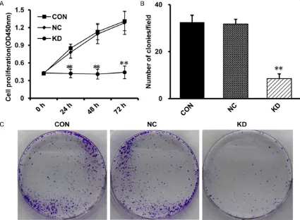
Figure 2. Inhibition of cell growth with HPV16E7 RNAi in CaSki cells. A. Cell proliferation was determined by MTT as-say. Compared with the CON group and the NC group, cell proliferation in the KD group is suppressed at each time point (48 h and 72 h). The data are means ± SD, n=3, **P<0.01 vs the NC group. B. Colony formation assay was
performed to comfirm the inhibitory effect of lentivirus-mediated RNAi. Compared with the CON group and the NC group, the clone number in the KD group was significantly decreased. The data are means ± SD, n=3, **P<0.01 vs
out using GRAPHPAD Prism Version 5.0 (Graph-Pad Software, La Jolla, CA, USA).
Results
HPV16E7-RNAi-LV down-regulates HPV16E7 expression in CaSki cells
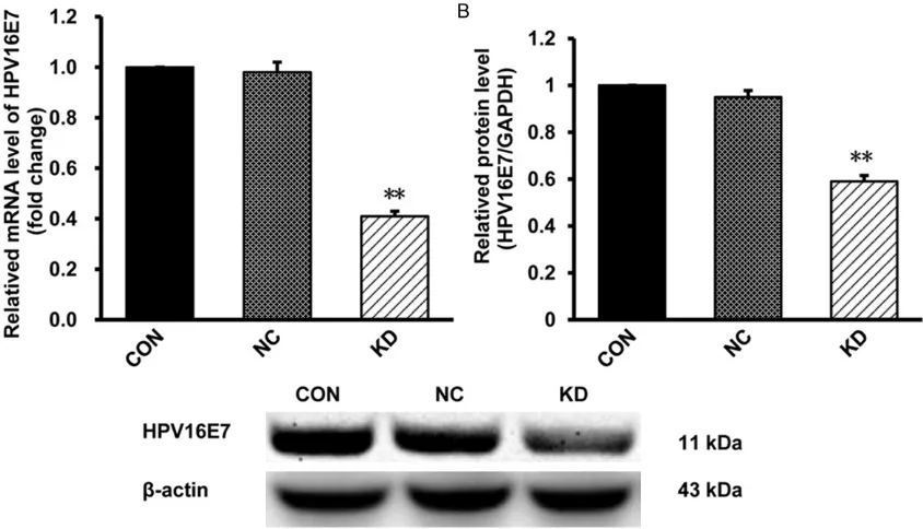
After lentivirus-mediated RNAi knockdown of HPV16E7, the efficiency of HPV16E7-RNAi-LV was examined by RT-PCR and western blot. Compared with NC group, the expression of HPV16E7 mRNA and protein in KD group was significantly decreased, respectively (Figure 1). The mRNA level was reduced by 60.4%, while the protein level decreased by 40.8%. There is no significantly difference between CON and NC groups. These results showed that HPV16E7 was down-regulated efficiently in the lentivirus-infected CaSki cells.
Knockdown of HPV16E7 inhibits proliferation and clonogenicity of CaSki cells
To determine the role of HPV16E7 on cell prolif-eration, MTT was performed. The optical den-sity (OD) of KD group was significantly lower than NC group at 48h and 72 h (Figure 2A) which suggested that CaSki cell proliferation was inhibited in KD group.
[image:5.612.93.520.72.402.2]Then, colony formation assay was performed to confirm the effect of HPV16E7-RNAi-LV on ell proliferation. Compared with NC group, the nu- mber of colonies in KD group was decreased obviously (Figure 2B, 2C) which demonstrated that HPV16E7-RNAi-LV resulted in suppression of CaSki cell proliferation. These findings declared that knocking down of HPV16E7 inhib-ited cell growth.
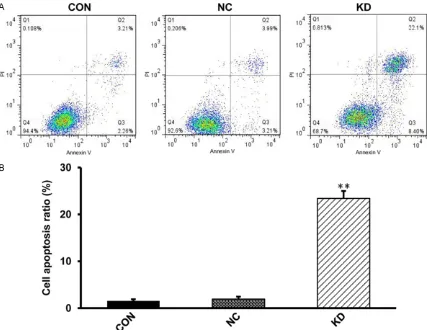
Figure 3. Apoptosis was detected by Annexin V-FITC. A. FCM data showed the apoptotic rate of CaSki cells infected
with lentivirus was significantly increased. B. The cell apoptosis ratio in different groups. The data are means ± SD,
Knockdown HPV16E7 suppress cervical cancer cell line growth
Knockdown of HPV16E7 induces the apoptosis of CaSki cells
In order to examine the apoptosis of CaSki cells after knocking down HPV16E7, flow cytometry was performed. Apoptotic rate of the CON and the NC group are 1.53 ± 0.37% and 1.93 ± 0.52%, while it is 23.4 ± 2.6% in KD group. Hence the percentage of apoptosis of CaSki cells in KD group was increased compared with CON and NC groups (Figure 3A, 3B). These data suggested that knockdown of HPV16E7 induced the apoptosis of CaSki cells which might be the cause of cell proliferation suppression.
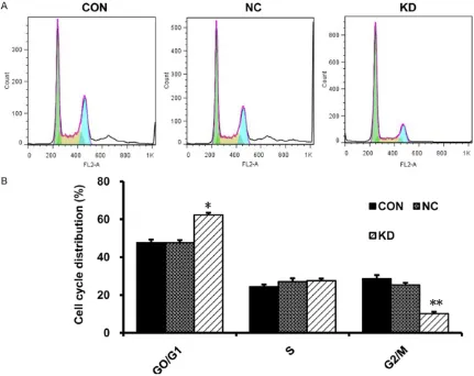
Knockdown of HPV16E7 arrests CaSki cells in the G0/G1 phase
To investigate the cell cycle distribution after knocking down the expression of HPV16E7,
flow cytometry showed an increased frequency in the proportion of G0/G1 phase (62.27 ± 0.68% vs 47.64 ± 1.37%) but a decreased fre-quency in the proportion of G2/M phase (10.17 ± 0.18% vs 25.31 ± 2.15%) in KD group com-pared with NC groups (Figure 4A, 4B). The results showed that knocking down HPV16E7 arrested CaSki cells in the G0/G1 phase.
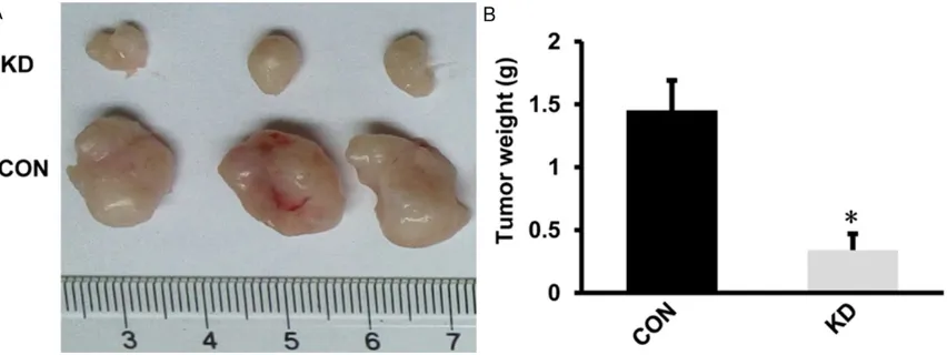
Inhibition of HPV16E7 reduces tumor forma-tion of CaSki cells in immune-deficient nude mice
[image:6.612.91.522.74.416.2]To further conform the effect of HPV16E7-RNAi-LV on tumorigenesis, tumor formation assay was performed in vivo. Mice were injected with CaSki cells with or without lentivirus-mediated RNAi knockdown of HPV16E7, xenografts were removed and weighed 4 weeks later. In vivo tumor formation assay, all 3 mice of CON group
formed xenografts while the 3 mice of KD group formed smaller xenografts (Figure 5A). The weight of tumor in KD group was lower than those from CON group (Figure 5B). It demon-strated that inhibition of HPV16E7 reduces tumor formation of cervical cancer cells in vivo. Discussion
The oncogenes in HPV16 are involved in cancer development and immortalization. Among these oncogenes in HPV16, HPV16E7 onco-gene plays an important role in tumorionco-genesis. Varying degrees of regulation in a range of cel-lular and molecular processes were led by HPV16E7 through its interaction with other pro-teins [17] such as major histocompatibility complex-1 (MHC-1) [18-23], tumor suppressor protein pRb [24] and cell kinase dual-specificity tyrosine phosphorylation-regulated kinase 1A (DYRK1A) [25] which resulting in carcinogene-sis. Furthermore, HPV16E7 has been reported to play an important role in the proliferation of various kinds of cells, especially cancer cells [14]. However, the role of HPV16E7-RNAi-LV in the inhibition of cell growth of cervical cancer has not been declared.
RNA interference (RNAi) is a widely used power-ful tool in inducing loss-of-function phenotypes. The combination of lentivirus and RNAi is a new method to treat cancers and has been shown highly efficiency in treating variously cancers, according to several reports [26, 27]. In our study, we down-regulate the HPV16E7 gene in transfected cervical cell line CaSki cells by con-structing an efficient and stable lentivirus
vec-tor. We decreased HPV16E7 expression in a cervical cancer cell line CaSki cells by lentivi-rus-mediated RNAi to study the effect of HPV16E7 silencing on the cell growth, apoptot-ic rate, cell cycle-progression in vitro and tumor formation in vivo.
Our results showed that after the delivery of lentivirus vector into CaSki cells, expression of HPV16E7 in the level of mRNA and protein were obviously decreased. Uncontrolled proliferation is one basic characteristic of tumor cells. In our study, HPV16E7 was knocked down by lentivi-rus in CaSki cells. MTT assay showed that knockdown of HPV16E7 significantly inhibited the viability of CaSki cells, and this inhibition was specific. Furthermore, this specific sup-pression was confirmed by the colony forma-tion assay in vitro which indicated knockdown of HPV16E7 blocked colony formation in CaSki cells. In addition, silencing of HPV16E7 reduced the tumor formation of cervical cancer in vivo. Therefore, inhibition of HPV16E7 can suppress the proliferation of cervical cancer cells in vitro and in vivo.
The suppression of proliferation could be the result of apoptosis. Hence, we performed apop-tosis assay. It demonstrated that the percent-age of apoptotic rate in lentivirus-mediated RNAi knockdown of HPV16E7 cervical cancer cells was increased compared with cells with-out transfection.
Proliferation and apoptosis are related to cell cycle distribution. When cells arrested in G0/ G1 phase, more apoptosis and less
[image:7.612.96.522.71.231.2]Knockdown HPV16E7 suppress cervical cancer cell line growth
tion were observed. To explore the relationship among cell cycle distribution, proliferation and apoptosis, we used flow cytometry to detect cell cycle. In our research, HPV16E7-RNAi-LV arrested CaSki cells in G0/G1 phase. As for its effect on cell cycle distribution, more apoptosis and less proliferation were found in our study. Previous research showed that the activation of HPV16E7 was able to boost cervical cancer cell growth which was regulated through interaction with other proteins, such as MHC-1 [28]. More- over, HPV16E7 interacted with RB which result-ed in blocking the activity of tumor suppressors [29]. Furthermore, HPV16E7 stimulated the S-phase genes cyclin A and cyclinE which con-tributed to tumorigenesis [8]. Hence, we specu-late that HPV16E7 gene silencing may inhibit proliferation of cervical cancer.
HPV16E7 may be a potential therapeutic target in treatment of cervical cancer. Zheng et al [30] reported that a novel vaccine consisting of modified HPV16E7 fused with human cytotoxic T-lymphocyte antigen 4 (CTLA4) resulted in re- tarded tumor growth and prolonged survival in mouse injected with TC-1 cervical cancer cells in hind flank. Therefore, starting with HPV16E7 will open a new horizon for the treatment of cer-vical cancer.
In conclusion, we demonstrate that lentivirus-mediated RNAi knockdown of HPV16E7 can significantly repress cell proliferation in vitro and in vivo, induce apoptosis in CaSki cells and arrest cells in the G0/G1 phase. This provides a pivotal therapeutic strategy for treatment of cervical cancer.
Disclosure of conflict of interest
None.
Address correspondence to: Ziyu Hu, Department of Obstetrics and Gynecology, People’s Hospital of Dongying, Dongying 257091, China. E-mail: huzi-yudongying@sina.com
References
[1] Jemal A, Bray F, Center MM, Ferlay J, Ward E, Forman D. Global cancer statistics. CA Cancer J Clin 2011; 61: 69-90.
[2] Cervical Cancer Treatment (PDQ(R)): Health Professional Version, in PDQ Cancer Informa-tion Summaries 2002: Bethesda (MD).
[3] Ghose S, Holloway L, Lim K, Chan P, Veera J, Vinod SK, Liney G, Greer PB, Dowling J. A re-view of segmentation and deformable registra-tion methods applied to adaptive cervical can-cer radiation therapy treatment planning. Artif Intell Med 2015; 64: 75-87.
[4] Morris M, Eifel PJ, Lu J, Grigsby PW, Levenback C, Stevens RE, Rotman M, Gershenson DM, Mutch DG. Pelvic radiation with concurrent chemotherapy compared with pelvic and para-aortic radiation for high-risk cervical cancer. N Engl J Med 1999; 340: 1137-1143.
[5] zur Hausen H. Papillomaviruses and cancer: from basic studies to clinical application. Nat Rev Cancer 2002; 2: 342-350.
[6] Walboomers JM, Jacobs MV, Manos MM, Bosch FX, Kummer JA, Shah KV, Snijders PJ, Peto J, Meijer CJ, Muñoz N. Human papilloma-virus is a necessary cause of invasive cervical cancer worldwide. J Pathol 1999; 189: 12-19. [7] Duensing S, Duensing A, Crum CP, Münger K.
Human papillomavirus type 16 E7 oncopro-tein-induced abnormal centrosome synthesis is an early event in the evolving malignant phe-notype. Cancer Res 2001; 61: 2356-2360. [8] Zerfass K, Schulze A, Spitkovsky D, Friedman
V, Henglein B, Jansen-Dürr P. Sequential acti-vation of cyclin E and cyclin A gene expression by human papillomavirus type 16 E7 through sequences necessary for transformation. J Vi-rol 1995; 69: 6389-6399.
[9] Jones DL, Alani RM and Munger K. The human papillomavirus E7 oncoprotein can uncouple cellular differentiation and proliferation in hu-man keratinocytes by abrogating p21Cip1-me-diated inhibition of cdk2. Genes Dev 1997; 11: 2101-2111.
[10] Funk JO, Waga S, Harry JB, Espling E, Stillman B, Galloway DA. Inhibition of CDK activity and PCNA-dependent DNA replication by p21 is blocked by interaction with the HPV-16 E7 on-coprotein. Genes Dev 1997; 11: 2090-2100. [11] Zerfass-Thome K, Zwerschke W, Mannhardt B,
Tindle R, Botz JW, Jansen-Dürr P. Inactivation of the cdk inhibitor p27KIP1 by the human papillomavirus type 16 E7 oncoprotein. Onco-gene 1996 13: 2323-2330.
[12] Malcolm KM, Gill J, Leggatt GR, Boyd R, Lam-bert P, Frazer IH. Expression of the HPV16E7 oncoprotein by thymic epithelium is accompa-nied by disrupted T cell maturation and a fail-ure of the thymus to involute with age. Clin Dev Immunol 2003; 10: 91-103.
[13] Hadis U, Leggatt GR, Thomas R, Frazer IH, Ko-vacs EM. IL-1 signalling determines the fate of skin grafts expressing non-self protein in kera-tinocytes. Exp Dermatol 2010; 19:723-729. [14] Ying XJ, Jin B, Chen XW, Xie J, Xu HM, Dong P.
expres-sion and inhibits cell proliferation in laryngeal squamous cell carcinoma Hep-2 cells in vitro. Biomed Res Int 2015; 2015: 150390.
[15] Thomas CE, Ehrhardt A and Kay MA. Progress and problems with the use of viral vectors for gene therapy. Nat Rev Genet 2003; 4: 346-358.
[16] McCormick F. Cancer gene therapy: fringe or cutting edge? Nat Rev Cancer 2001; 1: 130-141.
[17] Munger K, Basile JR, Duensing S, Eichten A, Gonzalez SL, Grace M, Zacny VL. Biological ac-tivities and molecular targets of the human papillomavirus E7 oncoprotein. Oncogene 2001; 20: 7888-7898.
[18] Cheng M, Chen Y, Xiao W, Sun R, Tian Z. NK cell-based immunotherapy for malignant dis-eases. Cell Mol Immunol 2013; 10: 230-252. [19] Jost S and Altfeld M. Control of human viral
in-fections by natural killer cells. Annu Rev Immu-nol 2013; 31: 163-194.
[20] Cooper MA, Colonna M and Yokoyama WM. Hidden talents of natural killers: NK cells in in-nate and adaptive immunity. EMBO Rep 2009; 10: 1103-1110.
[21] McCoy WHt, Wang X, Yokoyama WM, Hansen TH, Fremont DH. Cowpox virus employs a
two-pronged strategy to outflank MHCI antigen pre -sentation. Mol Immunol 2013; 55: 156-158. [22] Qiu T, Wang L, Liu XH, Weng XD, Kuang YL,
Chen ZY, Chen H, Zhu HC. Over-expressing transporters associated with antigen process-ing increases antitumor immunity response in prostate cancer. Cell Immunol 2012; 279: 167-173.
[23] Oliveira CC and van Hall T. Importance of TAP-independent processing pathways. Mol Immu-nol 2013; 55: 113-116.
[24] Zhang BY, Chen W and Roman A. The E7 pro-teins of low- and high-risk human papillomavi-ruses share the ability to target the pRB family member p130 for degradation. Proc Natl Acad Sci U S A 2006; 103: 437-442.
[25] Galceran J, de Graaf K, Tejedor FJ, Becker W. The MNB/DYRK1A protein kinase: genetic and biochemical properties. J Neural Transm Suppl 2003; 139-148.
[26] Li Z, Tian T, Hu X, Zhang X, Li L, Nan F, Chang Y, Wang X, Sun Z, Lv F, Zhang M. Targeting Six1 by lentivirus-mediated RNA interference inhibits colorectal cancer cell growth and invasion. Int J Clin Exp Pathol 2014; 7: 631-639.
[27] Chen J, Shi D, Liu X, Fang S, Zhang J, Zhao Y. Targeting SPARC by lentivirus-mediated RNA interference inhibits cervical cancer cell gro- wth and metastasis. BMC Cancer 2012; 12: 464.
[28] Guo H, Hu R, Guan X, Guo F, Zhao S, Zhang X. HPV16E7 silencing enhances susceptibility of CaSki cells to natural killer cells. Mol Med Rep 2014; 9: 1351-1354.
[29] Dyson N, Howley PM, Munger K, Harlow E. The human papilloma virus-16 E7 oncoprotein is able to bind to the retinoblastoma gene prod-uct. Science 1989; 243: 934-937.