Original Article
Downregulation of KIF20A induces cell cycle arrest
and apoptosis by suppressing PI3K/AKT
in human glioblastoma
Min Wang1,2, Kun Liu3, Xin-Ling Zhou4, Shu-Yu Mei1, Cui-Juan Zhang1, Ting-Guo Zhang1
1Department of Pathology, School of Medicine, Shandong University, Jinan 250012, Shandong, China;
Depart-ments of 2Pathology, 3Neurosurgery, 4Obstetrics and Gynecology, The Second People’s Hospital of Liaocheng,
Linqing 252601, Shandong, China
Received May 14, 2017; Accepted November 3, 2017; Epub December 15, 2017; Published December 30, 2017
Abstract: Background/Aim: Aberrant function of the kinesin family member 20A (KIF20A) has been reported to be vital in tumor genesis and development. However, the role of KIF20A in cell proliferation and invasion, as well as
the mechanism underlying cell cycle arrest, remains unclear. Materials and Methods: Here, we first measured the
expression of KIF20A in malignant astrocytoma and glioblastoma cell lines by immunohistochemistry and western blotting. Next, we knocked down KIF20A expression using siRNA to study its effect on cell proliferation and cycle.
Results: We found that KIF20A knock down significantly suppressed cell proliferation, migration and invasion of glio -blastoma cells. Importantly, KIF20A knock down induced cell cycle arrest in G0/G1 phase and promoted apoptosis by inactivating the PI3K/Akt pathway via c-Myc and activating the intrinsic apoptosis pathway. Conclusion: Taken together, our results suggest that KIF20A down-regulation can inhibit glioma tumorigenesis, which may provide a therapeutic target for treating glioma.
Keywords: KIF20A, glioblastoma, cell proliferation, cell migration, cell cycle, PI3K/Akt pathway
Introduction
Glioma represents approximately 30% of pri-mary brain tumors of glial cell origin [1]. In humans, astrocytoma is the most common form of glioma, accounting for about 80% of malignancies of the central nervous system [2]. According to the World Health Organization (WHO) for Tumors in the Central Nervous
System in 2007, the diffusely infiltrative astro -cytic tumors can be graded on a three-tiered system: diffuse astrocytoma (grade II), ana-plastic astrocytoma (grade III), and glioblasto-ma (grade IV) [3]. Despite advances in surgi- cal and non-surgical treatments of gliomas, patients with glioblastoma still have a poor prognosis, with a median overall survival of 12-15 months [4]. However, the molecular mechanisms underlying glioma development and progression are still not well understood. In light of this, we sought to identify novel thera-peutic targets to improve prognoses for glioma patients.
pro-teins in treated cells. Among these, 4 propro-teins from the kinesin family-KIF11, KIF20A, KIF22, and KIF23-were downregulated. Interestingly, all of these proteins are involved in the regula-tion of cell cycle, cell growth, and proliferaregula-tion [17]. However, little is known about the role KIF20A plays in cell cycle regulation in gliomas. The intracellular phosphoinositide 3 kinase/Akt (PI3K/AKT) signaling pathway is important in the regulation of cell cycle, which is directly related to cellular quiescence, proliferation, cancer, and longevity [18]. Activation of BCL2, BCL-2-associated X protein (BAX) and caspas-es, and inhibition of KIF20A, mitogen-activated protein kinase (MAPK), and PI3K/Akt signaling pathways contribute to cell apoptosis during cancer. Thus, targeting these factors may af- fect the molecular mechanisms mediating the anti-cancer effects of genistein [19].
In this study, we investigated if KIF20A is asso-ciated with malignant tumor progression and cell cycle regulation in glioma. Our results
dem-onstrate for the first time that knock down of
KIF20A induces cell cycle arrest in G0/G1 phase and promotes apoptosis by inactivating the PI3K/Akt pathway via its downstream effec-tor, c-Myc, and activating the intrinsic apopto-sis pathway. Our study indicates that KIF20A plays a critical role in astrocytoma tumorigene-sis, providing a promising therapeutic strategy for glioma.
Materials and methods
Assays for Cell Proliferation, Colony Formation, and Cell Migration and Invasion are described in the Supplementary Materials and Methods. Tissue samples
Ninety-four human primary astrocytoma tis-sues were obtained from Qi Lu Hospital of Shandong University from 2010-2013. Tissues
were formalin fixed followed by paraffin-embed -ding. Malignant astrocytomas included 11 dif-fuse astrocytomas (WHO grade II), 22 anaplas-tic astrocytomas (WHO grade III), and 33 glioblastomas (WHO grade IV). None of the patients received radiotherapy or chemothera-py before surgery. Ten cases of normal human brain tissues were obtained from patients who had received decompressive craniectomy at The Second People’s Hospital of Liaocheng
City. All samples were examined by two
patholo-gists to confirm histological diagnoses.
Ethics statement
The study was approved by both the Ethics Committee of the School of Medicine, Shan- dong University and the Ethics Committee of The Second People’s Hospital of Liaocheng, Shandong, China. Written informed consent was obtained from all participants involved in our study.
Immunohistochemistry
Paraffin embedded samples were heated for
50 min in an oven at 60°C. The slides were
then deparaffinized twice in xylol for 10 min,
rehydrated through graded ethanols, and incu-bated in 3% hydrogen peroxide solution for 10 min at room temperature. Antigen retrieval was performed by boiling the sections at 100°C for 4 min in 0.01 M citrate buffer (pH 6.0). Slides were incubated with KIF20A antibody (1:100, Abcam, Cambridge, UK) and Ki-67 (1:100, Abcam, Cambridge, UK) overnight at 4°C, and then incubated with biotinylated anti-mouse secondary antibody (PV-9000 2-step plus® reagent kit, Zhongshan Biotechnology, Beijing, China) at 37°C for 30 min. The slides were visu-alized with 3,3’-diaminobenzidin (DAB) sub-strate liquid and washed with deionized water before hematoxylin counterstaining.
Staining intensity was scored as follows: 0, absent; 1, weak; 2, moderate; 3, strong. The percentage of positive cells was scored as
fol-lows: 1 ≤ 25%; 2 ≤ 50%; 3 ≤ 75%; 4 > 75%. The final score was calculated by multiplying the
intensity score by the percentage score. A
com-posite score ≤ 6 was considered low KIF20A expression; a score > 6 was considered high
KIF20A expression [20]. The Ki-67 labeling index was calculated as the mean percentage of immune-positive cells. One thousand
neo-plastic cells were counted in the five
most-labeled areas [21].
Cell culture and transfection
assas, VA, USA). Cells were cultured in Dul-
becco’s modified Eagle’s medium (DMEM) sup -plemented with 10% fetal bovine serum (FBS)
in a humidified cell incubator in an atmosphere
of 5% CO2 at 37°C.
Small interfering RNA (siRNA) was synthesized by Gene-Pharma (Shanghai, China). Cells were transiently transfected with siRNA using Lipo- fectamine 2000 (Invitrogen, Carlsbad, CA, USA) in accordance with the manufacturer’s
proto-col. Transfection efficiency was verified by
real-time quantitative PCR (RT-qPCR) and western blot analyses.
RT-qPCR
Total RNA was extracted from glioblastoma cells by RNAiso Plusl (TaKaRa, Dalian, China) and cDNA was synthesized with the ReverTra Ace qPCR RT Kit (Toyoto, Osaka, Japan) accord-ing to the manufacturer’s protocol. RT-qPCR reaction was performed using the SYBR® Green Realtime PCR Master Mix assay kit (Toyoto, Osaka, Japan) under the following conditions of 95°C for 60 s, 40 cycles at 95°C for 15 s, 60°C for 15 s and 72°C for 45 s. Glyceralde- hyde phosphate dehyrdrogenase (GAPDH) was used as an endogenous control. The primer se- quences were as follows: KIF20A: 5’-GCCAA- CTTCATCCAACACCT-3’ (forward); 5’-GTGGACA- GCTCCTCCTCTTG-3’ (reverse); GAPDH: 5’-GAG- TCAACGGATTTGGTCGT-3’ (forward); 5’-GACAA- GCTTCCCGTTCTCAG-3’ (reverse).
Cell cycle assay
Glioblastoma cells were harvested, washed,
fixed overnight in 75% ethanol at 4°C following
transfection. Before analysis, RNase A was added in the water bath for 30 min at 37°C. The cell suspension was stained with propidi-um iodide in the dark. Cell cycle distribution
was determined by flow cytometry (FACSCalibur,
BD Biosciences). The results were quantitated with ModFit analytic software.
Apoptosis assay
After transfection, glioblastoma cells were har-vested and stained with the Annexin V-FITC/PI Apoptosis Detection Kit (BestBio, Shanghai, China) according to the manufacturer’s instruc-tions. Stained cells were immediately analyzed
by flow cytometry (FACSCalibur, BD
Bioscien-ces). The total apoptotic rate was the sum of the early apoptotic rate and the late apoptotic rate.
Western blot
Total protein was extracted from transfected glioblastoma cells after 48 h transfection. Lysates were isolated by electrophoresis and transferred to nitrocellulose membranes. The membranes were then incubated with antibod-ies against KIF20A (1:1,000, Abcam), phos-phorylated (p)Akt (Ser473, 1:5,000, Abcam), Akt (1:5,000, Abcam), Cyclin D1 (1:10,000, Ab- cam), Bcl-2 (1:1,000, Abcam), Bax (1:5,000), c-
Myc (1:5,000, CST, USA), and β-actin (1:1,000,
Beyotime, Beijing, China). The bands were ana-lyzed using ImageJ software.
Statistical analysis
Analysis was performed using the statistical software package for Social Sciences, version 20.0 (SPSS). Fisher’s exact test or chi-square test analysis was applied for the correlation between KIF20A expression and clinical-patho-logic parameters. The correlation between KIF- 20A and Ki-67 was determined by Spearman’s
correlation coefficient. Survival curves were
as-sessed by the Kaplan-Meier method, and the differences between the survival curves were analyzed by the log-rank test. Univariate and multivariate survival data were analyzed using the Cox proportional hazard regression model. Student’s t-test was used to analyze the differ-ences between two groups. p < 0.05 was
con-sidered statistically significant.
Results
KIF20A is a prognostic marker for malignant astrocytoma
We stained brain tissues with a KIF20A
anti-body to confirm that KIF20A is expressed in
malignant astrocytomas. Smooth muscle tis-sue was used as a negative control.We found positive KIF20A staining in 65 of the 94 cases (69.1%) of malignant astrocytomas and in 1 out of the 10 (10%) normal brain tissues. The intensity and percentage of KIF20A positive staining was enhanced as the pathological grade increased (Figure 1A-E). Consistently, the Kaplan-Meier survival curves
demonstrat-ed significant association between KIF20A
trocytoma patients (Figure S1A; p = 0.000). The univariate survival analysis and multivariate analysis showed that high KIF20A expression correlated with poorer overall survival (Table S1; p = 0.022; hazard ratio (HR) = 1.925; 95%
confidence interval (CI) = 1.098-3.375).
We also assessed KIF20A expression in glio-blastoma cell lines. KIF20A expression was
sig-nificantly higher in U251 and U87 cells com -pared to T98 and LN229 cells (Figure S1B, p <
0.0001). Moreover, KIF20A expression signifi -cantly correlated with WHO pathological grade (Table 1; p = 0.003) and Ki-67 index (Table 1; p = 0.002). In addition, KIF20A and Ki-67 immu-noreactivity were positively correlated (r = 0.320, p = 0.002).
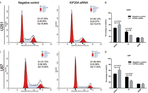
and from 48.33 ± 0.70% to 61.49 ± 3.55% in U87 cells (Figure 3A-D; p = 0.0032), while the percentage of cells in S phase was reduced from 35.53 ± 1.85% to 24.40 ± 0.41% in U251 cells (Figure 3A, 3B; p = 0.0005) and 38.08 ± 0.33% to 24.76 ± 2.16% in U87 cells (Figure 3C, 3D; p = 0.0005). These results suggest that knock down of KIF20A induces cell cycle arrest in G0/G1 phase, which may in part explain the growth inhibition of glioblastoma cells.
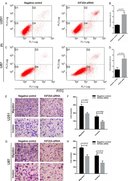
KIF20A knock down in glioblastoma cells induces apoptosis and inhibits migration and invasion
[image:4.612.89.381.71.418.2]Next, we measured apoptosis following KIF20A knock down. We found that the apoptosis rate
Figure 1. KIF20A is a prognostic marker for malignant astrocytoma. Immu-nohistochemical staining showed that KIF20A was located in the cytoplasm and nucleus in astrocytoma cells. A. KIF20A was weakly detected in human normal brain tissues. B. Weak KIF20A staining was noted in low grade glioma (WHO II). C, D. Strong KIF20A staining was noted in high grade glioma (WHO III and WHO IV). The intensity and percentage of positive staining of KIF20A was enhanced as the pathological grade increased. E. Smooth muscle tissue as negative control.
In vitro knockdown of KIF20A suppresses glioblas-toma cell proliferation
RT-qPCR and western blot analyses demonstrated that siRNA KIF20A transfection reduced KIF20A expression in the knock down group compared to the control group in both U251 and U87 cell lines (Figure S2A, S2B). The cell proliferation rate, as assessed by the CCK-8 assay, decreased in the KIF20A-siRNA transfect-ed U251 and U87 cells com-pared to the control group (Figure S2C, S2D; *p < 0.05, **p < 0.01, respectively). Furthermore, knock down of KIF20A in U251 and U87 cells decreased the number of colonies as evidenced by colony formation assays (Figure 2A-D; p = 0.0009 and 0.0020, respectively). Downregulation of KIF20A arrests cell cycle in G0/G1 phase
Table 1. Association of KIF20A expression with clinico-pathological parameters
Clinical-pathological
variable n
KIF20A expression p value Low (n, %) High (n, %)**
Age (years)
≤ 40 32 24 (75.0) 8 (25.0) 0.679
> 40 62 44 (71.0) 18 (29.0) Gender
Male 55 42 (76.4) 13 (23.6) 0.300 Female 39 26 (66.7) 13 (33.3)
Tumor size (cm)
< 5 42 29 (69.0) 13 (31.0) 0.521
≥ 5 52 39 (75.0) 13 (25.0) WHO grade
I 27 24 (88.9) 3 (11.1) 0.003*
III 32 25 (78.1) 7 (21.9)
IV 35 18 (51.4) 17 (48.6)
Ki-67 (%)
< 12 46 40 (87.0) 6 (13.0) 0.002*
≥ 12 48 28 (58.3) 20 (41.7)
*indicates p < 0.05, significant difference. **A composite score ≤ 6
was considered low KIF20A expression; a score > 6 was considered high KIF20A expression.
in U251 (Figure 4A, 4B; p = 0.0017) and U87 (Figure 4C, 4D; p = 0.0054) cells transfected with KIF20A siRNA was increased compared to control. The Transwell assay further showed
that KIF20A knock down significantly
decreas-ed migration and invasion in U251 (Figure 4E, 4F; p = 0.0061 and p = 0.0083, respectively) and U87 (Figure 4G, 4H; p = 0.0010 and p = 0.0075, respectively) cells compared to the control group.
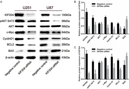
The effects of KIF20A knock down on critical cell cycle protein expression in cancer cells
To explore the downstream targets of KIF20A, we measured expression of several cell cycle regulated genes including phosphorylated AKT (PI3K/AKT signaling pathways), c-Myc, and Cyclin D1 (important for cancer formation, pro-gression), BCL2, and BAX (critical factors for cell apoptosis). siRNA mediated knock down of KIF20A decreased pAKT (Ser473) protein expression in U251 (Figure 5A, 5B) and U87 (Figure 5A, 5C) cells compared to the control group, which suggests that the decreased cell viability and cell cycle arrest in G0/G1 phase may occur via inactivation of the PI3K/Akt
path-way. We also found that KIF20A siNRA signifi
-cantly reduced c-Myc and Cyclin D1 pro-tein expression in U251 (Figure 5A, 5B) and U87 (Figure 5A, 5C) cells compared to the control group. In addition, we assessed the protein levels of apoptosis-related genes and found that siRNA mediated
KIF20A knock down significantly decreased BCL2 and significantly increased BAX
expression in U251 (Figure 5A, 5B) and U87 (Figure 5A, 5C) cells compared to the control group. These data indicate that downregulation of KIF20A inhibits cell apoptosis in glioblastoma cells.
Discussion
Glioma is a highly heterogeneous tumor, and its carcinomatous process is limited compared to other cancers. Many studies have focused on gene alternation in glio-ma to understand the molecular mecha-nisms of tumorigenesis and progression [22-24].
KIF20A is a member of the kinesin super-family-6 and has two microtubule-binding sites, including the N-terminal kinesin motor domain and the C-terminus Rab6-binding domain [25, 26]. KIF20A is involved in key cel-lular functions such as intracelcel-lular movement of organelles and vesicles, spindle formation, and cytokinesis [27, 28]. Accumulating evi-dence has shown that ectopic overexpression of KIF20A exists in various tumors, which indi-cates that KIF20A contributes to tumorigenesis [8-13]. In the current study, KIF20A was highly expressed in malignant astrocytomas, and this
elevated expression significantly correlated
with advanced pathological grade and high
Ki-67 index. These findings are consistent with
recent studies that have also demonstrated that high KIF20A expression is an independent prognostic factor for overall survival in glioma patients, which suggests that KIF20A is a tar-get for treating glioma [29, 30]. However, the previous research only explored the effects of KIF20A on glioblastoma cells without providing any new insight into the underlying molecular mechanism. In the present study, we are the
first to report variation in cell cycle distribution
A recent study demonstrated that silencing FOXM1 (a member of the Forkhead box family) and KIF20A similarly caused abnormal spindle morphology and chromosome alignment, which induces mitotic catastrophe in breast carcino-ma paclitaxel-resistant MCF-7 TaxR cells [10]. However, up-regulated KIF20A expression led to impaired mitosis and cytokinesis, aberrant ploidy, and genomic instability in hepatoma cells [16]. Thus, we speculate that KIF20A’s mechanism of action in glioblastoma cells is analogous with the above points.
KIF20A accumulates in mitotic cells where it localizes to the midzone of the spindle during anaphase. Cell cycle regulation is believed to be the foundation of tumor cell proliferation
and cell division. For the first time, we demon -strate that KIF20A knock down arrests glioblas-toma cells in G0/G1 phase, which indicates that reducing KIF20A expression can suppress cell cycle transition from G0/G1 to S phase, causing inhibition of cell proliferation and colo-ny formation. This result is consistent with some studies that showed that KIF20A down-regulation caused accumulation of MCF7 in breast carcinoma cells [31]. In contrast, silenc-ing KIF20A inhibited gastric cancer cell viability and induced cell cycle arrest in G2/M phase [17]. Thus, cell cycle arrest induced by KIF20A differs among cancers cells, which explains how silencing KIF20A can promote proliferation
of glioblastoma cells by influencing cell cycle
[image:6.612.89.520.70.450.2]phase distribution.
The molecular mechanisms of kinesin super-family members have been described in vari-ous types of tumors [32-34]. PI3K is a lipid kinase that generates second messengers involved in the regulation of cell functions including proliferation, apoptosis and survival, all of which play key roles in cell cycle progres-sion, prevention of apoptosis, and malignant transformation [35]. One of PI3K’s major effec-tors is protein kinase B (Akt). Our results indi-cate that downregulation of KIF20A not only decreased pAKT (Ser473), but also suppressed the levels of cell cycle correlated genes, includ-ing c-Myc and Cyclin D1. This suggests that KIF20A is an upstream regulator of the PI3K/ Akt pathway, and a decrease in KIF20A induces cell cycle arrest in G0/G1 phase by inactivating the Akt pathway via its downstream effector, c-Myc. The literature shows that Cyclin D1 is a G0/G1 related cell cycle regulatory factor [36, 37]; thus, a decrease in KIF20A expression arrests glioblastoma cells in G0/G1 phase
rath-er than G2/M phase. Moreovrath-er, we also found that BCL2 levels were reduced while BAX levels were increased in glioblastoma cells transfect-ed with KIF20A siRNA. These data suggest that KIF20A downregulation induces apoptosis through inactivating Akt and activating the downstream intrinsic apoptosis pathway via regulating the BCL-2/BAX ratio.
In conclusion, our results suggest that down-regulating KIF20A inhibits cell proliferation and invasion, induces cell cycle arrest in G0/G1 phase, and promotes apoptosis. The KIF20A mediated effects on these cell processes is likely occurring by inactivation of the PI3K/Akt pathway and its downstream effectors. Our
findings confirm that KIF20A is an attractive
therapeutic target for glioma. Disclosure of conflict of interest
[image:9.612.93.520.73.366.2]None.
Address correspondence to: Ting-Guo Zhang, De- partment of Pathology, School of Medicine, Shan- dong University, Wenhuaxi Road 44#, Jinan 2500- 12, Shandong, China. Tel: +8618560081178; Fax: +86-531-88383168; E-mail: ztguo@sdu.edu.cn
References
[1] Ho VK, Reijneveld JC, Enting RH, Bienfait HP, Robe P, Baumert BG, Visser O; Dutch Society for Neuro-Oncology (LWNO). Changing inci-dence and improved survival of gliomas. Eur J Cancer 2014; 50: 2309-2318.
[2] Taylor LP. Diagnosis, treatment, and prognosis
of glioma: five new things. Neurology 2010; 75:
S28-32.
[3] Louis DN, Ohgaki H, Wiestler OD, Cavenee WK, Burger PC, Jouvet A, Scheithauer BW and
Klei-hues P. The 2007 WHO classification of tu -mours of the central nervous system. Acta Neuropathol 2007; 114: 97-109.
[4] Takahashi S, Yamada-Okabe H, Hamada K, Ohta S, Kawase T, Yoshida K and Toda M. Downregulation of uPARAP mediates cytoskel-etal rearrangements and decreases invasion and migration properties in glioma cells. J Neu-rooncol 2011; 103: 267-276.
[5] Hirokawa N and Noda Y. Intracellular transport and kinesin superfamily proteins, KIFs: struc-ture, function, and dynamics. Physiol Rev 2008; 88: 1089-1118.
[6] Hirokawa N, Noda Y, Tanaka Y and Niwa S. Ki-nesin superfamily motor proteins and intracel-lular transport. Nat Rev Mol Cell Biol 2009; 10: 682-696.
[7] Liu X, Gong H and Huang K. Oncogenic role of kinesin proteins and targeting kinesin therapy. Cancer Sci 2013; 104: 651-656.
[8] Taniuchi K, Nakagawa H, Nakamura T, Eguchi H, Ohigashi H, Ishikawa O, Katagiri T and Naka-mura Y. Down-regulation of RAB6KIFL/KIF20A,
a kinesin involved with membrane trafficking
of discs large homologue 5, can attenuate growth of pancreatic cancer cell. Cancer Res 2005; 65: 105-112.
[9] Shi C, Huang D, Lu N, Chen D, Zhang M, Yan Y, Deng L, Lu Q, Lu H and Luo S. Aberrantly acti-vated Gli2-KIF20A axis is crucial for growth of hepatocellular carcinoma and predicts poor prognosis. Oncotarget 2016; 7: 26206-26219. [10] Khongkow P, Gomes AR, Gong C, Man EP,
Tsang JW, Zhao F, Monteiro LJ, Coombes RC, Medema RH, Khoo US and Lam EW. Paclitaxel targets FOXM1 to regulate KIF20A in mitotic catastrophe and breast cancer paclitaxel resis-tance. Oncogene 2016; 35: 990-1002. [11] Lu Y, Liu P, Wen W, Grubbs CJ, Townsend RR,
Malone JP, Lubet RA and You M. Cross-species comparison of orthologous gene expression in
human bladder cancer and carcinogen-in-duced rodent models. Am J Transl Res 2010; 3: 8-27.
[12] Yamashita J, Fukushima S, Jinnin M, Honda N, Makino K, Sakai K, Masuguchi S, Inoue Y and Ihn H. Kinesin family member 20A is a novel melanoma-associated antigen. Acta Derm Ve-nereol 2012; 92: 593-597.
[13] Claerhout S, Lim JY, Choi W, Park YY, Kim K, Kim SB, Lee JS, Mills GB and Cho JY. Gene
ex-pression signature analysis identifies vorino -stat as a candidate therapy for gastric cancer. PLoS One 2011; 6: e24662.
[14] Zhang W, He W, Shi Y, Gu H, Li M, Liu Z, Feng Y, Zheng N, Xie C and Zhang Y. High expression of KIF20A is associated with poor overall survival and tumor progression in early-stage cervical squamous cell carcinoma. PLoS One 2016; 11: e0167449.
[15] Tomita Y, Yuno A, Tsukamoto H, Senju S, Kuro-da Y, Hirayama M, Irie A, Kawahara K, YatsuKuro-da J, Hamada A, Jono H, Yoshida K, Tsunoda T, Kohrogi H, Yoshitake Y, Nakamura Y,
Shinoha-ra M and NishimuShinoha-ra Y. Identification of promis -cuous KIF20A long peptides bearing both CD4+ and CD8+ T-cell epitopes:
KIF20A-spe-cific CD4+ T-cell immunity in patients with ma -lignant tumor. Clin Cancer Res 2013; 19: 4508-4520.
[16] Gasnereau I, Boissan M, Margall-Ducos G, Couchy G, Wendum D, Bourgain-Guglielmetti F, Desdouets C, Lacombe ML, Zucman-Rossi J and Sobczak-Thepot J. KIF20A mRNA and its product MKlp2 are increased during hepato-cyte proliferation and hepatocarcinogenesis. Am J Pathol 2012; 180: 131-140.
[17] Yan GR, Zou FY, Dang BL, Zhang Y, Yu G, Liu X and He QY. Genistein-induced mitotic arrest of gastric cancer cells by downregulating KIF20A, a proteomics study. Proteomics 2012; 12: 2391-2399.
[18] Carnero A, Blanco-Aparicio C, Renner O, Link W and Leal JF. The PTEN/PI3K/AKT signalling pathway in cancer, therapeutic implications. Curr Cancer Drug Targets 2008; 8: 187-198. [19] Spagnuolo C, Russo GL, Orhan IE,
Habtemari-am S, Daglia M, Sureda A, Nabavi SF, Devi KP, Loizzo MR, Tundis R and Nabavi SM. Genistein and cancer: current status, challenges, and fu-ture directions. Adv Nutr 2015; 6: 408-419. [20] Chen J, Gomes AR, Monteiro LJ, Wong SY, Wu
LH, Ng TT, Karadedou CT, Millour J, Ip YC, Cheung YN, Sunters A, Chan KY, Lam EW and Khoo US. Constitutively nuclear FOXO3a local-ization predicts poor survival and promotes Akt phosphorylation in breast cancer. PLoS One 2010; 5: e12293.
tumor suppressor in glioma. BMC Cancer 2015; 15: 990.
[22] Wang S, Zhang S, Li J, Xu X, Weng Y, Zheng M, Ouyang L and Li F. CXCL12-induced upregula-tion of FOXM1 expression promotes human glioblastoma cell invasion. Biochem Biophys Res Commun 2014; 447: 1-6.
[23] Chen X, Ma WY, Xu SC, Liang Y, Fu YB, Pang B, Xin T, Fan HT, Zhang R, Luo JG, Kang WQ, Wang M and Pang Q. The overexpression of epithelial cell adhesion molecule (EpCAM) in glioma. J Neurooncol 2014; 119: 39-47.
[24] Liu M, Dai B, Kang SH, Ban K, Huang FJ, Lang FF, Aldape KD, Xie TX, Pelloski CE, Xie K, Sawa-ya R and Huang S. FoxM1B is overexpressed in human glioblastomas and critically regulates the tumorigenicity of glioma cells. Cancer Res 2006; 66: 3593-3602.
[25] Hirokawa N, Noda Y and Okada Y. Kinesin and dynein superfamily proteins in organelle trans-port and cell division. Curr Opin Cell Biol 1998; 10: 60-73.
[26] Echard A, Jollivet F, Martinez O, Lacapere JJ, Rousselet A, Janoueix-Lerosey I and Goud B. Interaction of a Golgi-associated kinesin-like protein with Rab6. Science 1998; 279: 580-585.
[27] Hill E, Clarke M and Barr FA. The Rab6-binding kinesin, Rab6-KIFL, is required for cytokinesis. EMBO J 2000; 19: 5711-5719.
[28] Allan VJ and Schroer TA. Membrane motors. Curr Opin Cell Biol 1999; 11: 476-482. [29] Duan J, Huang W and Shi H. Positive
expres-sion of KIF20A indicates poor prognosis of gli-oma patients. Onco Targets Ther 2016; 9: 6741-6749.
[30] Saito K, Ohta S, Kawakami Y, Yoshida K and Toda M. Functional analysis of KIF20A, a po-tential immunotherapeutic target for glioma. J Neurooncol 2017; 132: 63-74.
[31] Groth-Pedersen L, Aits S, Corcelle-Termeau E, Petersen NH, Nylandsted J and Jaattela M.
Identification of cytoskeleton-associated pro -teins essential for lysosomal stability and sur-vival of human cancer cells. PLoS One 2012; 7: e45381.
[32] Liu Z, Rebowe RE, Wang Z, Li Y, Wang Z, De-Paolo JS, Guo J, Qian C and Liu W. KIF3a pro-motes proliferation and invasion via Wnt sig-naling in advanced prostate cancer. Mol Cancer Res 2014; 12: 491-503.
[33] Xu H, Choe C, Shin SH, Park SW, Kim HS, Jung SH, Yim SH, Kim TM and Chung YJ. Silencing of KIF14 interferes with cell cycle progression and cytokinesis by blocking the p27 (Kip1) ubiquitination pathway in hepatocellular carci-noma. Exp Mol Med 2014; 46: e97.
[34] Huang W, Wang J, Zhang D, Chen W, Hou L, Wu X and Lu Y. Inhibition of KIF14 suppresses tu-mor cell growth and promotes apoptosis in hu-man glioblastoma. Cell Physiol Biochem 2015; 37: 1659-1670.
[35] Fresno Vara JA, Casado E, de Castro J, Cejas P, Belda-Iniesta C and Gonzalez-Baron M. PI3K/ Akt signalling pathway and cancer. Cancer Treat Rev 2004; 30: 193-204.
[36] Sha J, Li J, Wang W, Pan L, Cheng J, Li L, Zhao H and Lin W. Curcumin induces G0/G1 arrest and apoptosis in hormone independent pros-tate cancer DU-145 cells by down regulating Notch signaling. Biomed Pharmacother 2016; 84: 177-184.
Supplementary materials and methods
Cell proliferation assay
U251 and U87 cells were seeded in 96-well plates at a density of 2 × 10³ cells/well after transfection. The Cell Counting Kit-8 (CCK-8) assay (Dojindo, Kumamoto, Japan) was used to determine glioblastoma
cell proliferation. Cells were incubated with 10 μL CCK-8 per well for 1 h at daily intervals (24, 48, 72,
and 96 h), and absorbance was measured at 450 nm with a microplate reader. Colony formation assay
U251 and U87 glioblastoma cells transfected with siRNA for 48 h were seeded in 6-well plates at a
density of 1,000 cells per well. Cells were incubated for 10 days and then fixed and stained with crystal
violet. The visible colonies were counted. Cell migration and invasion assays
Migration assay was performed with a Transwell that included an 8.0 μm pore size polycarbonate mem -brane insert (Corning, New York, USA). The invasion assay was performed with the Transwell inserts pre-coated with Matrigel matrix (BD Science, Sparks, MD, USA). The glioblastoma cells (4 × 10⁴) were suspended in serum-free medium added to the upper chamber, while the lower chambers contained complete culture medium containing 10% FBS. The cells on the upper surface were removed with a cot-ton swab after several hours of incubation (18 h for U251 and U87 migration assays; 24 h for U251 and
U87 invasion assays), and the lower surface was fixed, stained and counted under the microscope
[image:12.612.91.521.382.617.2](Olympus, Japan).
Figure S1. Increased expression of KIF20A in glioblastoma. A. Kaplan-Meier analysis of total survival showed a
Figure S2. Verification of KIF20A expression and cell proliferation rate in siRNA mediated KIF20A knock down. A, B.
After transfection for 48 h, RT-qPCR and western blot were performed to analyze the expression of KIF20A between KIF20A siRNA and control groups in U251 and U87 cells. C, D. CCK-8 assay was used to measure proliferation.
Com-pared to the control group, the proliferation rate of KIF20A-siRNA transfected U251 and U87 cells was significantly
[image:13.612.92.520.224.523.2]decreased (*p < 0.05, **p < 0.01). N = 6 in each group. Data are expressed as mean ± SEM.
Table S1. Univariate and multivariate Cox proportional hazards regression analyses of overall survival after surgery
Variable Univariate analysis Multivariate analysis
HR CI (95%) p HR CI (95%) p
Overall survival
Age > 40 y 2.622 1.413-4.868 0.002*
Ki-67 ≥ 12% 6.754 3.756-12.143 0.000*