Original Article
Efficacy evaluation of 585 nm pulsed
dye laser in pathological scars
Gang Liu, Fang Wang, Lei Yan, Siyu Wang, Jun Xie, Ning Pan
Department of Dermatology, Sichuan Academy of Medical Science & Sichuan Provincial People’s Hospital, Chengdu 610072, China
Received June 13, 2015; Accepted December 19, 2015; Epub February 15, 2016; Published February 29, 2016
Abstract: Objective: The 585 nm pulsed dye laser (PDL) is a widely used method for treating pathological scars in clinical practice. The present paper evaluated the efficacy and safety of 585 nm PDL in pathological scar. Method: PubMed, Cochrane Library, CNKI Database and Wanfang Database were searched for relevant data, and meta-analysis was performed for homogeneous trials using Revman 5.2 software. Results: Seven trials with a total of 268 scars were included. Analysis results showed that the Vancouver scar scale (VSS) scores in the group of 585 nm PDL were significantly superior to those of the blank control group. Although 585 nm PDL was generally effective for improving scars, no explicit evidence was gained on its efficacy in terms of scar size, erythema, pliability and hard-ness. Conclusion: The present study indicated that 585 nm PDL is safe and effective in treating pathological scars.
Keywords: 585 nm pulsed dye laser (PDL), pathological scar, efficacy evaluation, systematic evaluation, meta-analysis
Introduction
Pathological scars are common dermatological disease consisting of hypertrophic scars and keloids [1]. The main cause for pathological scars is believed to be excess dermal fibrosis due to cell function disorder during regulation of wound healing [2, 3]. So far the pathogenesis of pathological scar as a type of dermal tumor is still unknown [4]. Besides the sensations of itch and pain and psychological stress, patho-logical scars occurring to joints also bring ab- out extremity dysfunction. Many techniques are applied to treat pathological scars [5], includ- ing surgical treatment, therapeutic irradiation, radioisotope therapy, pressure therapy, silicone therapy, cryotherapy, laser therapy and medica-tion. But any of the above therapies used alone has limited effect. With a lack of controlled pro-spective trials [6], the efficacy and safety of most therapies are not sufficiently confirmed by evidence-based medicine. There is little guid-ance that clinicians can resort to when admin-istering the treatment.
Alster et al. [7] first reported the use of flash-lamp-pumped pulsed dye laser (585 nm) in
1994 that improved the color and texture of hypertrophic scars after one or two treatments with a response rate of 57-83%. Since then many reports have been published on the appli-cation of 585 nm PDL in pathological scars with varying response rate [8, 9]. This technique has found extensive applications due to its conve-nience and invasiveness.
PDL is a vascular-specific laser that destroys scarred vessels via selective photothermolysis. Though believed to be a promising laser thera-py [10], PDL can cause purpura of varying de- gree in nearly every patient. Purpura is usually transient and spontaneously disappears within 2 weeks but may reduce patient compliance according to some clinicians. It is of high neces-sity to evaluate the efficacy of 585 PDL using high-quality randomized clinical trials and to perform comparative analysis in terms of its safety with other therapies.
Materials and methods
Inclusion criteria
All the inclusion criteria should be following below:
Design of the trial: Randomized or semi-ran-domized controlled trials on 585 nm PDL con-sisting of blank control or other single therapy were included.
Subjects: Patients with hypertrophic scars or keloids.
Interventions: 585 nm PDL alone was adminis-tered for the treatment group. The control gr- oups were divided into (1) blank control and (2) silicone therapy, pressure therapy, intra-lesion-al injection of corticintra-lesion-al hormone, cryotherapy and other laser therapy (595 nm PDL) used alone.
Primary outcome measures: (1) Vancouver scar scale (VSS); (2) Erythema; (3) Size, pliability, softness and texture.
Secondary outcome measures: (1) Patients’ su- bjective evaluation of the scars; (2) Improveme- nt of itch and pain; (3) Other evaluation scales; (4) Other.
Exclusion criteria
The combined use of 585 nm PDL and other therapies (including other laser therapies and non-laser therapies) was excluded. Surgical tr- eatment and comprehensive treatment were excluded.
Literature search
Cochrane Central Register of Controlled Trails (CCRT), MEDLINE Database, Embase, Chinese Biomedical Literature Database, Vendor Infor- mation Pages (VIP) Database and CNKI Data- base (1979 to 2014) were searched for rele-vant literature.
Screening of included trials
The titles and abstracts of the preliminarily included trials were reviewed by two resear- chers independently. For qualified trials the whole text was read to decide whether they conformed to inclusion criteria. Cross-check- ing was performed for the included trials. Divergence of opinions was resolved by
discus-sion between the two researchers or by con-sulting with the third party.
Quality evaluation
Cochrane Handbook for Systematic Reviews of Interventions (version 5.2) was used to eval-uate the quality of the included trials by 2 researchers from four dimensions indepen-dently. Divergence of opinions was resolved by discussion or by consulting with the third party. Three quality levels were set up, namely, A, B and C.
Data extraction
Data extraction table was specifically design- ed for the current study. One researcher was responsible for data extraction and input, and the other for cross-checking. Divergence of opinions was resolved by discussion. The direc-tors of the included trials were contacted for additional information. The extracted data cov-ered the following aspects: (1) General informa-tion: name, author and site of trials; (2) Features of trials: type, subjects, baseline comparability and control inventions of trials; (3) Measure- ments: outcome measures, follow-up visits and adverse reaction report.
Data analysis
If the data available from the included trials were not fit for meta-analysis, descriptive anal-ysis was performed instead. RevMan5.2 soft-ware was used for quantitative analysis. Clinical heterogeneity was evaluated in terms of age, scar type and course of disease of the subjec- ts. Trials presenting with clinical heterogeneity were described separately. For trials showing no clinical heterogeneity, chi-square test was performed to detect statistical heterogeneity. If no statistical heterogeneity was found betwe- en the trials (P≥0.1, I2≤50%), the fixed-effects
signifi-cant. The existence of publication bias was checked using funnel plot if a sufficient amount of primary indicators were included.
Results
General features of included trials
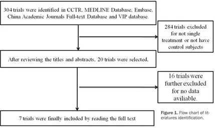
Using the search strategies, 304 trials were identified in CCTR, MEDLINE Database, Em- base, China Academic Journals Full-text Data- base and VIP database. After reviewing the titles and abstracts, 20 trials were selected, and 7 trials were finally included by reading the full text [11-17], the flow chart was shown in
Figure 1. All trials were written in English or Chinese, and 268 scars (hypertrophic scars and keloids) in 259 subjects were included without limitation by age and gender. Among them, 7 trials divided one scar into different
parts to respectively receiving PDL and other therapies.
Efficacy determination
[image:3.612.93.522.73.333.2](1) Primary indicators: VSS was used to evalu-ate the size of erythema and scars (thickness, volume, length and width) along with blood flow, pliability and softness; (2) Secondary indi-cators: Secondary indicators were measured in all trials, including the symptoms and subjec-tive evaluation of subjects; (3) Treatment time: All treatments lasted for 22 weeks to 2 years. The loss to follow-up rate was 5% in 1 trial [11] and 0% in the remaining trials; (4) Adverse reac-tions: Four trials reported adverse reactions, including intraoperative pain, purpura, pigmen-tation and blister [12-15]. One trial reported adverse reactions [13], and the remaining 2 tri-als reported no adverse reactions [11, 16].
[image:3.612.91.524.362.464.2]Figure 1. Flow chart of lit-eratures identification.
Table 1. Characteristics of included studies in the meta-analysis
Authors Publication year designStudy Random methods concealmentAllocation Blind method consistencyBaseline Dropout rate Class
Alster 1995 QRCT No description No description Double blind Yes 0% B
Wittenberg 1999 QRCT Random number table Yes Assessor-blinded Yes 5% A
Manuskiatti 2002 QRCT No description No description No description Yes 0% B
Bowes 2002 QRCT No description No description Assessor-blinded Yes 0% B
Chan 2004 QRCT No description No description No description Yes 0% B
Omranifard 2007 QRCT No description No description Assessor-blinded Yes 0% B
Quality of the included trials
Of 7 randomized controlled trials (RCT), 1 trial performed computer-assisted creation of ran-dom number table for ranran-dom allocation. Six QRCTs did not describe the method of random allocation. No allocation concealment was ad- opted except in 1 trial [12] (Table 1). One trial was double-blinded, and 3 trials were asses-sor-blinded; it was not certain whether blind method was used in 2 trials. All trials included follow-up procedures which lasted for 22 weeks to 2 years. The loss to follow-up rate was 0% in all except 1 trial where the loss to follow-up rate was 5%. It was not clarified in the latter whether intention-to-treat (ITT) analysis was performed. Result analysis
Five trials compared the primary indicators and secondary indicators between 585 nm PDL group and non-treatment group. Two trials re- ported VSS scores and the results indicated a statistically significant decline of VSS scores compared with the control.
As shown in Table 2, mitigation of erythema was compared in 2 trials. One trial reported an obvious mitigation of erythema at week 32 after treatment. But no difference of statistical significance was found between 585 nm PDL group and the control group. The other trial indi-cated an obvious mitigation of erythema after 1 or 2 treatments, showing a statistically signifi-cant difference compared with the control. Different measuring methods were used in the 2 trials and the first trial did not report the origi-nal data, which made quantitative aorigi-nalysis im- possible.
Height, length, width and volume of the scars were measured. Changes of the scar size were compared in 4 trials, and 3 of them described the height of the scars. One trial indicated a
marked decline of scars after 1 or 2 treatments compared with the control. Another trial report-ed an obvious decline of scar height at week 32 after treatment, showing statistically significa- nt difference compared with the control (P= 0.005), but no original data was included. One trial showed an insignificant decline of scar height between 585 nm PDL and blank control. The original data were incomplete in these 2 trials, therefore the data were not combined and analysis was carried out separately. One trial described the scar volume. Results of 40-week trial indicated no significant differ-ence in scar volume between 585 nm PDL group and the blank control. As to pliability and hardness, 4 trials compared scar pliability and hardness and 2 trials described scar hardness. In 1 trial, scar hardness reduced considerably at the end of 1 (P=0.000 7) or 2 treatments, showing a significant difference compared with the control. Another trial indicated a decline of scar hardness at week 32 after 585 nm PDL treatment compared with the baseline, but the difference was not significant (P=0.02); for the control group, a significant difference was no- ted at week 24 compared with the baseline (P=0.046), but this difference did not persist to week 32. Since this trial did not report the origi-nal data, it could not be combined with the above trials. Two trials described scar pliability; 1 trial reported no significant differences in pli-ability between the treatment group and the control group, but this trial did not report the original data. One trial involved the use of skin elasticity meter in the measurement of pliabili-ty, and a significant difference between the treatment group and the control group was observed only in 1 out of 5 measurements. The 2 trials were not combined due to the lack of original data.
[image:4.612.92.523.86.188.2]Two trials reported patients’ subjective evalua-tion. One trial indicated that the proportion of
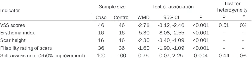
Table 2. Meta-analysis of efficacy evaluation of 585 nm pulsed dye laser in pathological scars
Indicator Sample size Test of association
Test for heterogeneity
Case Control WMD 95% CI P P I2
VSS scores 46 46 -2.78 -3.12, -2.46 <0.001 0.51 0%
Erythema index 16 16 -5.30 -8.08, -2.55 <0.001 -
-Scar height 16 16 -2.30 -3.40, -1.09 <0.001 -
patients reporting an improvement by 50% or above in self-evaluation did not differ signifi-cantly between the treatment group and blank control group. In another trial, the patients in treatment group reported an obvious improve-ment regarding itch, pain, sensitivity and scar size during subjective evaluation, and the dif-ference was significant compared with the con-trol. However, rate of obvious improvement sub-jectively evaluated by patients in terms of pin- prick sensation and color change did not differ considerably between the two groups. The ov- erall scores of the treatment group were much higher than those of the blank control.
One trial reported scores for itch and pain. According to this trial, 585 nm PDL and blank control did not differ significantly in their effects in improving the sensation of itch and pain. Publication bias of the included literature Funnel plot was used to test the publication bias of all included studies. Funnel plot shape of all included studies prompted no obvious asymmetry (Figure 2), suggesting no obvious publication bias.
Discussion
In the present study, we performed a meta-analysis to reveal efficacy and safety of 585 nm PDL in pathological scar and found 585 nm PDL is safe and effective in treating pathologi-cal scars.
Most of the seven trials included for systematic review were not sufficiently randomized. Ran- dom allocation was either implemented
improp-was used. In that case, implementation bias and measurement bias were inevitable. Only 1 trial had a loss to follow-up rate of 5%, and that of the remaining trials was controlled within 10%. In spite of the biases, the included trials generally had a high quality.
VSS is an important measuring tool for patho-logical scars from the dimensions of scar size, texture and hardness. As shown by systematic review of the included trials in these dimen-sions, 585 nm PDL is generally effective for improving VSS scores. Moreover, 585 nm PDL also improved scar size and pliability and ery-thema, but no consistent and explicit conclu-sion regarding the efficacy of 585 nm PDL was reached in any of these dimensions. For patho-logical scars with poor improvement in height, erythema or pliability, other therapies should be adopted in combination. Patients’ subjec-tive evaluation indicated that 585 nm PDL has no overall efficacy or efficacy in improving the sensation of itch and pain. This technique is not the patient-preferred therapy.
[image:5.612.89.378.74.263.2]Many studies have been carried out over 585 nm PDL. But meta-analysis is difficult because of the following problems: (1) The method for random allocation was not properly chosen for RCTs. Random number table was the most commonly used, or no description was provid-ed for the selectprovid-ed random method at all; (2) Most studies contained no control groups but only treatment effect observation, leading to poor reliability of the conclusions; (3) Different scar evaluation indicator systems and mea-surement methods were adopted. Scar assess-ment scales also varied greatly from one trial to
Figure 2. Funnel plot for publica-tion bias tests.
another, and some were even designed by the researchers. This makes the evaluation of the efficacy difficult. We suggest the use of VSS or newly designed scales, and for patients’ sub-jective evaluation, the Patient and Observer Scar Assessment Scaleare recommended. The later covers the subjective evaluation by both patients and doctors. Some studies have dem-onstrated that this scale is more reliable than VSS; (4) Current studies are less concerned with the psychological impact of 585 nm PDL, improvement of patients’ life quality or cost-benefit analysis; (5) Hypertrophic scars and keloids can be easily confused due to morpho-logical similarities. Although the two types of scars show distinct clinical and physiological features, no subgroup analysis was included in most literature; (6) many studies only provide diagrams of the results but no original data. In conclusion, 585 nm PDL is generally safe and effective for pathological scars. However, the findings need to be confirmed by more stud-ies given the limited number of included trials.
Disclosure of conflict of interest
None.
Address correspondence to: Ning Pan, Department of Dermatology, Sichuan Academy of Medical Science & Sichuan Provincial People’s Hospital, Chengdu, 610072, China. Tel: +86-02899897863; Fax: +86-02899897863; E-mail: panning404@126. com
References
[1] Menter A. Recognizing and managing comor-bidities and complications in hidradenitis sup-purativa. Semin Cutan Med Surg 2014; 33: S54-6.
[2] Tanaka A, Hatoko M, Tada H, Iioka H, Niitsu- ma K, Miyagawa S. Expression of p53 familyin scars. J Dermatol Science 2004; 34: 17-24. [3] Amadeu TP, Braune AS, Porto LC, Desmoulière
A, Costa AM. Fibrillin-1 and elastinare differen-tially expressed in hypertrophic scars and ke-loids. Wound Repair Regen 2004; 12: 169-174.
[4] Kia KF, Burns MV, Vandergriff T, Weitzul S. Pre-vention of scar spread on trunk excisions: a rater-blinded randomized controlled trial. JAMA Dermatol 2013; 149: 687-91.
[5] Liu A, Moy RL, Ozog DM. Current methods em-ployed in the prevention and minimizatio of surgical scars. Dermatol Surg 2011; 37: 1740-17446.
[6] Parodi A, Cozzani E. Cutaneous manifestations of lupus erythematosus. G Ital Dermatol Vene-reol 2014; 149: 549-54.
[7] Alester TS. Improvement of erythematous and hypertrophicscars by 585 nm flashlamp-pu- mped pulsed dyelaser. Ann Plast Surg 1994; 32: 186-190.
[8] Nouri K, Elsaie ML, Vejjabhinanta V, Stevens M, Patel SS, Caperton C, Elgart G. Comparison of the effects of short- and long-pulse dura-tions when using a 585 nm pulsed dye laser in the treatment of new surgical scars. Lasers Med Sci 2010; 25: 121-6.
[9] Nouri K, Rivas MP, Stevens M, Ballard CJ, Sing-er L, Ma F, Vejjabhinanta V, Elsaie ML, Elgart GW. Comparison of the effectiveness of the pulsed dye laser 585 nm versus 595 nm in the treatment of new surgical scars. Lasers Med Sci 2009; 24: 801-10.
[10] Asilian A, Darougheh A, Shariati F. New combi-nation of triamcinolone, 5-Fluorouracil, and pulsed-dye laser for treatment of keliod and hypertrophicscars. Dermatol Surg 2006; 32: 907-915.
[11] Alster TS, Williams CM. Treatment of keliod-sternotomy scars with 585 nm flashlamp-pumped plulsed-dyelaser. Lancet 1995; 345: 1198-200.
[12] Wittenberg GP, Fabian BG, Bogomilsky JL, Schultz LR, Rudner EJ, Chaffins ML, Saed GM, Burns RL, Fivenson DP. Prospective, Single-blind, Randomized, Controlled Study to Assess the efficacy of the 585-nm Flashlamp-Pumed-plused-dye Laser and silicone GelSheeting in Hypertrophic Scar Treatment. Arch Dermatol 1999; 135: 1049-55.
[13] Manuskiatti W, Fitzpatrick RE. Treatment Pe-sponse of keliodal and Hypertrophic Sternoto-my Scars. Arch Dermatol 2002; 138: 1149-55. [14] Bowes LE, Nouri K, Berman B, Jimenez G, Par-do R, Rodriguez L, Spencer JM. Treatment of Pigmented Hypertrophic Scars with the 585 nm Pulsed Dye Laser and the 532 nm Frequn-cy-Doubled Nd: YAG Laser in the Q-Switchedand Variable Pulsed Modes: A Comparative Study. Dermatol Surg 2002; 28: 714-719.
[15] Chan HH, Wong DS, Ho WS, Lam LK, Wei W. The use of Pulsed Dye Laser for the Prevention and Trentment of Hypertrophicscars in Chi-nese Persons. Dermatol Surg 2004; 30: 987-994.
[16] Omranifard M. Comparing the effects of con-ventional method, plused dye laser and erbi-um laser for the treatment of hyperteophic scars in Iranian petients. JRMS 2007; 12: 277-281.