Original Article
Bioinformatics analysis of T- and B-combined
epitopes of OMP31 protein of
Brucella
melitensis
in Xinjiang, China
Pan Pang1*, Fengbo Zhang1*, Bin Jia2*, Ming Li4, Jinwei Hu1, Ping Ji1, Rongjiong Zheng2, Jianbing Ding1,3, Yuexin Zhang2
Departments of 1Clinical Laboratory, 2Infectious Diseases, The First Affiliated Hospital of Xinjiang Medical Uni
-versity, Urumqi, Xinjiang, China; 3Department of Immunology, School of Preclinical Medicine of Xinjiang Medical
University, Urumqi, Xinjiang, China; 4Central Hospital of Karamay, Karamay, Xinjiang, China. *Equal contributors. Received June 19, 2017; Accepted August 31, 2017; Epub September 15, 2017; Published September 30, 2017
Abstract: Brucellosis is one of the major zoonoses in the word that widely inflicts health problems in animal and
human. In China, brucellosis is gradually increasing in recent years, especially in the xinjiang. Therefore, this study is to predict Brucella T-and B-combined epitopes of OMP31 protein by bioinformatics software. B-cell epitopes of OMP31 protein is predicted using DNAStar and IEDB software. The prediction of T-cell epitopes of OMP31 protein is performed by SYFPEITHI and ProPred MHC Class-II Binding Peptide Prediction Server. The results suggest that some position of OMP31 protein has several higher antigen indexes. The high-scoring of dominant B-cell epitopes were located at positions 25-31, 108-114, 201-207, 152-157, 166-172, 230-238, 123-127, 96-102 and 171-176. Five potential of dominant T-cell epitopes regions were revealed, located at positions 19-30, 92-106, 106-114, 137-179, 196-228. Finally, OMP31 protein has selected the three advantages of T- and B-combined epitopes, respectively are: 108-114, 152-157, 171-176. The bioinformatics research of OMP31 protein and T- and B-combined epitopes laid to the foundation of dominant epitopes vaccine.
Keywords:Brucella, bioinformatics, T-and B-combined epitopes, vaccine
Introduction
Brucellosis is a zoonotic chronic infectious dis-ease caused by Brucella. Brucella is Gram-negative intracellular pathogenic bacteria that infect a range of different mammals including human [1-3]. Brucella infection in human can cause a broad range of clinical manifestations that most commonly result in sacroiliitis as well as spondylitis and peripheral arthritis [4, 5]. The incidence of brucellosis rose sharply in many countries. At present, approximately 500,000 new brucellosis cases are reported annually worldwide [6]. The infection is endem-ic in Mediterranean, the Middle East, central Asia, Africa and parts of Latin America [7]. In China, brucellosis has a wide distribution in many provinces especially in xinjiang with high-er prevalence of husbandry animals. Previous study revealed that brucellosis has significantly increased in xinjiang [8]. Because it causes a
wide variety of signs and symptoms, which is hard to diagnosis is immediately and patients may not receive the proper treatment in time. It is impossible to cure the patients when the acute brucellosis turns into a chronic phase [9]. Therefore, early preventive measures of Br- ucella infection are particularly important.
brucello-sis in clinical examination and serological diag-nosis. Bowden et al [14] found that OMP31 has a good immunogenicity which can be used as a subunit vaccine candidate molecule for brucel-losis. Wang et al [15] had predicted the Bruce- lla melitensis BP26 and OMP31 proteins and founded the five T-cell antigen epitopes of BP-26 and five specific antigen epitopes of OMP31. Ghasemi et al [16] predicted the epitopes of BP26 in 93-111 and OMP31 in 48-74. The st- udy revealed that the antigen epitopes of OM- P31 as Brucella outer membrane proteins can be used for the development of Brucella sub-unit vaccine candidate molecules.
In summary, this study predicted and analyzed the T- and B-cells epitopes of OMP31 protein by bioinformatics software, especially its T- and B-combined epitopes, which may help develop a new vaccine to protect the host against Br-
ucella.
Materials and methods
Reagents
OMP31 protein amino acid sequence: Re- leased from GenBank in length of 240 bp of OMP31 protein of B.melitensis, the amino acid sequence is as the following:
MKSVILASIAAMFATSAMAADVVVSEPSAPTAA- PVDTFSWTGGYIGINAGYAGGKFKHPFSSFDKE- DNEQVSGSLDVTAGGFVGGVQAGYNWQLDNG- VVLGAETDFQGSSVTGSISAGASGLEGKAETKVE- WFGTVRARLGYTATERLMVYGTGGLAYGKVKS- AFNLGDDASALHTWSDKTKAGWTLGAGAEYAI- NNNWTLKSEYLYTDLGKRNLVDVDNSFLESKV- NFHTVRVGLNYKF.
secondary structure prediction and analysis
The secondary structure of the OMP31 protein was predicted by the improved self-opti- mized prediction method (SOPMA) software (http://npsa-pbil.ibcp.fr/cgi-bin/npsa_automat. pl?page=/NPSA/npsa_sopma.html). The inpu- ts of the amino acid sequence of the OMP31 protein, and four conformational states, includ-ing helices, sheets, turns and coils, were an- alyzed.
Tertiary structure prediction and analysis
OMP31 protein tertiary structure prediction and analysis was conducted by online PHYRE2
Protein Fold Recognition Server and Swiss model (http://swissmodel.expasy.org/interac-tive). RasMol version was used to analyze dif-ferent modes of the tertiary structure. The ter-tiary structure was displayed in the modes of Cartoon, Structure and Group.
T-and B-combined epitopes prediction and
analysis
The software which predict B-cell epitopes are DNAStar (V5.0) (http://www.dnastar.com) and the online prediction software IEDB (http:// tools.immuneepitope.org/bcell). The major his-tocompatibility complex class-II (MHC-II): hu- man leukocyte antigen (HLA)-DRB1*0301-re- stricted T-cell epitopes were predicted using online prediction software SYFPEITHI (http:// www.syfpeithi.de) and ProPred MHC Class-II Binding Peptide Prediction Server (http://www. imtech.res.in/raghava/propred/index.html). Using Gamier-Robson [17] and Chou-Fasman [18] method of DNAstar software, separately predict Alpha helix and beta Angle of OMP31 protein; Using Emini method of DNAStar soft-ware [19], according to the specific composi -tion of amino acid residues, predict the OMP31 protein molecular surface accessibility; Using Kyte-Doolittle [20] method of DNAStar soft-ware, according to the OMP31 amino acid mol-ecules composition, predict its hydrophobic region and hydrophilic region; Using Karplus-Schulz [21] method of DNAStar software pre-dict the OMP31 polypeptide skeleton flexibility; Using Jameson-Wolf [22] method of DNAStar software finish the prediction of antigenic determinant of OMP31 protein. IEDB [23] soft-ware is used for prediction and analysis of OMP31 protein, B-cell linear epitopes, beta cor-ner, surface accessibility, flexibility of skeleton, antigen index, and hydrophobicity. SYFPEITHI [24] and ProPred MHC Class-II Binding Peptide Prediction Server [25] are used for comprehen-sive predictionand analysis of OMP31 protein and dominant T-cell epitopes.
Results
Prediction of the secondary structure of OMP31 protein
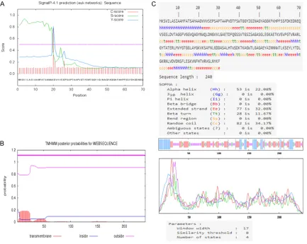
signal peptide. Each amino acid has an S and C value, the signal peptide of is high, and the cleavage sites of C value are higher. The Y score distinguishes between C score peaks by choos-ing the one where the slope of the S score is steep. We speculated that there is a signal pep-tide in OMP31 protein, and signal peppep-tide site is 1-19. When OMP31 protein matured, this sig-nal peptide site will be removed. In order to improve the confidence level of the prediction results, we abandoned the signal peptide site (Figure 1A). Transmembrane and non-trans-membrane regions of OMP31 protein were pre-dicted by TMHMM server v 2.0. The region of amino acids that start from 19 to 240 was an extracellular domain (Figure 1B), its secondary structure was predicted by SOPMA Server soft-ware. The results revealed that the proportion of β turns, random coils, α helices and extend -ed strands (β folds) account-ed for 11.67%, 34.17%, 22.08% and 32.08% of the secondary structure, respectively (Figure 1C).
Results of tertiary structure prediction of OMP31 protein
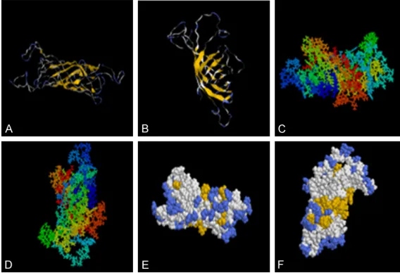
[image:3.612.93.526.73.418.2]ly fixed and often in the interior of the protein, is not easy to combine with antibodies and is not considered as an antigen epitope. While beta Angle and random curl at the surface of the pro-tein, loose structure is easy express on the sur-face of the protein, is easy to combine with anti-bodies and more likely to become the antigen epitopes. The front and back view of the tertiary structure using space-filling model showed that the yellow region was gathered and distributed substantially at the surface of the structure. When an antibody binds an antigen, the anti-gen epitopes need to be fully exposed to facili-tate better binding. Thus we speculated that the yellow and pale blue region in this model is the antigen epitopes that potentially binds to the antibody.
T-cell epitopes prediction of OMP31 protein
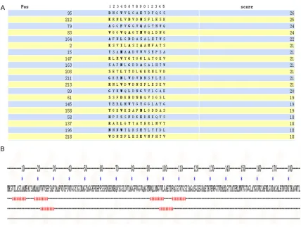
In order to improve the accuracy of prediction, we predicted the T-cell epitopes with SYFPEITHI and ProPred MHC Class-II Binding Peptide Prediction Server. HLA molecule selected type: HLA-DRB1*0301. We are using SYFPEITHI soft-ware to predict T-cell epitopes of OMP31 pro-tein. The regions with high scores are 95-110, 212-227, 79-94, 83-98, 164-179, 2-17, 15-30, 147-162, 163-178, 203-218, 211-226,
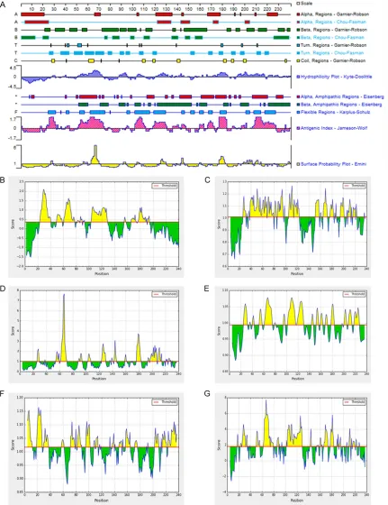
[image:4.612.89.373.72.266.2]213-73-94, 96-103, 110-117, 135-140, 146-166, 184-187, 192-195, 202-208, 212-219 and 226-240. In alpha helices and beta corner regions, the distribution of different seventeen corner regions and nine coil regions, corner regions are mainly distributed in: 26-29, 36-43, 48-55, 58-65, 67-74, 78-81, 89-92, 94-97, 106-109, 118-121, 152-155, 168-171, 176-180, 188-191, 196-199, 211-214, 218-221. coil regions are mainly distributed in: 28-30, 61-65, 109-111, 118-120, 140-142, 181-183, 188-190, 199-201, 236-237 (>1) (Figure 4A). Emini method of analysis showed that the sur-face probability plots were located at positions 62-69, 123-127 and 175-180 (Figure 4A). Kyte-Doolittle method of analysis showed that the hydrophilicity plot are located at positions 50-70, 85-95, 101-110, 118-131, 162-182, 190-217 and 218-228 (≥0.5) (Figure 4A). Ka- rplus-Schulz method of analysis showed that the flexible regions are located at positions 25-31, 40-42, 53-57, 61-75, 94-96, 102-115, 119-128, 143-145, 152-155, 159-163, 168-171, 178-183, 200-204, 208-214 and 218-227 (Figure 4A). Jameson-Wolf method of anal-ysis showed that the antigenic index are located at positions 21-32, 50-73, 100-110, 117-130, 162-169, 175-182 and 202-228 (Figure 4A). Figure 2. (A, B) Represents Tertiary structure prediction results of OMP31,
the α helixes are shown by the curved lines and the β folds are shown by the laminated structures. The structure of the OMP31 modeled using Phyre, an-alyzed by Rasmol software in Srick and group (C) the front view (D) the back view. The structure of the OMP31 modeled using Phyre, analyzed by Rasmol
software in Structure and spacefill (E) the front view (F) the back view.
Ma-genta area corresponded to the α helixes, pale blue area corresponded to
the turns. The laminated structures and the amino acids in the yellow area
corresponded to the flexible regions of the protein.
228, 89-104, 61-76, 145-160, 158-173, 58-73, 137-152, 196-211 and 218-233 (Figu- re 3A). ProPred MHC Class- II Binding Peptide Prediction Server software is used to pre-dict T-cell epitopes of OMP31 protein. The regions with high scores are 4-12, 19-26, 92- 100, 106-114, 22-30, 98-106 (Figure 3B).
B-cell epitopes prediction of OMP31 protein
Using DNAstar software pre
Using IEDB software predicted B-cell epitopes:
Through IEDB online software which analyze B-cell linear epitopes of OMP31 protein, the regions with high scores are 24-40, 50-58, 61-77, 82-85, 87-89, 91-93, 104-129, 155-157, 166-174, 176-190 and 192-194 (Figure 4B). Through IEDB online software which ana-lyze corner area of OMP31 protein, the regions with high scores are 108-114, 166-172, 48-54, 91-97, 104-110, 107-113, 42-48, 62-68, 25-31, 90-96, 89-95, 67-73, 40-46, 53-59, 38-44, 196-202, 68-74, 49-55, 61-67 and 118-124 (Figure 4C). Through IEDB online soft-ware which analyze surface accessibility of OMP31 protein, the regions with high scores are 65-70, 64-69, 177-182, 63-68, 178-183, 62-67, 142-147, 66-71, 124-129, 176-181, 179-184, 202-207, 175-180, 61-66, 200-205, 123-128, 209-214, 204-209, 26-31 and 67-72 (Figure 4D). Through IEDB online software which analyze the flexibility of OMP31 protein, the regions with high scores are 106-112, 64-70, 107-113, 63-69, 105-111, 65-71,
123-129, 122-128, 108-114, 121-127, 62-68, 66-72, 104-110, 109-115, 25-31, 124-130, 178-184, 177-183, 67-73 and 24-30 (Figure 4E). Through IEDB online software which ana-lyze antigen index of OMP31 protein, the regions with high scores are 19-25, 22-28, 4-10, 3-9, 18-24, 21-27, 20-26, 2-8, 94-100, 71-77, 230-236, 23-29, 5-11, 98-104, 232-238, 82-88, 97-103, 17-23, 1-7 and 201-207 (Figure 4F). Through IEDB online software which analyze the hydrophilicity of OMP31 pro-tein, the regions with high scores are 64-70, 66-72, 178-184, 65-71, 123-129, 67-73, 63-69, 62-68, 61-67, 68-74, 108-114, 25-31, 166-172, 103-109, 107-113, 104-110, 125-131, 208-214, 124-130 and 176-182 (Figure 4G).
T-and-B combined epitopes prediction results of OMP31 protein
[image:5.612.91.519.66.390.2]This research use DNAStar and IEDB software for comprehensive analysis of B-cell antigen Figure 3. T-cell epitope prediction results for the OMP31 protein. Selected from the twenty highest-scoring regions with SYFPEITHI software (A). Using ProPred MHC Class-II Binding Peptide Prediction Server software (B) to predict
Figure 4. DNAStar Server software to predict B-cell epitope of the OMP31 protein (A). IEDB online software was used
to predict the B-cell linear epitope (B), Beta-turn (C), surface accessibility (D), skeleton area flexibility (E), antigen
index (F), hydrophilicity (G).
epitopes. The B-cell epitopes position of
Using SYFPEITHI and ProPred MHC Class-II Binding Peptide Prediction Server software for comprehensive analysis of T-cell epitopes of OMP31 protein, T-cell epitopes are located at positions 19-30, 92-106, 106-114, 137-179 and 196-228 (Table 1).
According to the comprehensive analysis results of the regions with high scores and posi-tion of B-cell epitopes and T-cell epitopes of OMP31 protein, finally, we have selected the three advantages T-and-B combined epitopes of OMP31 protein: 108-114, 152-157 and 171-176 (Table 1).
Discussion
Brucellosis is considered as one of the great threat to public health which cannot be ignored in xinjiang region [26, 27]. xinjiang is one of the main pastoral areas in China, which is also a re-emerging epidemic area with a high inci-dence rate of brucellosis [28-30]. New effec-tive vaccines are required to protect humans and animals against Brucella. The epitope-based vaccines has gradually become the hotspot of current molecular vaccine [31], that can activate B cell or T cell to protect the host
[image:7.612.91.316.98.376.2]ing linear epitopes and conformational epit-opes mainly induce the humoral immune re- sponse. B-cell epitopes prediction analysis is mainly consisted of alpha helix, beta Angle, sur-face accessibility, hydrophilicity, flexibility, anti -gen index and so on. Predictions of B-cell epit-opes help develop vaccine and antigen-anti- body interactions at the molecular level [36-38]. B-cell epitopes from DNAStar and IEDB software comprehensive analysis are 25-31, 108-114, 201-207, 152-157, 166-172, 230-238, 123-127, 96-102 and 171-176. The sec-ondary and tertiary structures of OMP31 pro-tein were predicted. The secondary structure of OMP31 protein prediction was performed by SOPMA Server software. We found that the pro-portion of random coils, and β turns accounted for 34.17% and 11.67% (Figure 1C) respective-ly. Due to the presence of hydrogen bonds which act to maintain structural stability, the helices and β sheets are very regular struc -tures, and are not easily deformed in the sec-ondary structure of proteins. The β turn and random coil regions are located on the surface of the protein, its structure is loose, easily to be distorted, which is conducive to ligand binding and have a high possibility of forming epitopes Table 1. The T-and B-cell epitopes and T-B combined
epitopes prediction
Epitopes Position Sequence
B-cell epitopes 25-31 SGPSAPT
108-114 GSSVTGS 201-207 LLSGTLT
152-157 GTGGLA
166-172 ALGAAAS 230-238 HTVAVGLAT
123-127 GGLAG
96-102 AGVVLGA
171-176 ASALHT
T-cell epitopes 19-30 AADVVVSEPSAP
92-106 WQLDNGVVLGAETDF 106-114 FQGSSVTGS 137-179 RARLGYTATERLMVYGT
GGLAYGKVKSAFNLGDD ASALHTWSD 196-228 NNNWTLKSEYLYTDLGK
RNLVDVDNSFLESKVN T-B combined epitopes 108-114 GSSVTGS
152-157 GTGGLA
171-176 ASALHT
against pathogens [32]. How to find epitope-based vaccines has been challenging scien-tists for many years [33].
In order to check whether the signal peptide would present in whole OMP31 protein, we confirmed its position is 1-19 according to S value, C value and Y value by Signal-4.1 Server. In order to improve the confidence level of the prediction results, we aban-doned the signal peptide site of OMP31 pro-tein. In order to check the transmembrane of Brucella, TMHMM was used to predict whether the OMP31 protein was presented on transmembrane. In this study, TMHMM predicts that all the amino acid of OMP31 protein is on the outer membrane of Brucella. Outer membrane amino acids pro-vide the chance to contact to immune cells, which is suggested that the outer membrane amino acids could form epitopes more than other amino acids locations.
includ-[39]. On the other hand, α helices and β sheets are usually located inside the protein, which are difficult for ligand binding. Therefore, it is diffi -cult to form the antigen epitopes. We predicted the tertiary structure of OMP31 protein and found that most parts of OMP31 protein were flexible (Figure 2). This flexible structure is eas -ier to form antigen epitopes.
Cellular immunity play crucial role in the clear-ance of intracellular pathogens. Whether the vaccine could elicit cell mediated immune response regarded as the vital character of candidate vaccines. The immunogenic poten-tial of candidate vaccines needs to be indicat-ed in the context of HLA polymorphisms. Hence, our study preformed the T cell epitopes predic-tion taking into account MHC-II (major histo-compatibility complex II) molecules [40]. CD4+ T cells play a key role in controlling bacteria growth, through IFN-γ which induce CD4+ T cells in the body to control Brucella infections [41]. This study predicts the epitopes which elicit HLA-II restricted CD4+ T cell immune respon- se [42, 43]. HLA-II play an important role in immune cells, they can present the specific antigen to CD4+ T cells to induce the immune response, HLA-II molecules including HLA-DR, HLA-DQ, HLA-DP [44]. HLA-DR antigen encoded by HLA-DRA and HLA-DRB, and MHC-II can be used as antigen presenting or regulatory mole-cules participating in specific immune resp-onse and innate immune respresp-onse [45]. Ac- cording to the report of the frequency of HLA alleles in humans, HLA-DRB1 locus is one of the most common HLA-A alleles in Uygur of xinjiang. Allele HLA-DRB1*0301 in the Han, Uygur and Kazak has high frequency distribu-tion [46, 47]. This study has selected the xinjiang people’s alleles HLA-DRB1*0301 to analysis T-cell epitopes OMP31 protein by using SYFPEITHI and ProPred MHC Class-II Binding Peptide Prediction Server software and found five T-cells dominant epitopes, which respec -tively are 19-30, 92-106, 106-114, 137-179, 196-228 (Table 1).
A strong protective epitope-based vaccine sh- ould contain T- and B-combined dominant epit-opes, that can induce the host to produce long-lasting immune response, in order to effectively eliminate pathogenic microorganisms [48]. Liu et al [49] found that a length of T-B combined epitopes of echinococcus granulosus recombi-nant ferritin antigen epitope by using
bioinfor-matics software. Wang et al [50] found that Em95 protein has a good immunogenicity by predicting the echinococcus granulosus Em95 protein of T-B combined epitopes. Zhang et al [51] found that the recombinant protein rAg85B has a good immunogenicity by predicting T-B combined epitopes of Mycobacterium tubercu -losis Ag85B protein, which help develop tuber-culosis dominant epitope vaccine. In this study, based on the T-cell and B-cell epitopes informa-tion obtained, as well as the secondary and ter-tiary structure, using bioinformatics software we analyzed the T-B combined epitopes of OMP31 protein. The results show that there are potential T-B combined epitopes of OMP31 pro-tein. The T-B combined epitopes were 108-114, 152-157, 171-176. These positions have proper epitopes characteristics, so could help stimu-late the cellular and the humoral immunity to eliminate pathogens for construction of multi-valent vaccine against Brucella infection. To sum up, using the bioinformatics software, this study found out the advantage T- and B- cell epitopes and ultimately selected the four T-B combined dominant epitopes of Brucella OMP31 protein. This result laid foundation for constructing multivalent epitope vaccine to pro-tect the body from Brucella infection.
Acknowledgements
We would like to thank Ahmed Alamer for his help to modify this article. This work was sup-ported by National Natural Science Foundation of China (No.81560322) and Natural Science Foundation of xinjiang (No.2015211C100). Disclosure of conflict of interest
None.
Address correspondence to: Jiangbing Ding, Depart-
ment of Immunology, School of Preclinical Medicine, xinjiang Medical University, 393 xinyi Road, Uru- mqi 830011, xinjiang, China. Tel: 86-9914362403; E-mail: [email protected]; Yuexin Zhang, De-
partment of Infectious Diseases, the First Affiliated
Hospital of xinjiang Medical University, 393 xinyi Road, Urumqi 830011, xinjiang, China. Tel: 86- 9914365503; E-mail: [email protected]
References
[2] Elmardi ME, Al Hebaishi Y, Al Kassab S, Dagriri K, Al Ghamdi S, Ahmed A and Al Fagih A. Im-plantable cardioverter device infection due to Brucella melitensis in two patients. Hellenic J Cardiol 2016; [Epub ahead of print].
[3] Zhang K, Wang H, Guo F, Yuan L, Zhang W, Wang Y and Chen C. OMP31 of Brucella meli-tensis 16M impairs the apoptosis of macro-phages triggered by TNF-alpha. Exp Ther Med 2016; 12: 2783-2789.
[4] Elzein FE and Sherbeeni N. Brucella septic ar-thritis: case reports and review of the litera-ture. Case Rep Infect Dis 2016; 2016: 4687840.
[5] Pang P, Zhu Y, Jia B, Yang Y, Hu J, Zhang Y, Zhang F and Ding J. [Increased serum levels of soluble programmed death-1 (1) and sPD-ligand 1 in patients infected with Brucella]. xi Bao Yu Fen Zi Mian Yi xue Za Zhi 2016; 32: 1675-1678.
[6] Pappas G, Papadimitriou P, Akritidis N, Chris-tou L and Tsianos EV. The new global map of human brucellosis. Lancet Infect Dis 2006; 6: 91-99.
[7] Ilhan Z, Solmaz H and Ekin IH. In vitro antimi-crobial susceptibility of Brucella melitensis iso-lates from sheep in an area endemic for hu-man brucellosis in Turkey. J Vet Med Sci 2013; 75: 1035-1040.
[8] Wang W, Liao Q, Wu x, Hou S, Wang Y, Wu J, Shen C, Chen S, Allain JP and Li C. Potential risk of blood transfusion-transmitted brucello-sis in an endemic area of China. Transfusion 2015; 55: 586-592.
[9] Rajala EL, Grahn C, Ljung I, Sattorov N, Boqvist S and Magnusson U. Prevalence and risk fac-tors for Brucella seropositivity among sheep and goats in a peri-urban region of Tajikistan. Trop Anim Health Prod 2016; 48: 553-558. [10] Golshani M, Rafati S, Dashti A, Gholami E,
Sia-dat SD, Oloomi M, Jafari A and Bouzari S. Vac-cination with recombinant L7/L12-truncated Omp31 protein induces protection against Bru-cella infection in BALB/c mice. Mol Immunol 2015; 65: 287-292.
[11] Zheng WY, Wang Y, Zhang ZC and Yan F. Immu-nological characteristics of outer membrane protein omp31 of goat Brucella and its mono-clonal antibody. Genet Mol Res 2015; 14: 11965-11974.
[12] Zygmunt MS, Bundle DR, Ganesh NV, Guiard J and Cloeckaert A. Monoclonal
antibody-de-fined Specific C epitope of brucella O-polysac -charide revisited. Clin Vaccine Immunol 2015; 22: 979-982.
[13] Tiwari S, Kumar A, Thavaselvam D, Mangalgi S, Rathod V, Prakash A, Barua A, Arora S and Sathyaseelan K. Development and compara-tive evaluation of a plate enzyme-linked
immu-nosorbent assay based on recombinant outer membrane antigens Omp28 and Omp31 for diagnosis of human brucellosis. Clin Vaccine Immunol 2013; 20: 1217-1222.
[14] Bowden RA, Estein SM, Zygmunt MS, Dubray G
and Cloeckaert A. Identification of protective
outer membrane antigens of Brucella ovis by passive immunization of mice with monoclonal antibodies. Microbes Infect 2000; 2: 481-488. [15] Wang W, Wu J, Qiao J, Weng Y, Zhang H, Liao Q,
Qiu J, Chen C, Allain JP and Li C. Evaluation of humoral and cellular immune responses to BP26 and OMP31 epitopes in the attenuated Brucella melitensis vaccinated sheep. Vaccine 2014; 32: 825-833.
[16] Ghasemi A, Ranjbar R and Amani J. In silico analysis of chimeric TF, Omp31 and BP26 frag-ments of Brucella melitensis for development of a multi subunit vaccine candidate. Iran J Ba-sic Med Sci 2014; 17: 172-180.
[17] Yao W, Li Y, Yuan J, Wang L, Li Q, Song M, Lu G and Zhang L. [Structure, Immunogenicity and Clinical Value of Chlamydiaphage Capsid Pro-tein 3]. Bing Du xue Bao 2015; 31: 420-424. [18] Saxena A, Tamuly S and Saxena MK. Cloning,
sequencing, and in silico characterization of Omp 28 of Salmonella Typhi (strain MTCC 733) to develop r-DNA vaccine for typhoid fever. J Nat Sci Biol Med 2012; 3: 133-138.
[19] Yu SL, Yu DM, Zhang DH, Jiang JH, Chen J, Deng L and Zhang xx. [Functional analysis of hepatitis B virus immune escape mutants with insertion mutations in the surface antigen]. Zhonghua Gan Zang Bing Za Zhi 2013; 21: 510-513.
[20] Koz S, Sahin I, Kayabas U and Kuzucu C. Bru-cella and peritoneal dialysis related peritonitis: case report and review of literature. Clin Nephrol 2014; 82: 283-286.
[21] Wan CS, Zhou Y, Yu Y, Peng LJ, Zhao W and Zheng xL. B-cell epitope KT-12 of enterohem-orrhagic Escherichia coli O157:H7: a novel pep-tide vaccine candidate. Microbiol Immunol 2011; 55: 247-253.
[22] Tae H, Shallom S, Settlage R, Hawkins GN, Ad-ams LG and Garner HR. Complete genome se-quence of Brucella suis VBI22, isolated from bovine milk. J Bacteriol 2012; 194: 910. [23] Gonzalez-Diaz H, Perez-Montoto LG and Ubeira
FM. Model for vaccine design by prediction of B-epitopes of IEDB given perturbations in pep-tide sequence, in vivo process, experimental techniques, and source or host organisms. J Immunol Res 2014; 2014: 768515.
[24] Dong HL and Sui YF. Prediction of
[25] Wang L, Pan D, Hu x, xiao J, Gao Y, Zhang H, Zhang Y, Liu J and Zhu S. BiodMHC: an online server for the prediction of MHC class
II-pep-tide binding affinity. J Genet Genomics 2009;
36: 289-296.
[26] Zhang Y, Li T, Zhang J, Li Z, Zhang Y, Wang Z, Feng H, Wang Y, Chen C and Zhang H. The Bru-cella melitensis M5-90 phosphoglucomutase (PGM) mutant is attenuated and confers pro-tection against wild-type challenge in BALB/c mice. World J Microbiol Biotechnol 2016; 32: 58.
[27] Lei S, Zhong Z, Ke Y, Yang M, xu x, Ren H, An C, Yuan J, Yu J, xu J, Qiu Y, Shi Y, Wang Y, Peng G and Chen Z. Deletion of the small RNA chaper-one protein Hfq down regulates genes related to virulence and confers protection against wild-type brucella challenge in mice. Front Mi-crobiol 2015; 6: 1570.
[28] Zhang F, Li Z, La x, Ma x, Zhang Y, Ji P, Jiang M, Hu J, Zhang Z, Lu x and Ding J. Multiple-locus variable-number tandem-repeat analysis of Brucella isolates from patients in xinjiang Chi-na. Int J Clin Exp Med 2015; 8: 15716-15723. [29] Liu Q, Wei J, Sun Q, Wang B, Wang Y, Hu Y and
Wu W. Detection of Brucellosis in Sika Deer (Cervus nippon) through Loop-mediated
Iso-thermal Amplification (LAMP). J Wildl Dis 2017;
53: 612-615.
[30] Jia B, Zhang F, Pang P, Zhang T, Zheng R, Zhang W, Zhang Y and Ding J. Brucella endocarditis: Clinical features and treatment outcomes of 10 cases from xinjiang, China. J Infect 2017; 74: 512-514.
[31] Parvege MM, Rahman M, Nibir YM and Hos-sain MS. Two highly similar LAEDDTNAQKT and LTDKIGTEI epitopes in G glycoprotein may be useful for effective epitope based vaccine de-sign against pathogenic Henipavirus. Comput Biol Chem 2016; 61: 270-280.
[32] Ma x, Zhou x, Zhu Y, Li Y, Wang H, Mamuti W, Li Y, Wen H and Ding J. The prediction of T- and B-combined epitope and tertiary structure of the Eg95 antigen of Echinococcus granulosus.
Exp Ther Med 2013; 6: 657-662.
[33] Vider-Shalit T, Raffaeli S and Louzoun Y. Virus-epitope vaccine design: informatic matching the HLA-I polymorphism to the virus genome. Mol Immunol 2007; 44: 1253-1261.
[34] EL-Manzalawy Y, Honavar V. Building classifier
ensembles for B-cell epitope prediction. Meth-ods Mol Biol 2014; 1184: 285-294.
[35] Singh H, Ansari HR and Raghava GP. Improved method for linear B-cell epitope prediction us-ing antigen’s primary sequence. PLoS One 2013; 8: e62216.
[36] Malm M, Rollman E, Ustav M, Hinkula J, Krohn K, Wahren B and Blazevic V. Cross-clade
pro-tection induced by human immunodeficiency
virus-1 DNA immunogens expressing
consen-sus sequences of multiple genes and epitopes from subtypes A, B, C, and FGH. Viral Immunol 2005; 18: 678-688.
[37] Sun P, Ju H, Zhang B, Gu Y, Liu B, Huang Y, Zhang H and Li Y. Conformational B-cell epit-ope prediction method based on antigen pre-processing and mimotopes analysis. Biomed Res Int 2015; 2015: 257030.
[38] Wang A, Jiang M, Li N, Zhang G, Qi Y, Liu Y, xi Y and Zhou J. [Prediction of B cell epitopes of hu-man papillomavirus type 16 L1 protein]. xi Bao Yu Fen Zi Mian Yi xue Za Zhi 2016; 32: 442-445.
[39] Li Y, Liu x, Zhu Y, Zhou x, Cao C, Hu x, Ma H, Wen H, Ma x and Ding JB. Bioinformatic predic-tion of epitopes in the Emy162 antigen of Echi-nococcus multilocularis. Exp Ther Med 2013; 6: 335-340.
[40] Zhang xW. A combination of epitope prediction
and molecular docking allows for good identifi -cation of MHC class I restricted T-cell epitopes. Comput Biol Chem 2013; 45: 30-35.
[41] Ibana JA, Cutay SJ, Romero M and Schust DJ. Parallel expression of enzyme inhibitors of CD8T cell activity in tumor microenvironments and secretory endometrium. Reprod Sci 2016; 23: 289-301.
[42] Vitry MA, Hanot Mambres D, De Trez C, Akira S, Ryffel B, Letesson JJ and Muraille E. Humoral immunity and CD4+ Th1 cells are both neces-sary for a fully protective immune response upon secondary infection with Brucella meli-tensis. J Immunol 2014; 192: 3740-3752. [43] Vitry MA, De Trez C, Goriely S, Dumoutier L,
Akira S, Ryffel B, Carlier Y, Letesson JJ and Mu-raille E. Crucial role of gamma interferon-pro-ducing CD4+ Th1 cells but dispensable func-tion of CD8+ T cell, B cell, Th2, and Th17 responses in the control of Brucella melitensis infection in mice. Infect Immun 2012; 80: 4271-4280.
[44] Karosiene E, Rasmussen M, Blicher T, Lund O, Buus S and Nielsen M. NetMHCIIpan-3.0, a
common pan-specific MHC class II prediction
method including all three human MHC class II isotypes, HLA-DR, HLA-DP and HLA-DQ. Immu-nogenetics 2013; 65: 711-724.
[45] Sun C, Wei L, Luo F, Li Y, Li J, Zhu F, Kang P, xu R, xiao L, Liu Z and xu P. HLA-DRB1 alleles are associated with the susceptibility to sporadic Parkinson’s disease in Chinese Han popula-tion. PLoS One 2012; 7: e48594.
[46] Wang W, Hu FY, Wu xT, An DM, Yan B and Zhou D. Genetic predictors of Stevens-Johnson syn-drome and toxic epidermal necrolysis induced by aromatic antiepileptic drugs among the Chi-nese Han population. Epilepsy Behav 2014; 37: 16-19.
Allele polymorphism and haplotype diversity of HLA-A, -B and -DRB1 loci in sequence-based typing for Chinese Uyghur ethnic group. PLoS One 2010; 5: e13458.
[48] Sekhavati MH, Heravi RM, Tahmoorespur M,
Yousefi S, Abbassi-Daloii T and Akbari R. Clon -ing, molecular analysis and epitopics predic-tion of a new chaperone GroEL Brucella meli-tensis antigen. Iran J Basic Med Sci 2015; 18: 499-505.
[49] Liu x, Zhao H, Cao W, Liu Y, Zhang C, Lan x, Peng S, Wen H, Ding J and Ma x. Bioinformatic prediction of the antigenic epitopes of recom-binant ferritin of Echinococcus granulosus. Mol Med Rep 2016; 13: 888-894.
[50] Wang H, Zhang F, Ma x, Ma H, Zhu Y, Liu x, Zhu M, Wen H and Ding J. Prokaryotic expression
and identification of B- and T-cell combined
epitopes of Em95 antigen of Echinococcus multilocularis. Int J Clin Exp Pathol 2014; 7: 5117-5122.