Original Article
Pretreatment of
Lycium barbarum
polysaccharide
reduces H
2O
2-induced myocardial apoptosis
Hao Pan1, Yan Zhao2, Ningfu Wang1
1Department of Cardiology, Hangzhou First People’s Hospital, Hangzhou, Zhejiang, China; 2Department of
Cardiol-ogy, Zhejiang Provincial People’s Hospital, Hangzhou, Zhejiang, China
Received November 29, 2016; Accepted March 14, 2017; Epub May 15, 2017; Published May 30, 2017
Abstract: The study is aimed to explore the effects of Lycium barbarum Polysaccharide (LBP), the main active compound of Lycium barbarum, on H2O2-induced oxidative stress in myocardial cells, thereby to investigate the pos-sibility of LBP to be an effective prophylactic agent for cardiac vascular disease (CVD). In the study, rat myocardial cells were randomly divided into control group, H2O2 model group and three LBP+H2O2 groups. Myocardial viability,
cell apoptosis andoxidative stress parameters were detected. Real-time fluorescent quantitative PCR (RT-PCR) and Western blot were applied to investigate the influences of LBP pretreatment on apoptosis regulator proteins B-cell
lymphoma 2 (Bcl-2) and Bcl-2-associated X (Bax) expression and NF-E2 p45-related factor 2 (Nrf2)/antioxidant
responsive element (ARE) signaling pathway. The results revealed that LBP pretreatment significantly attenuated
the increased myocardial apoptosis in H2O2-induced oxidative stress by activating the Nrf2 signaling pathway and endogenous antioxidants. It suggested that LBP has great potential to be an antioxidant to protect people from oxidative stress induced CVD.
Keywords:Lycium barbarum polysaccharide, cardiovascular disease, apoptosis, oxidative stress
Introduction
CVD, including atherosclerosis, hypertension, ischemic heart disease and hyperlipemia etc., are becoming one of the biggest health killers worldwide in recent decades [1]. In China, CVD has been the second highest cause of death only after cancer. In the pathological processes of numerous CVD, excessive generation of oxy-gen free radicals is oxy-generally triggered when the anti-oxidant defenses are restrained, result-ing in imbalanced status of oxidation and anti-oxidation in body [2]. As big family members of oxygen free radicals, reactive oxygen species (ROS) play an important part in not only normal signal and physiological mechanisms but also pathological processes, however, when the level of ROS overwhelm the body’s capability of regulation, oxidative stress, a condition consid-ered as possible underlying pathogenic mecha-nism in progression of CVD, ensues [1, 3]. When the stress level exceeds the certain limit, irreversible damages i.e. cell apoptosis and necrosis will be caused [2]. Myocardial
apopto-sis originates from the lost of myocardial cells, with recurrent lost, cellular progressive function
subsequently deteriorates due to drop of cell
count and functional degradation of the rest cells, resulting in necrosis and entering to the terminal phase of CVD, heart failure [4]. It admits of no delay to research the more in-depth pathogenesis of CVD and discover safer and more effective medicines.
Red-colored fruits of Lycium barbarum (family
Solanaceae), commercially called goji berry, have been used as a traditional Chinese herbal medicine for thousands of years [5]. With a large variety of biological effects and pharma-cological functions, Lycium barbarum fruits contribute to preventing and treating many chronic diseases, such as diabetes, hyperlipid-emia, cancer, hepatitis, hypo-immunity func-tion, thrombosis and male infertility [6]. Lycium barbarum polysaccharide (LBP), a soluble poly-saccharide extracted from Lycium barbarum, is studied to be one of the active ingredients
H2O2 with and without LBP, plus a normal con-trol group. RT-PCR and Western blot were
applied to investigate the influences of LBP on
apoptosis regulator proteins Bcl-2 and Bax expression and Nrf2/ARE signaling pathway. Materials and methods
Animals and treatments
Myocardial cell line of neonatal Wistar rats were purchased from Jining Shiye Co. Ltd
(Shanghai, China). α-sarcomericactin immuno -cytochemical method was applied to identify the myocardial cells. Normal myocardial cells were cultured for 2-3 days, proliferated to reach the density of 80%-95%. Cells that pulsed at the rate 120-140 times per minute were select-ed for the use of experimental myocardial cells. Cells were divided into groups: normal control group, H2O2 group, and three H2O2+LBP experi-mental groups. Cells in LBP+H2O2 groups were
pretreated with respectively 100 μg/mL, 200 μg/mL and 400 μg/mL of LBP (Jianglai
Biote-chnology Co. Ltd., Shanghai, China) for 24 h, then treated with 50 µmol/L H2O2 for 3 h for the following experiments. Cells in H2O2 group were treated with 50 µmol/L H2O2 (final
con-centration).
Methyl thiazolyl tetrazolium (MTT) assay
The cell metabolic activity was assessed by using MTT formazan powder (Sigma-Aldrich Co.
LLC., USA). Briefly, myocardial cells were seed -ed in 96-well plates at density of 5×10⁴ cells per well, and then were incubated at 37°C for 48 h. Then after different preconditions depending on experimental groups, the cells were washed with phosphate-buffered saline (PBS) (Whiga Technology Co. Ltd., Guangdong, China) 3 times. With addition of 20 µL 5 g/L MTT dye and 100 µL serum free DMEM culture
Myocardial cells were seeded in six-well plate, grouped by different experimental treatments. Detection of the level of ROS, lactate dehydro-genase (LDH), malondialdehyde (MDA) and super oxide dismutase (SOD) were conducted according to their corresponding test kits (Nanjing Jiancheng Bioengineering Institute, Nanjing, Jiangsu, China), the OD values were read at 420 nm on a spectrophotometer (Sigma-Aldrich Co. LLC., USA). The contents of ROS, LDH, MDA and SOD in each sample were calculated. Each test was run three times.
Flow cytometry
Myocardial cells were seeded in 6-well plates at density of 2×10⁴ cells/well, digested and col-lected with EDTA free trypsin (Whiga Technology Co. Ltd., Guangdong, China), stained by Anne- xin V-FITC and Propidium iodide (Keygen Biote- chnology Co. Ltd., Jiangsu, China), then incu-bated at room temperature for 5-15 min in dark place. The cultures were analyzed by
FACSCalibur flow cytometry (Becton, Dickinson
and Company, USA) with excitation wavelength 488 nm and emission wavelength 530 nm. The test was run three times, the proportion of apoptotic and necrotic cells in every group were calculated.
Real-time quantification PCR (RT-PCR)
RT-PCR and SYBR Green I chemistry (Trans-
Start Top Green qPCR, SuperMix, TransGen
concentra-tion of the extracted RNA were measured on a UV spectrophotometer. cDNA was synthesized
by reverse transcription, then fluorescence
qu-antitative detection of the target genes was performed afterwards. GAPDH, as the internal
control, was used to monitor RT-PCR efficiency.
All RT reactions were performed in triplicate. Primers for each gene were designed by Shanghai Sangon Biotech Co. Ltd. (Shanghai,
China). The specific primer sequences were
listed as the follows: 5’ TCATGGGCTGGACAC- TGGAC 3’ and 5’ CAGCCACCCTGGTCTTGGAT 3’ for Bax (product: 67 bp); 5’ GCCTGAGAGCA- ACCCAATGC 3’ and 5’ CGGAGGGTCAGATGGAC- CAC 3’ for Bcl-2 (product: 129 bp); 5’ AG- CACTCCGTGGAGTCTTCC 3’ and 5’ GGAGAA- TGTGCTGGCTGTGC 3’ for Nrf2 (product: 130 bp); 5’ CTACACGGCCCTGGAAGAGG 3’ and 5’ TGAGGCCCATACCAGAAGGC 3’ for HO-1 (prod-uct: 144 bp) and 5’ TGGCCTTCCGTGTTCCTACC 3’ and 5’ TTCAGTGGGCCCTCAGATGC 3’ for GAPDH (product: 121 bp).
Western blot
Myocardial cells were seeded at a density of 2×10⁷ cells/well in 6-well plates, collected in
2-3 days and then grouped by different experi-mental treatments. Each group of cells was harvested and washed twice with PBS and pro-tein lysed in ice-cold radio immunoprecipita- tion assay buffer (Whiga Technology Co. Ltd., Guangdong, China) with freshly added 0.01%
protease inhibitor phenylmethanesulfonyl
flu-oride and incubated on ice for 30 min. Cell lysis was centrifuged at 10,000×g for 5 min at 4°C,
and the supernatant (20-30 μg of protein) was
run on 10% sodium dodecyl sulfate polyacryl-amide gel electrophoresis gel and transferred electrophoretically to a nitrocellulose mem-brane (Millipore, Shanghai, People’s Republic of China), then detected with Bax, Bcl-2, Nrf2, HO-1 and NQO1 proteins. Protein loading was estimated using mouse anti-glyceraldehyde 3-phosphate dehydrogenase (antiGAPDH) mo- noclonal antibody. Blots were visualized using enhanced chemiluminescence (Thermo Fisher
Scientific Inc, NY, USA).
Statistical analysis
[image:3.612.90.520.73.281.2]All values were expressed as mean ± S.D. Differences between groups were assessed by means of variance analysis and student’s t-test.
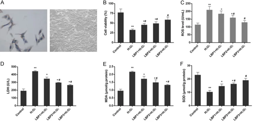
Figure 1. Morphological observation of cells and changes in cell proliferation and oxidative stress parameters in control, H2O2 (50 µmol/L) and LBP (100, 200 and 400 μg/mL)+H2O2 (50 µmol/L) groups. A: Myocardial cells were
identified by α-sarcomeric actin immunocytochemical strain (×400). B: The proliferation of myocardial cells was improved with LBP pretreatment in H2O2-induced injury. C: The intracellular ROS level was decreased with LBP pre-treatment in H2O2-induced injury. D: The content of LDH was reduced with LBP pretreatment in H2O2-induced injury. E: The content of MDA was decreased with LBP pretreatment in H2O2-induced injury. F: The activity of SOD was in-creased LBP pretreatment in H2O2-induced injury. Data were presented as mean ± SD, n=3, *P<0.05 and **P<0.01
vs. control, #P<0.05 and ##P<0.01 vs. H
1A).
LBP pretreatment improved cell viability in H2O2-induced injury
The changes of cell viability under the different conditions of H2O2 and LBP pretreatment were detected with MTT assay in vitro. In condition of 50 µmol/L of H2O2 without any pretreatment, a
significant decrease of cell proliferation was
observed compared to control group (P<0.01). The study found that LBP pretreatment of
myocardial cells for 24 h positively
influenc-ed on H2O2-induced decrease of cell viability, from 21.57%±2.25% in H2O2 group to 45.53%± 3.35%, 49.13%±4.22% and 58.70%±6.86% in H2O2+LBP groups with respectively 100, 200
and 400 μg/mL of LBP (P<0.05), and the effect was concentration dependent (Figure 1B).
LBP pretreatment decreased level of ROS, LDH and MDA and increased SOD activity in H2O2-induced injury
Treatment of H2O2 to myocardial cells obviously increased the content of ROS, LDH and MDA and reduced the level of SOD by over 50% in comparison with control group (P<0.01) (Figure 1C-F). In the H2O2-induced injury, with 100, 200
and 400 μg/mL LBP pretreatment, the activity
of SOD was improved while the contents of ROS, LDH and MDA were all reduced (P<0.05). The effects of LBP pretreatment on these parameters were positively correlated with the concentration of LBP.
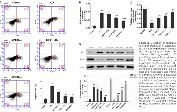
LBP pretreatment reduced myocardial apopto-sis in H2O2-induced injury
A remarkable increase of apoptosis rate from
6%±1% to 23.4%±1.8% was detected with flow
cytometry in myocardial cells under the condi-tion of 50 µmol/L of H2O2 as compared with
the expression of apoptosis-related Bax/Bcl-2 protein and Nrf2 pathway in this study. Treatment of 50 µmol/L H2O2 to myocardial cells evidently increased the expression of Bax and suppressed the expression of Bcl-2 in com-parison with control one (P<0.01). Expression of Bax mRNA and protein was down-regulated while that of Bcl-2 was up-regulated by LBP pre-treatment in a dose-dependent manner (P<0.05) (Figure 2B-D). Significant decrease
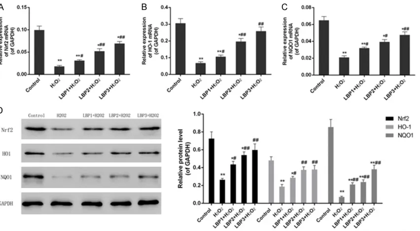
was observed in protein and mRNA expres-sions of Nrf2, HO-1 and NQO1 under condition of H2O2 without LBP pretreatment as compared with control group (P<0.01). The research found
that LBP pretreatment significantly improved
the expression of Nrf2, HO-1 and NQO1 mRNA and proteins through a positive regulatory way in a dose-dependent manner (P<0.05) (Figure 3A-D).
Discussion
Our study provided evidences of protective effect of LBP pretreatment on cardiomyocyte apoptosis in H2O2-induced injury by regulating apoptosis related genes and Nrf2 pathway. In our established model of H2O2-induced injury,
significant decrease of proliferation, increase
of apoptosis rate along with a series of changes in oxidative stress parameters were detected. Myocardial cells pretreated with proper con-centration of LBP were rescued from severe H2O2-induced oxidative stress by improving cell viability and reducing cell apoptosis.
Based on previous studies, oxidative stress has been reported to be involved in a variety of phases of apoptotic pathway and development of CVD [9]. Excessive ROS distorts the delicate
equilibrium between free radical production
Figure 2. Detection of apoptosis rate and expression of apoptosis-related mRNA/proteinsin control, H2O2 (50 µmol/L) and LBP (100, 200 and 400 μg/mL)+H2O2 (50 µmol/L) groups. A: Flow cytometry found LBP pretreatment reduced myocardial apoptosis rate in H2O2 -induced injury. B: LBP pretreat-ment down-regulated the expres-sion of pro-apoptotic Bax mRNA. C: LBP pretreatment up-regulated the expression anti-apoptotic Bcl-2 mRNA in H2O2-induced injury. D: Expression of Bax proteins was increased when that of Bcl-2 pro-teins was decreased with LBP pre-treatment in H2O2-induced injury. Data were presented as mean ± SD, n=3, *P<0.05 and **P<0.01
vs. control, #P<0.05 and ##P<0.01
Oxidative actions lead to DNA damage, intra- cellular caspases activation as well as incre- ase of membrane brittleness and decrease of
fluidity in cell membrane lipid [10]. In hypoxic
condition, structure and function of mitochon-dria can be also damaged by disturbance of oxidative phosphorylation, increasing exhaust of high energy phosphate compound and reduction of the activity of energy-dependent ionic pump, resulting in accelerate of mitochon-dria-dependent cell apoptosis [11]. If the condi-tion of myocardial apoptosis persists, progres-sive loss of myocardial cells are ensued, and even develop into heart failure [4].
A lot of work has been done in the area of exploring an effective antioxidant to eliminate the damage of ROS to myocardial cells and avoid induced CVD. In the treasure house of Chinese herbal medicine, there are a lot of potential natural antioxidants. Previous rese- arches have demonstrated that LBP possesses numerous biological activities and pharmaco-logical functions, such as enhancing exercise endurance capacity, reducing fatigue and exhibiting antioxidant activity in vitro and in vivo
[6]. In H2O2-induced injury of rat myocardial
cells, we found LBP pretreatment increased cell proliferation, and reduced apoptosis rate and ROS content closely to normal level, which suggests that LBP pretreatment is able to improve heart function in oxidative stress and potentially protect against heart failure. More- over, the result shows LBP pretreatment also inhibited the increase in the level of LDH and MDA, and restrained the reduction in the activ-ity of SOD. As three good indicators of oxidative stress in cells, the level of LDH, MDA and SOD
indirectly reflect the peroxidatic reaction in the
[image:6.612.96.522.72.306.2]study. LDH, an enzyme found in almost every living cell, is released extensively during tissue damage including heart failure. According to the leakage rate of it, the integrity of envelope can be determined [12]. As the end-product of polyunsaturated fatty acids oxidation, the con-tent of MDA indicates the level of lipid peroxida-tion in cells [13]. SOD, a group of enzymes which catalyzes the reaction of superoxide anion clearance, can protect the organism from oxygen radical damage [14]. The amounts of SOD in cells indirectly reveal the capability of cellular oxygen radical clearance and lipid per-oxidation resistance. In the condition of H2O2 -induced oxidative stress, the enhancement of
Figure 3. Expression of Nrf2 pathway related mRNA/proteinsin control, H2O2 (50 µmol/L) and LBP (100, 200 and 400 μg/mL)+H2O2 (50 µmol/L) groups. A: LBP pretreatment up-regulated the expression of Nrf2 mRNA in H2O2 -induced injury. B: LBP pretreatment up-regulated the expression of HO-1 mRNA in H2O2-induced injury. C: LBP pre-treatment up-regulated the expression of NQO1 mRNA in H2O2-induced injury. D: Increasing expression of proteins in Nrf2 pathway was observed. Data were presented as mean ± SD, n=3, *P<0.05 and **P<0.01 vs. control, #P<0.05
and ##P<0.01 vs. H
myocardial membrane permeability makes LDP which is inside of membrane began to be leaked into nutrient solution, resulting in an remarkable increase of LDH level detected in the test.
It is well accepted that apoptosis is regulated by a series of intracellular apoptosis regulation genes including pro-apoptotic genes and anti-apoptotic genes. Bcl-2 family, which includes anti-apoptotic genes like Bcl-2, Bcl-xl, Mcl-1 and Bcl-2 and pro-apoptotic proteins such as
Bax, Bad, Bak and Bid, is the first apoptosis
regulating gene family ever found. The interac-tion of anti-apoptotic genes and pro-apoptotic genes can be affected by different levels of ROS, so the relative ratio of pro-apoptotic genes and anti-apoptotic genes of a cell is the key
fac-tor to influence cellular survive or perish. As the
center of apoptosis regulation in Bcl-2 family, the expression of Bcl-2 and Bax proteins is used as the markers for apoptosis in the study
[15]. Our findings suggested that LBP pretreat -ment obviously increased the expression of Bcl-2 and inhibited the expression of Bax, lead-ing to improvement of the imbalance between Bax and Bcl-2 caused by H2O2-induced oxida-tive stress. The anti-apoptotic functions of LBP pretreatment made contributions, at least in part, to its protective effects on heart failure induced by oxidative stress.
Nrf2 is a key transcription factor of cell anti-oxidative stress system, and Nrf2 nuclear translocation combined with anti-oxidative re- sponse element ARE on the nucleic acid
sequence is a key link in the process of activa -tion of Nrf2 signaling pathways [16]. The activa-tion of Nrf2 signaling pathway can start the transcription of the downstream phase II
detox-ification enzymes and antioxidant enzymes
such as Heme oxygenase-1 (HO-1), NAD (P) H:
quinine oxidoreductase (NQO1) and glutathi -one-S-transferses (GST) etc. so as to reduce the cell injuries caused by reactive oxygen and electronic materials, keep the cells in a stable state, and maintain the body’s redox in dynam-ic balance [17]. In the study, it was observed that LBP pretreatment activated Nrf2 pathway and up-regulated the expression of Nrf2, HO-1 and NQO1, which explained the protective effects of LBP pretreatment on cardiomyocyte apoptosis in H2O2-induced injury.
Conclusion
These results demonstrate that LBP has a pro-tective effect on H2O2-induced myocardial apoptosis in vitro. LBP pretreatment decreased apoptosis rate through activating Nrf2 signal-ing pathway and down-regulatsignal-ing the ratio of Bax/Bcl-2. LBP could be a potential prophylac-tic agent for CVD, but the further in vitro and in vivo experiments are required to explore the
internal mechanism in the near future. Acknowledgements
This work was supported by grants from the Nanjing Medical University Science and Te- chnology Development Fund 07NMUZ031.
Disclosure of conflict of interest
None.
Address correspondence to: Yan Zhao, Department of Cardiology, Zhejiang Provincial People’s Hospital, 158 Shangtang Road, Hangzhou 310014, Zhejiang, China. Tel: +86 (571) 8766-6666; Fax: +86 (571) 8533-5800; E-mail: [email protected]
References
[1] Khurana S, Piche M, Hollingsworth A, Venkata-raman K and Tai TC. Oxidative stress and car-diovascular health: therapeutic potential of polyphenols. Can J Physiol Pharmacol 2013; 91: 198-212.
[2] Radi E, Formichi P, Battisti C and Federico A. Apoptosis and oxidative stress in neurodegen-erative diseases. J Alzheimers Dis 2014; 42 Suppl 3: S125-152.
[3] Singh R, Devi S and Gollen R. Role of free radi-cal in atherosclerosis, diabetes and dyslipidae-mia: larger-than-life. Diabetes Metab Res Rev 2015; 31: 113-126.
[4] Gaballa MA and Goldman S. Ventricular re-modeling in heart failure. J Card Fail 2002; 8: S476-485.
[5] Li XM, Ma YL and Liu XJ. Effect of the Lycium barbarum polysaccharides on age-related oxi-dative stress in aged mice. J Ethnopharmacol 2007; 111: 504-511.
[6] Shan X, Zhou J, Ma T and Chai Q. Lycium barbarum polysaccharides reduce exercise-in-duced oxidative stress. Int J Mol Sci 2011; 12: 1081-1088.
organization. J Cell Sci 2007; 120: 838-848. [11] Chen ZH, Saito Y, Yoshida Y and Niki E. Effect
of oxygen concentration on free radical-in-duced cytotoxicity. Biosci Biotechnol Biochem 2008; 72: 1491-1497.
[12] Kerr JF, Wyllie AH and Currie AR. Apoptosis: a basic biological phenomenon with wide-rang-ing implications in tissue kinetics. Br J Cancer 1972; 26: 239-257.
[13] Davey MW, Stals E, Panis B, Keulemans J and Swennen RL. High-throughput determination of malondialdehyde in plant tissues. Anal Bio-chem 2005; 347: 201-207.
tion of inflammation-associated carcinogene -sis. Pharm Res 2010; 27: 999-1013.
[17] Li L, Dong H, Song E, Xu X, Liu L and Song Y. Nrf2/ARE pathway activation, HO-1 and NQO1 induction by polychlorinated biphenyl
quinone is associated with reactive oxygen