Original Article
miRNA-134 suppresses angiotensin II-induced vascular
smooth muscle cell dysfunction by targeting
angiotensin II type 1 receptor (AT1R)
Yuan Zhang1,2, Xin Lu3, Zhi-Liang Li1, Feng Ding2, Hao Cheng2
1Department of Cardiology, Zhujiang Hospital of Southern Medical University, Guangzhou 510280, China; 2Department of Cardiology, Inner Mongolia People’s Hospital, Hohhot 010017, China; 3Department of Electrocardiogram, The Affiliated Hospital of Inner Mongolia Medical University, Hohhot 010050, China
Received January 15, 2016; Accepted April 6, 2016; Epub May 15, 2017; Published May 30, 2017
Abstract: MicroRNA-134 is a multifunctional post-translational modulator that participates in a variety of diseases. However, the relationship between Ang II, miRNA-134 and AT1R has yet to be fully explored in cardiovascular dis-eases. In this study, we hypothesized that a post-translational mechanism of microRNAs regulated the expression of AT1R in VSMCs in the present of Ang II. To identify differentially expressed miRNAs in response to Ang II in cultured
VSMCs, we performed microarray assay and found that miRNA-134 expression was significantly suppressed in Ang
II-treated VSMCs. Next, bioinformatic analysis showed the potential miRNA-134 binding sites within the 3’-UTR of
AT1R in human sapiens. Luciferase assays verified significantly reduced luciferase activity in miRNA-134-transfect -ed wild-type VSMCs compar-ed with NC cells. In addition, miRNA-134 could inhibit Ang II-induc-ed cell proliferation, secretory and mitochondrial dysfunction in VSMCs. Furthermore, overexpressed miRNA-134 could reverse Ang II-induced increase of ACE and AT1R and decrease of AT2R in VSMCs. In conclusion, these results demonstrated that miRNA-134 played a protective effect on Ang II-induced VSMCs dysfunction in vitro, and the underlying mechanism was mediated, at least partially, through the inhibition of AT1R signaling.
Keywords: MiRNA-134, vascular smooth muscle cell, angiotensin II, AT1R
Introduction
Vascular smooth muscle cells (VSMCs) dys-function are a common pathological basis for hypertension, atherosclerosis and vascular restenosis. Moreover, various cardiovascular diseases risk factors lead to damage to the blood vessel wall and cellular infiltration [1, 2]. The renin-angiotensin system (RAS) has been implicated in the pathogenesis of cardiovascu-lar diseases through its primary effector mole-cule angiotensin II (Ang II), which has a major impact on the cardiac and vascular function through regulation of systemic hemodynamics and blood volume [3]. Recent reports have demonstrated that Ang II can induce VSMCs proliferation, migration and hypertrophy [4-6]. Importantly, Ang II induces the expression of cytokines such as interleukin-6 (IL-6), mono-cyte chemoattractant protein-1 (MCP-1) and osteopontin in VSMCs, which have a crucial
tar-get mRNAs to inhibit translation or promote mRNAs degradation. The key features of miR-NAs regulate cell proliferation and differentia-tion of various cell types. A growing number of studies have demonstrated that the pathogenic change in various tissues has been linked to miRNAs [11-14]. Interestingly, miRNAs are ex-pressed and play many important biological functions in the cardiovascular system, such as miRNA-181b, miRNA-599 and miRNA-322 which regulate VSMCs proliferation and differ-entiation [15-17]. However, the underlying mole-cular mechanisms of miRNAs in Ang II-mediated VSMC dysfunction by targeting AT1R remain to be determined.
In this study, we hypothesize that a post-trans-lational mechanism might exist for AT1R signal-ing, which could be regulated by miRNAs in Ang II-mediated VSMCs dysfunction. By using PicTar, TargetScan, and miRBase database and microarray assay, we found that miRNA-134 was a regulator of AT1R through the predicted binding sites in its 3’-UTR. Therefore, the aim of the present study was to explore the effects of miRNA-134 on the Ang II-induced VSMCs dys-function and also to investigate the role of the AT1R signaling in this process.
Materials and methods
Cell culture
The human vascular smooth muscle cells (VSMCs) were obtained from the Cell Resource Center, Shanghai Institutes for Biological Sciences (SIBS, China), and maintained in RPMI-1640 (Invitrogen, USA) supplemented with 10% FBS (Invitrogen, USA) at 37°C in a humidified incubator (Thermo, USA), 5% CO2, 95% air atmosphere.
Cell viability detection by MTT
VSMCs proliferation was monitored by a 3-(4, 5-dimethylthiazol-2-yl)-2, 5-diphenyltetrazolium bromide (MTT) Cell Proliferation/Viability Assay kit (R&D SYSTEMS) in according to the guide- lines.
Nitric oxide quantification
VSMCs were plated and treated in 96-well plates and were stimulated with Ang II or trans-fected with miRNA-134. Twelve hours later cen-trifuged to obtain the supernatant, and the
level of nitric oxide was measured by nitrite pro-duction using the Griess reagent (Invitrogen, USA) at 540 nm using an ELISA reader (BioTek, USA) according to the manufacturer’s instruct- ions.
Detection of Ca2+ concentrations
VSMCs were plated and treated in 12-well plates and were incubated with Ang II or trans-fected with miRNA-134 to detect changes in Ca2+ levels. Cells were harvested and washed twice, and re-suspension in Indo 1/AM (3 μg/ ml) at 37°C for 30 min and analyzed by flow cytometry.
Determination of the mitochondrial membrane potential
The mitochondrial membrane potential was assessed using a fluorometric probe, DiOC6 (Molecular Probes). Briefly, cells were plated in 6-well culture dishes. After reaching conflu -ence, cells were treated with Ang II or transfect-ed with miRNA-134. After incubation, cells were stained with DiOC6 (40 nM) for 15 min at 37°C. Cells were collected, washed twice in PBS, and analyzed by FACScan flow cytometry.
Luciferase reporter gene activity assay
The 3’-UTR of AT1R gene containing the predi-cated target sites for miRNA-134 was obtained by PCR amplification. The fragment was insert -ed into the multiple cloning sites in the pMIR-REPORT luciferase microRNA expression re-porter vector (Ambion, Austin, USA). VSMVs were co-transfected with 100 ng of luciferase reporters containing AT1R 3’-UTR and miRNA-134 mimics by Lipofectamine 2000 (Invitrogen, CA, USA). We harvested the cell lysates after 12 hours transfection and measured the luciferase activity with a dual luciferase re- porter assay kit according to manufacturer’s instruction.
Transfection of miRNA-134 mimics and inhibi-tor
-es of 2’ -OMe-miRNA-134 inhibitor and 2’-Ome-scramble oligonucleotides were as follows: 5’- CCCCUCUGGUCAACCAGUCACA-3’; and 5’-ACA- UCAGAGGUCUUGACCUAG-3’. Cells were trans-fected using Lipofectamine2000 (Invitrogen, CA, USA) at a final concentration of 50 nM. At 24 h post-transfection, the culture medium was changed. After 12 hours, cells were harvested for analysis.
Real time-polymerase chain reaction
RNA extraction was performed according to the TRIzol manufacturer’s protocol (Invitrogen, Carlsbad, CA, USA). Synthesis of cDNAs was performed by reverse transcription reactions with 4 μg of total RNA using moloney murine leukemia virus reverse transcriptase (Invitro- gen) with oligo dT (15) primers (Fermentas) as described by the manufacturer. miRNA-134 level was quantified by the mirVana qRT-PCR miRNA detection kit (Ambion, Austin, USA) in conjunction with real-time PCR with SYBR Green. After circle reaction, the threshold cycle (Ct) was determined and relative miRNA-134 level was calculated based on the Ct values and normalized to U6 level in each sample. PCR with the following primers: ACE, Forward 5’-CCCATCTGCTAGGGAACATGT-3’ and Reverse 5’-GGTGTCCCTGCTTTATCA-3’; AT1R, Forward 5’- CCATCACCAGATCAAGTGCA-3’ and Reverse 5’- TGGGGCAGTCATCTTGAATTCT-3’; AT2R, Forward 5’-CAGTTGACGTGATGCACAGG-3’ and Reverse 5’-CGGTTGAAGTCGTGGAGCCC-3’; GAPDH, For- ward 5’-GCACCGTCAAGCTGAGAAC-3’ and Re- verse 5’-TGGTGAAGACGCCAGTGGA-3’.
Western blotting
VSMCs were homogenized and extracted in NP-40 buffer, followed by 5-10 min boiling and centrifugation to obtain the supernatant. Samples containing 30 μg of protein were separated on 10% SDS-PAGE gel, transferred to nitrocellulose membranes (Bio-Rad Labo- ratories, Hercules, CA, USA). After saturation with 5% (w/v) non-fat dry milk in TBS and 0.1% (w/v) Tween 20 (TBST), the membranes were incubated with primary antibodies: ACE, AT1R and AT2R (Santa Cruz Biotechnology, CA, USA). After three washes with TBST, The membranes were next incubated with the appropriate HRP (horseradish peroxidase)-conjugated antibody visualized with chemiluminescence (Thermo, USA).
Statistical analysis
The data from these experiments were report-ed as mean ± standard errors of mean (SEM) for each group. All statistical analyses were per-formed by using PRISM version 4.0 (Graph- Pad). Inter-group differences were analyzed by one-way ANOVA, and followed by Tukey’s multi -ple comparison test as a post test to compare the group means if overall P < 0.05. Differences with P value of < 0.05 were considered statisti-cally significant.
Results
Identification of Ang II-regulated miRNAs in human VSMCs
To identify differentially expressed miRNAs in response to Ang II in cultured VSMCs, we per-formed microarray assay with small RNA librar-ies generated using total RNA extracted from control group or Ang II-stimulated (10 nM) VSMCs for 12 hours. We found that miRNA- 134 and miRNA-185 was significantly lowly expressed in Ang II-treated group as compared to control group (Figure 1A). Among the Ang II-induced miRNAs, we concluded that miRNA-134 and miRNA-185 might be closely related to VSMCs dysfunction in the present of Ang II. miRNA-134 and miRNA-185 have been report-ed to be expressreport-ed as a highly conservreport-ed clus-ter and can modulate diverse functions in dif-ferent cell systems [18, 19]. Therefore, we fur -ther investigated the functional roles of miRNA-134 and miR-185 in VSMCs when they were exposed to Ang II. Time course and concentra-tion dependent experiments showed that Ang II markedly suppressed 134 and miRNA-185 expression in VSMCs (Figure 1B and 1C). Subsequently, pretreatment with AT1R blocker Losartan (10 μM) for 12 hours significantly re-versed Ang II-induced down-regulated miRNA-134 and miRNA-185 in VSMCs (Figure 1D). However, miRNA-134 in response to Ang II- treated in VSMCs was more sensitive to miRNA-185, and we finally focused on miRNA-134 in our study.
Ang II-stimulated RAS components in human VSMCs
induced by Ang II in VSMCs were not fully under-stood. In the present study, the mRNA and pro-tein expression of RAS components, such as angiotensin-converting enzyme (ACE), AT1R and AT2R, were measured by real-time PCR and western blotting respectively. The results showed that the mRNA and protein expression of ACE and AT1R were significantly increased in a time dependent manner when the VSMCs were exposed to Ang II at concentration of 10 nM (Figure 2A and 2B). In contrast to that the mRNA and protein expression of AT2R were sig-nificantly decreased when the VSMCs were exposed to Ang II after 6 hours (Figure 2C). These results demonstrated that RAS signaling might be involved in VSMCs dysfunction in vitro.
miRNA-134 targets AT1R
Because each miRNA target prediction algo-rithm (PicTar, TargetScan and miRBase) has its own advantages, targets theoretically predict-ed by multiple bioinformatics software and hav-ing conserved seed sequences are more likely to be true targets. Therefore, we used these bioinformatics tools and selected the targets that were predicted by at least two databases and conserved across rat, mouse and human. We found the potential miRNA-134 binding sites within the 3’-UTR of AT1R in human sapi-ens (Figure 3A). To verify if AT1R is a direct tar-get of miR-134, we cloned the 3’-UTR of the wild-type or mutant-type AT1R gene and co-Figure 1. Unsupervised hierarchical clustering of differentially expressed miRNAs (> 2-fold) in VSMC treated with
[image:4.612.92.524.72.391.2]transfected it along with miRNA-134 or NC oli-gonucleotides into VSMCs. Luciferase assays were performed 24 hours post-transfection. Results showed significantly reduced luciferase activity in miRNA-134-transfected cells com-pared with NC cells. In contrast, co-transfection miRNA-134 into AT1R mutant-type 3’-UTR cells, the luciferase activity did not show significant difference compared with NC group (Figure 3B). Next, we observed that transfection of VSMCs with miR-134 mimic oligonucleotides or miR-134 inhibitors significantly downregulated or upregulated, respectively, both AT1R mRNA (Figure 3C) and protein (Figure 3D) levels.
Overexpressed miRNA-134 protects against Ang II-induced VSMCs dysfunction
To investigate the potential roles of miRNA-134 on Ang II-induced the proliferation of VSMCs, we first examined the effect of miRNA-134 on cell survival by MTT assay. The results
indicat-ed that treatment of VSMCs with Ang II inducindicat-ed cell proliferation in a time-dependent manner, and the number of VSMCs was markedly increased in Ang II-treated group. However, co-incubated with Ang II and miRNA-134 mimics in VSMCs, the cell viability was significantly decreased as compared to Ang II single treat-ment group. Moreover, miRNA-134 mimics sin-gle treatment could also inhibit the cell prolif-eration compared with control group (Figure 4A). To assess Ang II-induced secretory dys-function of VSMCs and the modulation effect of miRNA-134, we measured the levels of NO and p-eNOS in VSMCs. As shown in Figure 4B and
[image:5.612.93.520.71.401.2]results suggested that miRNA-134 could mod-ulate the Ang II-induced secretory dysfunction of VSMCs. Furthermore, Ang II-induced mito-chondrial dysfunction and the potential protec-tive effects of miRNA-134 was investigated. As shown in Figure 4D and 4E, treatment of VSMCs with Ang II induced the loss of the mito-chondrial membrane potential and increase of Ca2+ releases as compared to control group. Ang II combination with miRNA-134 transfec-tion could significantly improve mitochondrial membrane potential and suppress Ca2+ releas-es in VSMCs (Figure 4D and 4E).
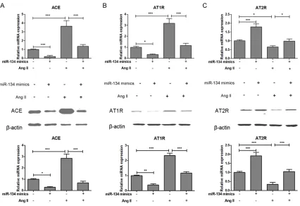
Overexpressed miRNA-134 inhibits RAS com-ponents in human VSMCs
To determine whether RAS components were involved in miRNA-134 inhibition of VSMC pro-liferation, we used real-time PCR and western blotting to examine RAS components mRNA and protein expression in miRNA-134 overex-pressed VSMCs. The mRNA and protein
expres-sion of ACE and AT1R were significantly de-creased in miRNA-134 overexpressed VSMCs as compared to control group. Moreover, over-expressed miRNA-134 could reverse Ang II-induced increase of ACE and AT1R in VSMCs (Figure 5A and 5B). In contrast to that the mRNA and protein expression of AT2R were significantly increased in miRNA-134 overex -pressed VSMCs as compared to control group. As expected, overexpressed miRNA-134 could reverse Ang II-induced decrease of AT2R in VSMCs (Figure 5C). These results demonstrat-ed that miRNA-134 playdemonstrat-ed a protective effect on Ang II-induced VSMCs dysfunction in vitro, and the underlying mechanism was mediated, at least partially, through the inhibition of AT1R signaling.
Discussion
[image:6.612.97.522.69.379.2]hypertension and restenosis. Moreover, it has been recognized as a central regulator of VSMCs proliferation and migration [4, 21]. One classical mechanism by which Ang II promotes cardiovascular remodeling and VSMCs apopto-sis is through the AT1R and AT2R [22, 23]. However, the underlying molecular mecha-nisms of miRNAs in Ang II-mediated VSMC dys-function by targeting AT1R remain to be deter-mined. miRNAs function as critical regulators of protein expression by binding to the 3’-UTRs of target mRNAs result in inhibition of protein translation in mammalian cells. Interestingly, miRNAs have been implicated in the pathogen-esis of vascular disease and have been sug-gested as potential therapeutic targets. The goal of our study was to evaluate the ability of miRNA-134 to regulate Ang II-mediated VSMC dysfunction by targeting AT1R. The results dem-onstrated that miRNA-134 expression was sig-nificantly suppressed in Ang II-treated VSMCs.
However, overexpressed miRNA-134 could in- hibit Ang II-induced cell proliferation, secretory and mitochondrial dysfunction in VSMCs and reverse Ang II-induced increase of ACE and AT1R and decrease of AT2R in VSMCs.
miRNA-134, located on 14q32.31, has been reported to be dysregulated in several malig-nant tumors and acts as a tumor suppressor in breast cancer [18], non-small cell lung cancer (NSCLC) [24], ovarian cancer [25] and osteosar -coma [26]. Recent studies have shown that miRNA-134 plays a role in vascular biology and are involved in biochemical and molecular pathways dysregulated during vascular injury and may regulate lipid accumulation and proin-fiammatory cytokine secretion [27, 28]. There-fore, we analyzed the functional relevance of miR-134 in Ang II-induced VSMC dysfunction. Our studies suggested that treatment of VSMCs with Ang II induced cell proliferation in a
time-VSMCs were transfected with miRNA-134 mimics or inhibitor for 12 hours, the mitochondrial membrane potential (D) and the release of Ca2+ (E) were examined by flow cytometry. Values were expressed as mean ± SEM, n = 3 in
[image:8.612.93.519.122.414.2]each group. **P < 0.01, ***P < 0.001.
dependent manner, and the number of VSMCs was markedly increased in Ang II-treated group. We established that directly transfecting miRNA-134 into VSMCs could improve Ang II-induced cell proliferation, secretory and mito-chondrial dysfunction. Collectively, these stud-ies indicated that miRNA-134 was downregu-lated in many pathophysiological processes, and our study extended these observations by showing that overexpressed miRNA-134 plays an important role in Ang II-induced VSMCs dysfunction.
Previous studies indicate that miRNA-134 by targeting angiopoietin-like 4 in THP-1 macro-phages regulates lipid accumulation and inflam -matory response [28], modulates the prolifera -tion of human cardiomyocyte progenitor cells by targeting Meis2 [29] and regulates isch -emia/reperfusion injury-induced neuronal cell death by regulating CREB signaling [30]. In this study, we found that miRNA-134, as a multi-functional signal miRNA, could target AT1R via the potential miRNA-134 binding sites within the 3’-UTR of AT1R in VSMCs. To our knowl-edge, the biological effects of Ang II are medi-ated by AT1R and AT2R, and Ang II-induced VSMC proliferation is mediated mainly by AT1R, rather than AT2R [4, 22]. Other studies in mac -rophages have shown that AT1R antagonists inhibit the effect of Ang II, but AT2R antagonists the opposite has happened [31]. In the present study, overexpressed miRNA-134 could reverse Ang II-induced increase of ACE and AT1R and decrease of AT2R in VSMCs. These results demonstrated that miRNA-134 played a protec-tive effect on Ang II-induced VSMCs dysfunc-tion in vitro, and the underlying mechanism was mediated, at least partially, through the inhibi-tion of AT1R signaling.
Disclosure of conflict of interest
None.
Address correspondence to: Dr. Zhi-Liang Li, The Department of Cardiology, Zhujiang Hospital of Southern Medical University, Guangzhou 510280, China. Tel: (+86) 13809774642; E-mail: xinlu_088@ 163.com
References
[1] Ponticos M and Smith BD. Extracellular matrix synthesis in vascular disease: hypertension,
and atherosclerosis. J Biomed Res 2014; 28: 25-39.
[2] Shi N and Chen SY. Mechanisms simultane-ously regulate smooth muscle proliferation and differentiation. J Biomed Res 2014; 28: 40-46.
[3] Mascitelli L and Pezzetta F. Renin-angiotensin system and cardiovascular risk. Lancet 2007; 370: 24; author reply 24-25.
[4] Yang LX, Liu G, Zhu GF, Liu H, Guo RW, Qi F and Zou JH. MicroRNA-155 inhibits angiotensin II-induced vascular smooth muscle cell prolifera-tion. J Renin Angiotensin Aldosterone Syst 2014; 15: 109-116.
[5] Remus EW, Lyle AN, Weiss D, Landazuri N, Weber M, Searles C and Taylor WR. miR181a protects against angiotensin II-induced osteo-pontin expression in vascular smooth muscle cells. Atherosclerosis 2013; 228: 168-174.
[6] Li L, Gao P, Zhang H, Chen H, Zheng W, Lv X, Xu T, Wei Y, Liu D and Liang C. SIRT1 inhibits an-giotensin II-induced vascular smooth muscle cell hypertrophy. Acta Biochim Biophys Sin (Shanghai) 2011; 43: 103-109.
[7] Jin W, Reddy MA, Chen Z, Putta S, Lanting L, Kato M, Park JT, Chandra M, Wang C, Tangirala
RK and Natarajan R. Small RNA sequencing
reveals microRNAs that modulate angiotensin II effects in vascular smooth muscle cells. J Biol Chem 2012; 287: 15672-15683.
[8] Sansom SE, Nuovo GJ, Martin MM, Kotha SR, Parinandi NL and Elton TS. miR-802 regulates human angiotensin II type 1 receptor expres-sion in intestinal epithelial C2BBe1 cells. Am J Physiol Gastrointest Liver Physiol 2010; 299: G632-642.
[9] Berk BC. Angiotensin type 2 receptor (AT2R): a challenging twin. Sci STKE 2003; 2003: PE16.
[10] Bartel DP. MicroRNAs: target recognition and regulatory functions. Cell 2009; 136: 215-233.
[11] Pan ZW, Lu YJ and Yang BF. MicroRNAs: a novel class of potential therapeutic targets for cardiovascular diseases. Acta Pharmacol Sin 2010; 31: 1-9.
[12] Urbich C, Kuehbacher A and Dimmeler S. Role
of microRNAs in vascular diseases, inflamma -tion, and angiogenesis. Cardiovasc Res 2008; 79: 581-588.
[13] Shukla GC, Singh J and Barik S. MicroRNAs: Processing, Maturation, Target Recognition and Regulatory Functions. Mol Cell Pharmacol 2011; 3: 83-92.
[14] da Costa Martins PA, Leptidis S, Salic K and De Windt LJ. MicroRNA regulation in cardiovascu-lar disease. Curr Drug Targets 2010; 11: 900-906.
muscle cells proliferation through activation of PI3K and MAPK pathways. Int J Clin Exp Pathol 2015; 8: 10375-10384.
[16] Xie B, Zhang C, Kang K and Jiang S. miR-599 Inhibits Vascular Smooth Muscle Cells Pro- liferation and Migration by Targeting TGFB2.
PLoS One 2015; 10: e0141512.
[17] Zeng Y, Liu H, Kang K, Wang Z, Hui G, Zhang X, Zhong J, Peng W, Ramchandran R, Raj JU and Gou D. Hypoxia inducible factor-1 mediates ex-pression of miR-322: potential role in prolifera-tion and migraprolifera-tion of pulmonary arterial smooth muscle cells. Sci Rep 2015; 5: 12098.
[18] O’Brien K, Lowry MC, Corcoran C, Martinez VG,
Daly M, Rani S, Gallagher WM, Radomski MW,
MacLeod RA and O’Driscoll L. miR-134 in ex -tracellular vesicles reduces triple-negative breast cancer aggression and increases drug
sensitivity. Oncotarget 2015; 6: 32774-32789. [19] Imam JS, Buddavarapu K, Lee-Chang JS,
Ganapathy S, Camosy C, Chen Y and Rao MK. MicroRNA-185 suppresses tumor growth and progression by targeting the Six1 oncogene in
human cancers. Oncogene 2010; 29:
4971-4979.
[20] Lu Y, Li S, Wu H, Bian Z, Xu J, Gu C, Chen X and
Yang D. Beneficial effects of astragaloside IV
against angiotensin II-induced mitochondrial dysfunction in rat vascular smooth muscle cells. Int J Mol Med 2015; 36: 1223-1232.
[21] Oishi Y, Manabe I, Imai Y, Hara K, Horikoshi M,
Fujiu K, Tanaka T, Aizawa T, Kadowaki T and Nagai R. Regulatory polymorphism in tran-scription factor KLF5 at the MEF2 element al-ters the response to angiotensin II and is as-sociated with human hypertension. FASEB J 2010; 24: 1780-1788.
[22] Diep QN, Li JS and Schiffrin EL. In vivo study of AT(1) and AT(2) angiotensin receptors in apop-tosis in rat blood vessels. Hypertension 1999; 34: 617-624.
[23] Tan NY, Li JM, Stocker R and Khachigian LM. Angiotensin II-inducible smooth muscle cell apoptosis involves the angiotensin II type 2 re-ceptor, GATA-6 activation, and FasL-Fas en-gagement. Circ Res 2009; 105: 422-430.
[24] Li J, Wang Y, Luo J, Fu Z, Ying J, Yu Y and Yu W. miR-134 inhibits epithelial to mesenchymal
transition by targeting FOXM1 in non-small cell
lung cancer cells. FEBS Lett 2012; 586: 3761-3765.
[25] Shuang T, Wang M, Shi C, Zhou Y and Wang D. Down-regulated expression of miR-134 con-tributes to paclitaxel resistance in human ovar-ian cancer cells. FEBS Lett 2015; 589: 3154-3164.
[26] Bao Y, Peng L, Ma J, Liu K and Li W. Decreased miR-134 expression and its tumor-suppressive function in human osteosarcoma. Genet Mol Res 2015; 14: 16771-16781.
[27] Thomas RA, Scicchitano MS, Mirabile RC, Chau NT, Frazier KS and Thomas HC. MicroRNA changes in rat mesentery and serum associ-ated with drug-induced vascular injury. Toxicol Appl Pharmacol 2012; 262: 310-320.
[28] Lan G, Xie W, Li L, Zhang M, Liu D, Tan YL, Cheng HP, Gong D, Huang C, Zheng XL, Yin WD and Tang CK. MicroRNA-134 Actives lipo-protein lipase-mediated Lipid Accumulation
and Inflammatory Response by Targeting
Angiopoietin-Like 4 in THP-1 Macrophages. Biochem Biophys Res Commun 2016; 472: 410-7.
[29] Wu YH, Zhao H, Zhou LP, Zhao CX, Wu YF, Zhen LX, Li J, Ge DX, Xu L, Lin L, Liu Y, Liang DD and Chen YH. miR-134 Modulates the Proliferation of Human Cardiomyocyte Progenitor Cells by Targeting Meis2. Int J Mol Sci 2015; 16: 25199-25213.
[30] Huang W, Liu X, Cao J, Meng F, Li M, Chen B and Zhang J. miR-134 regulates ischemia/re-perfusion injury-induced neuronal cell death by regulating CREB signaling. J Mol Neurosci 2015; 55: 821-829.
[31] Yang LX, Ye JS, Guo RW, Liu H, Wang XM, Qi F and Guo C. The effect of the expression of an-giotensin II on extracellular matrix metallopro-teinase inducer (EMMPRIN) in macrophages is