Original Article
An allele specific locked nucleic acid real time
quantitative PCR for detection of HBV
rtA181V and rtN236T mutations
Jing Chen1,3, Qing Wang2,3, Huijuan Chen2,3, Yongbin Zeng2,3
1Center of Liver Diseases, 2Department of Laboratory Medicine, The First Affiliated Hospital of Fujian Medical
University, Fuzhou, China; 3First Clinical College, Fujian Medical University, Fuzhou, China
Received December 5, 2016; Accepted June 18, 2017; Epub August 15, 2017; Published August 30, 2017 Abstract: Background: Drug-resistant mutations of hepatitis B virus (HBV) are the major causes for unsuccessful therapy for chronic hepatitis B infection (CHB). Adefovir dipivoxil (ADV) has been widely used in patients who failed to lamivudine (LMV) treatment. Early detection of ADV-resistant mutationsis of great clinical significance. In this study, we established an easy-to-use approach, real time allele specific locked nucleic acid quantitative PCR (RT-AS-LNA-qPCR) for early quantification of the rtA181V and rtN236T mutations associated with resistance to ADV and focused on its performance evaluation. Methods: Four recombinant plasmids for rtA181 and rtN236 mutations were con-structed. The assay was established and evaluated with standard recombinant plasmids and 102 serum samples from patients who experienced with ADV. Results: The linear range of the assay for the detection of rtA181V and rtN236T was between 1×10⁹ copies/μl and 1×10² copies/μl. Sensitivity of the assay was 10-5 in the wild-type
back-ground of 1×10⁷ copies/μl. The detection sensitivity of the assay was 0.03% in the detection of clinical samples.
RT-AS-LNA-qPCR had a high concordance with direct sequencing in detecting mutations associated with resistance to ADV. RT-AS-LNA-qPCR was more sensitive than direct sequencing in detecting minor variants which could detect these mutations earlier. Conclusions: RT-AS-LNA-qPCR assay was able to sensitively and specifically detect the rtA181V and rtN236T mutations associated with resistance to ADV. This easy-to-use approach may be a useful tool for monitoring ADV resistance mutations in patients with chronic HBV infection and for optimization of ADV therapy. Keywords: Hepatitis B virus, mutation, adefovir dipivoxil, locked nucleic acid, polymerase chain reaction
Introduction
Approximately 240 million people worldwide are chronically infected with hepatitis B virus
(HBV) and more than 75% of those reside in Asia-Pacific area, especially in China [1].
Ch-ronic HBV carriers may lead to chCh-ronic hepati-tis, cirrhosis, and hepatocellular carcinoma
(HCC) [1, 2]. Oral Nucleoside/nucleotideana -logues such as lamivudine (LAM), adefovir
dip-ivoxil (ADV), entecavir (ETV), telbivudine (LdT) and tenofovir (TDF), are widely used for the treatment of CHB [3]. Although recent treat -ment guidelines recommend that entecavir and
tenofovir are the first line treatment for CHB,
ADV is still widely prescribed in some develop-ing countries, such as China, due to the effect on lamivudine-resistant mutations occurring upon prolonged treatment and the economic considerations in rural areas in China. Un-
and increases overtime. The most important
mutations associated with ADV resistance are
rtA181V and rtN236T, respectively found within
the Band D functional domains of the HBV
reverse transcriptase [4]. Researches have
indicated that the cumulative annual incidence of genotypic resistance to ADV was estimated
to be 3, 9, 18 and 28% after 2, 3, 4 and 5 years of treatment, respectively [5]. Consequently,
early and periodic detection of HBV drug-resis-tant mudrug-resis-tants has received much attention due to its importance for the strategic treatment of chronic hepatitis B virus-infected patients. Many available assays such as line probe assay
(LiPA), next-generation sequencing (NGS),
se-lective real-time PCR (sPCR), mass
spectromet-ric analysis (MS), coamplification at lower dena
-turation temperature (COLD)-PCR etc. for
minority populations in less than 20% of a total viral population [9]. INNO-LiPA and MS are
capable of detecting minor variants, but more strict experiment conditions and instruments
are required [7].
Herein our aim was to establish an easy-to-use
approach, real time allele specific locked nucle
-ic acid quantitative PCR (RT-AS-LNA-qPCR) for early quantification of the rtA181V and rtN236T
mutations associated with resistance to ADV and focused on its performance evaluation. Materials and methods
Subjects
A total of 102 chronic HBV patients who were undergoing ADV mono-therapy were recruited in the Center of Liver diseases of the First
Affiliated Hospital of Fujian Medical University from October 2014 to May 2016. Of these patients, 71 (69.6%) were males and 31 (30.4%)
were females with an average age of 46.59 ± 12.95 years old. Serum samples were collect-ed at baseline and every 12 weeks. All the recruited patients met the following criteria: I) presence of HBV DNA inserum and an elevation
of serum ALT at least 2 times higher than the
normal level during the year prior to the initia-tion of the study; II) hepatitis B surface antigen (HBsAg) positive inserum for at least six mo- nths. All investigations were conducted with the approval of the ethics committee of the First
Affiliated Hospital of Fujian Medical University
and according to the tenets of the Declaration of Helsinki. Written informed consent was also obtained before collecting serum samples and studying the clinical feature of each patient.
with the principle of allele-specific PCR
(AS-PCR), general principles for LNA primer design
rules provided by Exiqon (http://www.exiqon.
com/oligo-tools) and the HBV reverse transcrip-tase polymorphism analysis of the most com-mon genotypes (B and C) in China obtained
from GenBank. Briefly, the forward primers,
KF181 and KF236, at nucleotide positions
767~787 and 595~614, were for the amplifica -tion of both wild-type and mutant DNA of
rtA181 and rtN236, respectively. The reverse primers modified with a LNA (LNA primer) at the 3’-end terminal position (L181O, L181A) were designed to detect wild type and the mutant type of rtA181 mutations, respectively. Similarly, LNA primers for wild type and
rtN-236mutations (L236O, L236A) were also
designed (Table 1). Blast analysis was
per-formed to demonstrate the specificity of the
primers.
Standard plasmid DNA construction
Four clinical samples sequenced harboring wild-type and mutant DNA sequences of rtA181
and rtN236 were used as templates for
plas-mids construction. Target DNA was amplified
with primers (KF, L) listed in Table 1. Then PCR products were cloned into pMD 18-T vector (Takara, Japan) and transformed into E. coli
DH5α competent cell (Takara, Japan). The posi
-tive plasmids were sequenced to confirm the presence of the expected sequences and then isolated using TIANprepMini Plasmid kit
(Tian-gen Biotech Company, Beijing, China). Plasmids
were quantified by NanoDrop 2000 spectro
-photometer (Thermo Fisher Scientific, USA). The corresponding copy number was calculat
-ed and 10-fold serially dilut-ed from 1×10¹⁰
cop-Table 1. Primers for RT-AS-LNA-qPCR
Primers Sequences (5’-3’) Positions (nt) Length (bp) Amplicon size (bp) KF181 GGGACTCAAGATGTTGTACAG 767~787 21
181LO CCTCAGTCCGTTTCTCYTGG+C 651~671 21 137
181LA CCTCAGTCCGTTTCTCYTGG+T 651~671 21 137 KF236 CACYTGTATTCCCATCCCAT 595~614 20
236LO CAWCKTTTKGTTTTRTKAGGG+T 839~860 22 266
236LA CAWCKTTTKGTTTTRTKAGGG+G 839~860 22 266
KF181/KF236 indicate the same common forward primers to each reaction;
181LO/181LA/236LO/236LA indicate specific RT-AS-LNA-qPCR reverse primers. Y indicates C/T; K indicates G/T; W indicates A/T; R indicates A/G; +C/+T+G
indicate LNA nucleosides.
HBV DNA extraction from serum
samples
HBV DNA was extracted from
200 μl of serum using QIAamp
DNA Blood Minikit (Qiagen, Hi-
lden, Germany) according to the
manufacturer’s protocol and st- ored at -20°C until used. Primers
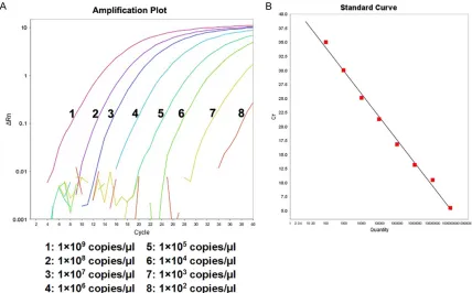
[image:2.612.92.369.86.187.2]Figure 1. Amplification plot and standard curve of RT-AS-LNA-qPCR for rtA181. A. The concentration from left to right were 1×10⁹ copies/μl to 1×10² copies/μl, respectively. B. Standard curve of rtA181: Y=-3.651X+45.843
(R2=0.987). The amplification efficiency was 94.904%.
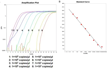
Figure 2. Amplification plot and standard curve of RT-AS-LNA-qPCR for rtA181V. A. The concentration from left to right were 1×10⁹ copies/μl to 1×10² copies/μl, respectively. B. Standard curve of rtA181V: Y=-4.093X+41.928
[image:3.612.93.521.407.673.2]Figure 3. Amplification plot and standard curve of RT-AS-LNA-qPCR for rtN236. A. The concentration from left to right were 1×10⁹ copies/μl to 1×10² copies/μl, respectively. B. Standard curve of rtN236: Y=-4.048X+44.374
(R2=0.996). The amplification efficiency was 96.807%.
Figure 4. Amplification plot and standard curve of RT-AS-LNA-qPCR for rtN236T. A. The concentration from left to right were 1×10⁹ copies/μl to 1×10² copies/μl, respectively. B. Standard curve of rtN236T: Y=-3.548X+39.439
[image:4.612.98.521.398.667.2]ies/μl to 1×101 copies/μl using Easy Dilution
Buffer (Takara, Japan) to generate standard
co-ncentrations. RT-AS-LNA-qPCR
RT-AS-LNA-qPCR was performed on the ABI
7500 Instrument (Applied Biosystems) using
the SYBR ® Premix Ex Taq™ (Takara, Japan). Amplification was carried out in a final volume of 25 ul containing 12.5 μl of SYBR Green Mix (Takara, Japan), 0.7 μl of each primer (10 μM), 0.5 μl of 50×ROX II reference dye (Takara, Japan), 8.6 μl of ddH2O and 2.0 μl of DNA tem -plate. Real-time PCR conditions were set as fol-low: 30 s at 94°C, followed by 40 cycles of denaturation for 5 s at 94°C and extension for
30 s at 60°C. All samples were amplified in
duplicate. In each run, negative and positive controls and a standard curve were included.
Direct sequencing
All clinical samples were subjected to direct
sequencing. The HBV DNA fragments contain
-ing the polymerase RT (reverse-transcriptase) domain were amplified using HBV sequencing
kit (Shenyou, Shanghai) according to the
manu-facturer’s protocol. The PCR products used for direct sequencing were purified by the High PCR Product Wizard kit (Qiagen GmbH, Hilden, Germany) and sequenced with the ABI Prism 3130 Genetic Analyzer (Life Technologies,
USA).
Statistical analysis
Data are expressed as the mean ± SD. Data was analyzed by the SPSS version 21.0
soft-ware (SPSS Inc, USA) and GraphPad
Pr-resultsor (II) sequencing showed a mixture of wild-type and mutant sequences but RT-AS-LNA-qPCR showed only a wild-type or a mutant sequence. Results were considered complete
discordance if one test showed a wild type and the other showed a mutant. All statistical tests were two-sided and the level of statistical
sig-nificance was set at P<0.05.
Results
Identification of recombinant plasmids by sequencing
Recombinant wild-type and mutant plasmids of rtA181 and rtN236 containing inserts
span-ning the target region and specific single-base mutations were successfully constructed. The recombinant clones were picked out and seque-nced. Then, the obtained DNA sequences were subjected to BLAST alignment against HBV genome database. The BLAST results showed the DNA sequence of the recombinant plas -mids were consistent with the HBV reference
sequence.
Performance of RT-AS-LNA-qPCR
Linear range: The 1×10¹⁰ copies/μl~1×101
cop-ies/μl of each recombinant plasmid was used
to test the linear range and detection limit of
RT-AS-LNA-qPCR. As can be seen from Figures 1-4 (Figure 1 was taken as the typical
repre-sentative, and similarly for the other figures),
there was an excellent linear correlation between the cycle number and the HBV DNA
copy number from the concentration of 1×10⁹
copies/μl to 1×10² copies/μl.
Specificity test: The cross-reactivity test was
Figure 5. Cross-reactivity test of RT-AS-LNA-qPCR for rtA181. A. Specific -ity of mutant primer rtA181V (KF181/181LA). No nonspecific amplification was observed. B. Specificity of wild-type primerrtA181 (KF181/181LO). Nonspecific amplification was detected when the concentration of mutant template was 1×10⁶ copies/μl and higher, but the corresponding mismatch
products decrease significantly.
USA). Concordance was as- sessed by Cohen’s Kappa test. Results were considered
complete concordance if RT-AS-LNA-qPCR and direct se-quencing showed the identical
results. Results were consid-ered partial concordance if (I)
RT-AS-LNA-qPCR provided
ad-ditional information compared
to that provided by sequenc
-ing, meaning that RT-AS-LNA-qPCR showed a mixture of wild-type and mutant seque-nces, whereas sequencing
[image:5.612.91.375.71.171.2]Figure 6. Cross-reactivity test of RT-AS-LNA-qPCR for rtN236. A. Specificity of mutant primer rtN236T (KF236/236LA). No nonspecific amplification was observed. B. Specificity of wild-type primer rtN236 (KF236/236LO). Nonspecific amplification was detected when the concentration of mutant template was 1×10⁶ copies/μl and higher, but the corresponding mismatch
[image:6.612.91.287.270.423.2]products decrease significantly.
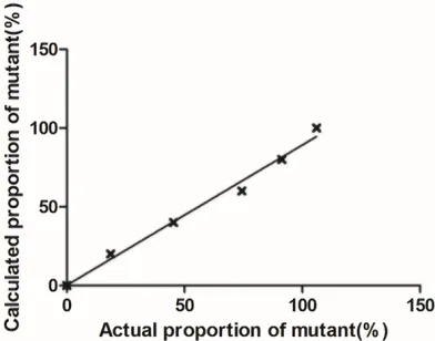
Figure 7. The correlation between actual and calcu -lated proportion of mutant plasmid DNA at concen-tration of 1×10⁷ copies/μl for rtA181. There was a
significant linear correlation (R2=0.9809, P=0.0001)
between actual proportion of mutant and calculated proportion of correlation for HBV standard plasmid DNA at concentration of 1×10⁷ copies/μl.
Figure 8. The correlation between actual and calcu -lated proportion of mutant plasmid DNA at concen-tration of 1×10⁷ copies/μl for rtN236. There was a
significant linear correlation (R2=0.9882, P<0.0001)
between actual proportion of mu-tant and calculated proportion of correlation for HBV standard plasmid DNA at concentration of 1×10⁷ copies/μl.
te i.e., different concentra-tions of wild-type and mutant-type recombinant plasmid
(1×10⁷ copies/μl~1×102
cop-ies/μl) were amplified with mutant specific and wild-type specific primers, respectively. Nonspecific amplification phe -nomenon was not observed
by mutant specific primers set
(KF181/181LA) (Figure 5). However, nonspecific amplification with the wild-type specific primers set (KF181/181LO) was detected at the con
-centration higher than or equal to 1×10⁶
cop-ies/μl. But, the observed copy number was not
in accordance with the excepted, representing at least 4 logs less than the expected copy
number. Interestingly, no nonspecific
amplific-ation was observed when the mismatch tem-plate was below 106 copies/μl. Similar results
were also observed while performed for rtN236 (Figure 6).
Accuracy test: A mixture of known quantity of
wild-type and mutant standard plasmids were used to generate different proportions of mu-
tant DNA, and then quantitative analysis of both wild-type and mutant DNA by RT-AS-LNA-qPCR was performed. Actual proportion of mutant had a significant linear correlation with
calculated proportion of correlation for HBV standard plasmid DNA at concentration of
1×10⁷ copies/μl (Figures 7 and 8).
Reproducibility test: The High (1×10⁷ copies/
μl), medium (1×10⁵ copies/μl) and low (1×10³
copies/μl) concentrations of YMDD and YIDD
recombinant plasmids were used as templates for 20 separate and simultaneous
measure-ments by RT-AS-LNA-qPCR. For the rtA181, the intra-assay coefficient of variation (CV) was 2.47%, 2.51% and 8.94% for wild-type plasmids and 3.61%, 1.61% and 5.82% for mutant-type
plasmids. For the rtN236, the intra-assay CV
was 3.95%, 3.35% and 7.39% for wild-type plasmids and 3.22%, 5.26% and 8.12% for
mutant-type plasmids.
The quantitative assay was also performed for
[image:6.612.91.287.527.681.2]inter-assay CV was 4.13%, 7.35% and 12.8% for wild-type plasmids and 5.9%, 7.87% and 8.61%
for mutant-type plasmids. For rtN236, the
inter-assay CV was 7.04%, 5.63% and 9.12% for wild-type plasmids and 4.53%, 8.79% and 13.85%
for mutant-type plasmids.
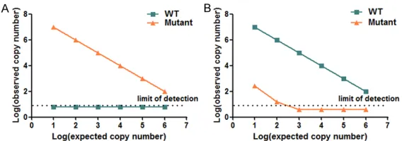
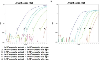
Sensitivity test: We tested the quantitative sen
-Figure 9. Sensitivity of RT-AS-LNA-qPCR in 1×10⁷ copies/μl wild-type DNA background. From left to right were 1×10⁷
copies/μl to 1×10² copies/μl mutant plasmids balanced mixing with 1×10⁷ copies/μl wild-type DNA respectively,
[image:7.612.100.525.71.325.2]100% pure wild-type DNA and no-template control. A. rtA181; B. rtN236.
Table 2. Sensitivity comparison of RT-AS-LNA-qPCR and sequencing on different proportions of
clinical samples Proportions of mutations
(rtA181V/rtN236T) RT-AS-LNA-qPCR Sequencing
50% WT/M WT/M
25% WT/M WT/M
20% WT/M WT/M
10% WT/M WT/M
5% WT/M WT
1% WT/M
-0.5% WT/M
-0.05% WT/M
-0.04% WT/M
-0.03% WT/M
-0.02% WT/M
-WT: Wild type; M: mutant; -: not performed.
of mutant plasmids DNA mixed with 1×10⁷
cop-ies/μl concentration of wild-type DNA (Figure 9). The results showed RT-AS-LNA-qPCR could accurately and steadily detect 1×10² copies/μl
of mutant DNAin the 1×10⁷ copies/μl concen
-tration of wild-type DNA, indicating that the sensitivity of the assay was 10-5 in the wild-type
background of 1×10⁷ copies/μl.
Comparison of RT-AS-LNA-qPCR and direct
sequencing
Sensitivity comparison using standard recom -binant plasmids: Plasmids containing different
proportions (50%, 25%, 20%, 15%, 10%, 5%, 1%, 0.5%, 0.05%, 0.04%, 0.03%) of mutations (rtA181V and rtN236T) were used to compare the sensitivity of RT-AS-LNA-qPCR and se-quencing. RT-AS-LNA-qPCR could measure mu-tations at a proportion as low as 0.02% (detec
-tion limit) of the total popula-tion, while sequenc -ing analysis can only measure at a proportion
of 10% (Table 2).
Sensitivity comparison using serum samples
The RT-AS-LNA-qPCR assay was evaluated
[image:7.612.91.307.429.597.2]both RT-AS-LNA-qPCR assay and sequencing.
As shown in Table 3, for rtA181, among the 102 samples analyzed, 83 wild-type, 5 mutant-type and 14 mixtures of wild mutant-types and mutants
were detected by RT-AS-LNA-qPCR. Compared the results obtained from RT-AS-LNA-qPCR with that from sequencing, the complete coinci
-dence rate was 88.2% (90/102), partial coinci
-dence rate was 11.8% (12/102), and no com -plete discordance was observed. While for rtN236, 83 wild-type, 6 mutant-type and 13 mixtures of wild types and mutants were de-
tected by RT-AS-LNA-qPCR (Table 4). The com
-plete coincidence rate was 91.2% (93/102), partial coincidence rate was 8.8% (9/102), and
no complete discordance was observed. All the partial coincidence in results between the two
methods were confirmed by subclone sequenc -ing, and the established method is consistent
with the subclone sequencing finding (data not
shown). Discussion
Adefovir dipivoxil, a nucleotide analogue, has become a treatment option for CHB due to its effect on lamivudine-resistant mutations occur-ring upon prolonged treatment and the
eco-nomic considerations in China. However, long-term therapy with ADV may lead to treatment failure because of the development of ADV
resistance. Two mutations in the HBV poly
-merase gene, rtA181V and rtN236T, have been reported to confer resistance to ADV [4]. HBV
drug resistance does not happen overnight. Instead, the emergence of a resistant strain is
a stepwise process [2]. Previous research doc -umented that minor pre-existing mutants could be gradually selected to become the dominant
species and finally precede the occurrence of virological or biochemical breakthrough [10]. Therefore, developing a rapid, simple and reli -able method to detect the ADV-resistant muta-tions as early as possible is of great clinical importance in clinical management of CHB. Many assays recently have been described with their respective advantages and disadvan-tages. Locked nucleic acid (LNA) is an RNA homologue with a methylene bridge between the ribose 2-oxygen and 4-carbon atoms. LNA increases the melting temperature of LNA-DNA hetero duplexes by up to 3°C per LNA base and, thus, enhances the discriminatory power be- tween match and mismatch under stringent
annealing conditions [11]. Because of the increased Tm to improve the mismatch discrim
-ination ability of PCR, LNA-modified nucleotides
have been widely used for a variety of
applica-tions, including SNP genotyping [12]. In this study, LNA primers for quantitative amplifica -tion of HBV DNA, rtA181 and rtN236 muta-tions were designed, and the results showed that they could detect more than 102 copies/μl of
variants. This study appears, to our knowledge, to be the first application of LNA PCR to quan
-tify rtA181V and rtN236T mutations associat -ed with ADV resistance. By using this method, it
was possible to detect 0.001% of mutated
strains in a wild type plasmid population. While
the sensitivity was 0.03% when the assay was performed in the clinical sample. To assess the
feasibility of this assay, wild type and mutant type of HBV rtA181 and rtN236 were tested in 102 patients with unsatisfactory response to
ADV by direct sequencing. The results showed
that the two assays had a high concordance.
Moreover, RT-AS-LNA-qPCR assay could detect
rtA181 and rtN236 mutations as well as some
ADV mutations which were not identified by direct sequencing, thus supporting the new
approach with a high concordance and a higher
sensitivity. Beyond that, RT-AS-LNA-qPCR pro -Table 3. Concordance between
RT-AS-LNA-qPCR and direct sequencing (rtA181)
RT-AS-LNA-qPCR WTDirect sequencingWT+Mut Mut Total
WT 83 0 0 83
WT+Mut 2 2 10 14
Mut 0 0 5 5
Total 85 2 15 102
Complete concordant results are shown in bold. Partial
[image:8.612.91.287.98.177.2]concordant results are shown in italics. The two assays showed a high concordance (Kappa=0.623, P<0.001).
Table 4. Concordance between RT-AS-LNA-qPCR and direct sequencing (rtN236)
RT-AS-LNA-qPCR Direct sequencing Total
WT WT+Mut Mut
WT 83 0 0 83
WT+Mut 3 4 6 13
Mut 0 0 6 6
Total 86 4 12 102
Complete concordant results are shown in bold. Partial
[image:8.612.91.289.255.336.2]vides a low-cost, user-friendly method that can be easily implemented in clinical laboratories.
A limitation also exists for the RT-AS-LNA-qPCR
assay though advantages of the assay over existing assays. We acknowledge that the
spec-ificity of wild-type primer is a little poor, the amplification of the mutant template with the
wild-type primer occurred at a mutant viral
con-centration above 1×10⁶ copies/μl. However,
comparison of the perfect match primer, the
nonspecific products amplified with mismatch
primer declined 4 orders of magnitude. No
non-specific products occurred when mutant DNA level was below 1×10⁶ copies/μl. To some
extent, the error can be neglected because we focus mainly on minor mutants in high wild-type background and two separate PCR including wild-type and mutant reaction tubes were run in parallel.
In conclusion, the RT-AS-LNA-qPCR assay
developed in this study was able to sensitively
and specifically detect the rtA181V and rtN236T mutations associated with resistance
to ADV. We believe that this easy-to-use ap- proach may be a useful tool for monitoring ADV resistance mutations in patients with chronic HBV infections and for optimization of ADV therapy.
Acknowledgements
The study was supported by the grants from
National Natural Science Foundation of China (81601834, 81572067), Fujian provincial Health and Family Planning Commission Fo- undation for Young Scientists (2015-1-50) and Fujian provincial health system foundation for middle-aged backbone teacher (2014-ZQN- ZD-13).
Disclosure of conflict of interest
None.
Address correspondence to: Yongbin Zeng, Depart- ment of Laboratory Medicine, The First Affiliated Hospital of Fujian Medical University, Fuzhou, China. Tel: 86-591-83340702; Fax: 86-591-83340702; E-mail: [email protected]
References
[1] Sarin SK, Kumar M, Lau GK, Abbas Z, Chan HL, Chen CJ, Chen DS, Chen HL, Chen PJ, Chien
RN, Dokmeci AK, Gane E, Hou JL, Jafri W, Jia J, Kim JH, Lai CL, Lee HC, Lim SG, Liu CJ, Locar -nini S, Al Mahtab M, Mohamed R, Omata M, Park J, Piratvisuth T, Sharma BC, Sollano J, Wang FS, Wei L, Yuen MF, Zheng SS and Kao JH. Asian-Pacific clinical practice guidelines on the management of hepatitis B: a 2015 up-date. Hepatol Int 2016; 10: 1-98.
[2] Zeng Y, Yang B, Wu Y, Chen J, Shang H, Chen X, Su M, Wu S, Lin J and Ou Q. Clinical signifi -cance of periodic detection of hepatitis B virus YVDD mutation by ultrasensitive real-time am-plification refractory mutation system quanti -tative PCR during lamivudine treatment in pa-tients with chronic hepatitis B. J Med Microbiol 2015; 64: 237-242.
[3] European Association For The Study Of The Liver. EASL clinical practice guidelines: Man-agement of chronic hepatitis B virus infection. J Hepatol 2012; 57: 167-185.
[4] Zhao WF, Shao YL, Chen LY, Wu JH, Zhu YL, Gan JH and Xiong H. Establishment of a new quantitative detection approach to adefovir-re -sistant HBV and its clinical application. World J Gastroenterol 2010; 16: 1267-1273.
[5] Niesters HG, Zoulim F, Pichoud C, Buti M, Sha -piro F, D’Heuvaert N, Celis L, Doutreloigne J and Sablon E. Validation of the INNO-LiPA HBV DR assay (version 2) in monitoring hepatitis B virus-infected patients receiving nucleoside analog treatment. Antimicrob Agents Che-mother 2010; 54: 1283-1289.
[6] Lupo J, Larrat S, Hilleret MN, Germi R, Boyer V, Nicod S, Bargues G, Leroy V, Seigneurin JM, Zarski JP and Morand P. Assessment of selec-tive real-time PCR for quantitation of lamivu -dine and adefovir hepatitis B virus-resistant strains and comparison with direct sequencing and line probe assays. J Virol Methods 2009; 156: 52-58.
[7] Lowe CF, Merrick L, Harrigan PR, Mazzulli T, Sherlock CH and Ritchie G. Implementation of next-generation sequencing for hepatitis B vi -rus resistance testing and genotyping in a clin-ical microbiology laboratory. J Clin Microbiol 2016; 54: 127-133.
[8] Liu C, Lin J, Chen H, Shang H, Jiang L, Chen J, Ye Y, Yang B and Ou Q. Detection of hepatitis B virus genotypic resistance mutations by coam-plification at lower denaturation temperature-PCR coupled with sanger sequencing. J Clin Microbiol 2014; 52: 2933-2939.
[10] Pallier C, Castera L, Soulier A, Hezode C, Nord-mann P, Dhumeaux D and Pawlotsky JM. Dy-namics of hepatitis B virus resistance to lami-vudine. J Virol 2006; 80: 643-653.
[11] Rupp J, Solbach W and Gieffers J. Single-nucle -otide-polymorphism-specific PCR for quantifi -cation and discrimination of Chlamydia pneu-moniae genotypes by use of a “locked” nucleic acid. Appl Environ Microbiol 2006; 72: 3785-3787.