Original Article
Hydroxysafflor yellow A attenuates allergic airway
inflammation by suppressing the activity of nuclear
factor-kappa B in ovalbumin-induced asthmatic mice
Hongmei Piao1*, Qianfei Xue2*, Jingzhi Jiang3, Mingyu Zheng4, Hong Cui3, Yan Jin4, Jinshi Yang4, Zhongjian Aan1, Yunho Choi5, Guanghai Yan3
1Department of Respiratory Medicine, Yanbian University Hospital, Yanji, Jilin, China; 2Department of Respiratory
Medicine, Second Hospital, Jilin University, Changchun, Jilin, China; 3Department of Anatomy and Histology and
Embryology, Yanbian University Medical College, Yanji, Jilin, China; 4College of Pharmacy, Yanbian University, Yanji,
Jilin, China; 5Department of Anatomy, Medical School, Institute for Medical Sciences, Chonbuk National University,
Jeonju, Jeonbuk, Republic of Korea. *Equal contributors.
Received February 27, 2016; Accepted August 15, 2016; Epub November 15, 2016; Published November 30, 2016
Abstract: Objective: This study is to investigate the effects of hydroxysafflor yellow A (HSYA) on the airway inflam -mation in ovalbumin (OVA)-induced asthmatic mice. Methods: After drug administration, the airway hyper-respon-siveness (AHR), and cell differentials and cytokine levels in bronchoalveolar lavage (BAL) fluid, were assessed. Lung tissue was detected with histological examination. Protein expression levels were determined with Western blot analysis. NF-κB/DNA binding activity was assessed with the electrophoretic mobility shift assay (EMSA). Re -sults: The AHR, cell chemotaxis, and inflammatory responses were substantially enhanced in OVA-induced asth -matic mice, which would be prevented by HSYA or BAY 11-7085. Moreover, levels of total and OVA-specific IgE, and inflammation-related cytokines, in the BAL fluid were significantly increased in OVA-induced asthmatic mice, which would be decreased by HSYA or BAY 11-7085. On the other hand, the nuclear NF-κB p65 level was significantly increased, while the cytosolic NF-κB p65 level was significantly decreased, by the OVA challenge. Moreover, EMSA showed increased NF-κB/DNA binding activity in OVA-induced asthmatic mice. Furthermore, immunostaining con -firmed the nuclear translocation of the p65 subunit. However, the treatment of HSYA or BAY 11-7085 significantly inhibited the nuclear translocation of NF-κB p65 in OVA-induced asthmatic mice. In addition, the treatment of HSYA or BAY 11-7085 significantly reduced the phosphorylation and degradation of IκB-α in lung of OVA-induced asth -matic mice. Conclusion: HSYA protects against the airway inflammation in OVA-induced asth-matic mice, through down-regulating inflammation-related cytokines and inhibiting NF-κB activity. Our findings support the utility of HSYA in the treatment of asthma in clinic.
Keywords: Asthma, hydroxysafflor yellow A (HSYA), airway hyper-responsiveness (AHR), airway inflammation, nuclear factor-kappa B (NF-κB)
Introduction
Bronchial asthma is a kind of chronic airway
disease characterized by eosinophilic inflam -mation, reversible airway obstruction, increas- ed mucus production, and airway hyper-respon-siveness (AHR) [1, 2]. These symptoms are ma-
inly caused by various inflammatory factors,
including T-helper2 (Th2) cells, B cells, mast cells, eosinophils, and cytokines and chemo-kines. In particular, interleukin (IL)-4, IL-5, and IL-13 produced by Th2 cells could activate eosinophils, which are related to the AHR and
airway inflammation [3]. Moreover, Th2 cell dif -ferentiation has also been associated with the pathogenesis and aggravation of asthma. The- refore, it is of great importance to investigate the Th2 cell activation for the development of disease treatment.
Ubiquitous eukaryotic transcription factor
nuclear factor-kappa B (NF-κB) plays key roles in various inflammatory and immune respons
-es, including asthma. Mammalian NF-κB family
as homo- or heterodimers bound to the IκB inhibitory complex. In response to pro-inflam
-matory stimuli, IκB-α would be phosphorylated
and degraded, leading to the nuclear
transloca-tion of NF-κB, which promotes the transcriptransloca-tion
of target cytokines and growth factors [4].
Allergen challenge could enhance the NF-κB
activity in lung and increase the numbers of air-way epithelial cells and macrophages in pati- ents with asthma [5]. It has been shown that,
the antisense NF-κB p65 pretreatment signifi -cantly inhibits the established asthmatic reac-tion in a murine model [6, 7]. Thus, inhibireac-tion of
NF-κB in lung might be a new strategy of the
management of asthma.
Safflower (Carthamus tinctorius L.) is a tradi-tional Chinese medicine extensively used to promote the blood circulation in patients suf-fering from trauma, and cardiovascular and cerebrovascular diseases. Among over 50
com-pounds in safflower, hydroxysafflor yellow A
(HSYA) has been recognized as the active ingre-dient [8]. HSYA is able to reduce platelet aggre-gation, suppress oxidative stress, and inhibit
inflammation in animal models [9, 10].
More-over, HSYA has been shown to alleviate the
lipo-polysaccharide-induced inflammatory response
and attenuate the development of
bleomycin-caused fibrosis in the lung tissue in murine
models [11-13]. However, the effects of HSYA
on the airway inflammation in asthma have
rarely been evaluated.
In this study, the effects of HSYA on the inflam -matory responses in asthmatic mice were in- vestigated. Mouse model of asthma was estab-lished by ovalbumin (OVA) induction. After drug
administration, the airway inflammation and
AHR in these mouse models were assessed, analyzed, and compared.
Materials and methods
Study animals
Totally 35 female BALB/c mice (specific patho -gen-free), 7-w old, were purchased from the House Section of the Yanbian University Health Science Center (Yanji, Jilin, China). These mice were maintained in a facility under standard condition for 1 w before experiments, and pro-vided with water and standard chow ad libitum. The animal experimental procedures were per-formed in compliance with the guideline ap-
proved by the Institutional Animal Care and Use Committee of Yanbian University School of Medical Sciences.
Animal modeling and grouping
Mice were intraperitoneally immunized with 10
μg OVA (Sigma, St. Louis, MO, USA) and 1.0 mg
aluminum hydroxide adjuvant (Imject® Alum; Pierce, Rockford, IL, USA). After 10 d, a booster
injection of 10 μg OVA and 1.0 mg aluminum
hydroxide was administered. These immunized mice were challenged with 1% OVA aerosol in PBS for 20 min on days 17-19.
These mouse models were randomly divided into the following groups: (1) the control group (n = 7), in which the models were treated with aerosolized saline; (2) the HSYA-treated groups (n = 7 per group), in which the mice were sub-jected to intraperitoneal administration of HSYA (dissolved in saline; Santa Cruz, Santa Cruz, CA, USA) at 30 and 60 mg/kg body weight, respectively, on days 21-23, beginning at 1 h before provocation; and (3) the BAY-treated group (n = 7), in which the mice were
intraperi-toneally treated with the NF-κB inhibitor BAY
11-7085 (20 mg/kg body weight; dissolved in
DMSO-PBS; Sigma), on days 21 and 23.
AHR assessment
Airway responsiveness was measured at 2 d after the last OVA challenge. Mice were placed in a barometric plethysmographic chamber (Buxco Electronics, Wilmington, NC, USA), and baseline reading was taken for 3 min. Aeroso- lized methacholine (Mch) was nebulized thro- ugh the main chamber inlet, with increasing concentrations (2.5-50 mg/mL), over 3 min. Bronchopulmonary resistance was expressed as the increase in enhanced pauses (Penh) compared to baseline, where the baseline Penh (for the saline challenge) was considered as 100%.
Differential cell counting in bronchoalveolar
lavage (BAL) fluid
Mice were anesthetized immediately following the airway responsiveness assessment, and the trachea was cannulated while gently mas-saging the thorax. The lung tissue was lavaged
with 0.7 Ml PBS, and the BAL fluid sample was
(Baxter Diagnostics, Deerfield, IL, USA). After
centrifugation, the cell pellets were re-sus-pended in PBS, and the Cytospin preparations
of BAL cells were stained with Diff-Quik solution
(International Reagents, Kobe, Japan). Cell dif-ferentials were enumerated based on the
cel-lular morphology and the staining profile.
Enzyme-linked immunosorbent assay (ELISA)
The BAL fluid sample was collected and centri
-fuged, and the levels of total and OVA-specific IgE, tumor necrosis factor (TNF)-α, IL-1β, IL-4,
IL-5, IL-13, eotaxin, intercellular adhesion mol-ecule 1 (ICAM-1), and vascular cell adhesion molecule 1 (VCAM-1) in the supernatant were
determined with the ELISA kits (R & D Systems,
Minneapolis, MN, USA), according to the manu-facturer’s instructions. The lower limits of detection for the cytokines were as follows (pg/
mL): TNF-α, 5.1; IL-1β, 2.0; IL-4, 3.3; IL-5, 5.0;
IL-13, 1.5; eotaxin, 3.0; ICAM-1, 0.017; and VCAM-1, 20.
Histological examination
The lung tissue was extracted and fixed in 10% formalin. After embedded in paraffin, the tissue was cut into 4-μm sections, which were sub -jected to the hematoxylin-eosin (H & E) and periodic acid-Schiff (PAS) staining. The
peri-bronchial and perivascular inflammation
degree was evaluated on a subjective scale of 0 to 3, as previously described [14]. The airway
mucus expression level was quantified by
counting the numbers of PAS-positive and -neg-ative epithelial cells in bronchioles [15]. Western blot analysis
Cytosolic and nuclear protein was extracted from the lung tissue as previously described [16]. After protein concentration determination,
30 μg protein was subjected to SDS-PAGE, and
then electronically transferred onto
polyvinyli-dene difluoride membrane. After blocking with
5% non-fat dry milk in PBS for 1 h, the mem-brane was incubated with rabbit anti-mouse
anti-NF-κB p65 primary antibody (1:1000 dilu
-tion; Santa Cruz), rabbit anti-mouse anti-IκB-α
primary antibody (1:1000 dilution; Santa Cruz),
rabbit anti-mouse anti-phosphorylated (p)-IκB-α
primary antibody (1:1000 dilution; Santa Cruz),
and rabbit anti-mouse anti-β-actin primary anti -body (1:1000 dilution; Santa Cruz),
respective-ly, at 4°C for overnight. Then the membrane was incubated with HRP-conjugated sheep anti-rabbit secondary antibody (1:2000 dilu-tion; Santa Cruz) at 37°C for 1 h. The protein bands were detected with the ECL detection system (Amersham Biosciences, Piscataway, NJ, USA), and the relative protein expression levels were calculated accordingly.
Electrophoretic mobility shift assay (EMSA)
For the EMSA, 1 mM PMSF was first added
to the lung nuclear extract. An oligonucleoti-
de containing the κ-chain binding site (κB,
5’-CCGGTTAACAGAGGGGGCTT-TCCGAG-3’) was used as probe. Two complimentary strands
were annealed and labeled with [α-32P]dCTP. The 20-μL reaction system consisted of 10,000 cpm labeled probe, 10 μg nuclear extract, and 2 μL binding buffer [10 mM Tris-HCl, 500 mM KCl, 10 mM EDTA, 50% glycerol, 100 ng poly (dI∙dC), and 1 mM dithiothreitol, pH 7.6. After
30 min at room temperature, the reaction
mix-ture was subjected to 4% SDS-PAGE in 0.5× Tris-borate buffer. DNA-protein interaction was specific for NF-κB as demonstrated in the com -petition EMSA using 50-fold excess unlabeled oligonucleotide.
Statistical analysis
Data were expressed as mean ± SD. Analysis of
variance was performed for group comparison,
with the Dunnett’s post-hoc test. P < 0.05 was
considered as statistically significant.
Results
HSYA inhibits AHR, cell chemotaxis, and
inflammatory response in OVA-induced asth -matic mice
Effects of HSYA on AHR, chemotaxis, and
infla-mmatory response in the OVA-induced
asth-matic mouse models were first investigated. AHR is a functional consequence of inflamma -tion that underlies asthma [17]. Our results showed that, compared with the control group, the AHR in response to inhaling Mch was sub-stantially enhanced in the OVA-challenged mice (Figure 1A). However, the treatment of HSYA
or BAY 11-7085 significantly prevented the AHR
in response to inhaling Mch (Figure 1A),
Next, the number of the inflammatory cells in the BAL fluid was counted, at 48 h after the last
OVA challenge. Our results showed that,
[image:4.612.90.517.73.543.2]com-pared with the control group, the OVA challenge resulted in dramatically increased eosinophils, as well as slightly increased neutrophils and
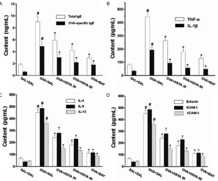
Figure 1. Effects of HSYA on AHR, cell chemotaxis in BAL fluid, and inflammatory response in OVA-induced asthmatic mice. A. The AHR in response to inhaling Mch was assessed in the saline-induced mice administered with saline (SAL+SAL), OVA-induced asthmatic mice administered with saline (OVA+SAL), and OVA-induce mice administrated with 30 mg/kg HSYA (OVA+HSYA 30), 60 mg/kg HSYA (OVA+HSYA 60), or BAY 11-7085 (OVA+BAY), respectively. Bronchopulmonary resistance was expressed as the increase in enhanced pauses (Penh) compared to baseline. Baseline Penh of the saline-treated control group was 100%. B. After drug administration with HSYA or BAY 11-7085, differential cell counts in the BAL fluid in OVA-induced asthmatic mice were analyzed. EOS, eosinophil; NEU, neutrophil; MAC, macrophage; LYM, lymphocyte. C. Lung tissue sections were subjected to the H & E and PAS stain-ing, respectively. Scale bar, 50 μm. Representative picture from three independent experiments was shown for each group. D. Statistical analysis of the inflammation scores based on H & E staining. E. Statistical analysis of the airway mucus expression according to PAS staining. Compared with the OVA+SAL group, *P < 0.05; compared with
lymphocytes, in the BAL fluid (Figure 1B). However, treatment of HSYA or BAY 11-7085
significantly attenuated the OVA-induced recr-uitment of eosinophils in the BAL fluid (P < 0.05), indicating reduced cell chemotaxis into the airway.
Besides, compared with the control group, the lung tissue from the OVA-challenged mice showed widespread perivascular and
peribron-chiolar inflammatory cell infiltration (Figure 1C
and 1D). Moreover, PAS staining showed that, the percentage of mucus-producing goblet
cells in OVA-exposed mice was significantly
higher than the control group (Figure 1C and
1E). However, administration of HSYA or BAY
11-7085 significantly decreased the number of infiltrating inflammatory cells and the degree of
goblet cell hyperplasia in these OVA-induced asthmatic mice (Figure 1C-E). Taken together,
these results indicate that HSYA could efficient
-ly attenuate the allergic airway inflammation
and mucus hypersecretion in OVA-induced asthmatic mice.
HSYA attenuates IgE release into BAL fluid in OVA-induced asthmatic mice
To investigate the effects of HSYA on the
release of total and OVA-specific IgE into the BAL fluid in OVA-induced asthmatic mice, ELISA
[image:5.612.93.523.71.428.2]was performed. Our results showed that, com-pared with the control mice, the IgE levels in
the BAL fluid were significantly increased in the
OVA-challenged mice (P < 0.05) (Figure 2A). However, administration of HSYA or BAY
11-7085 significantly reduced the total and OVA-specific IgE levels in the BAL fluid in the
OVA-exposed mice (Figure 2A). These results suggest that, the HSYA treatment could
attenu-ate the release of total and OVA-specific IgE into the BAL fluid in OVA-induced asthmatic
[image:6.612.84.516.67.548.2]mice.
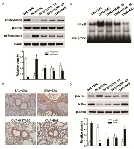
Figure 3. Effects of HSYA on NF-κB activation and IκB-α phosphorylation in OVA-induced asthmatic mice. A. Levels of the NF-κB p65 subunit in cytosol and nucleus were assessed with the Western blot analysis, for the saline-induced mice administered with saline (SAL+SAL), OVA-induced asthmatic mice administered with saline (OVA+SAL), and OVA-induce mice administrated with 30 mg/kg HSYA (OVA+HSYA 30), 60 mg/kg HSYA (OVA+HSYA 60), or BAY 11-7085 (OVA+BAY), respectively. B. NF-κB/DNA binding activity was assessed with EMSA. C. Immunohistochemical staining was performed to detect the nuclear translocation of p65. D. IκB-α phosphorylation and degradation were evaluated by the Western blot analysis. Compared with the OVA+SAL group, *P < 0.05; compared with the SAL+SAL
HSYA reduces inflammation-related cytokine levels in BAL fluid in OVA-induced asthmatic
mice
Allergic asthmatic inflammation is caused by the pro-inflammatory cytokines (such as TNF-α and IL-1β) and Th2 cytokines (such as IL-4, IL-5,
and IL-13) [18]. To assess the effects of HSYA
on the pulmonary inflammation in the asthmat
-ic m-ice, the cytokine levels in the BAL fluid were
measured with ELISA. Our results showed that, compared with the control mice, the levels of
TNF-α, IL-1β, IL-4, IL-5, and IL-13 in the BAL fluid were significantly increased in the
OVA-challenged mice (Figure 2B and 2C). However,
the cytokine levels in the BAL fluid were signifi -cantly decreased by the administration of HSYA or BAY 11-7085 (Figure 2B and 2C).
Chemokines and leukocyte-endothelial adhe-sion molecules are important for the
recruit-ment and migration of leukocytes to the inflam -matory sites [19]. The levels of eotaxin, ICAM-1,
and VCAM-1 in the BAL fluid were then mea -sured. Our results showed that, compared with the control mice, the levels of eotaxin, ICAM-1,
and VCAM-1 in the BAL fluid were significantly
increased in the OVA-challenged mice, which
could be significantly reduced by the treatment
of HSYA or BAY 11-7085 (Figure 2D). These results suggest that, the HSYA treatment could
significantly reduce the BAL fluid levels of in-flammation-related cytokines, indicating atten
-uated pulmonary inflammation, in the
OVA-induced asthmatic mice.
HSYA inhibits nuclear translocation of NF-κB and suppresses IκB-α phosphorylation in
OVA-induced asthmatic mice
NF-κB plays a key role in the allergic lung inflam -mation, which activates the transcription of
various pro-inflammatory mediators [4].
Whe-ther the attenuating effects of HSYA on
pulmo-nary inflammation in the asthmatic mice invo-lved NF-κB was next investigated. Our results
from the Western blot analysis showed that,
the OVA challenge significantly increased the nuclear NF-κB p65 level, while significantly decreased the cytosolic NF-κB p65 level, in the
cells from the extracted lung tissue (Figure 3A). Moreover, the EMSA results showed an increased binding activity of the lung nuclear
extract to the NF-κB consensus sequence for
the OVA-induced mice, compared with the
con-trol group (Figure 3B). Furthermore, the
immu-nostaining results confirmed the nuclear trans -location of the p65 subunit (Figure 3C). However, the treatment of HSYA or BAY 11-7085
significantly inhibited the nuclear translocation of NF-κB p65 in the OVA-induced asthmatic
mice.
Next, the effects of HSYA on IκB-α phosphoryla -tion were evaluated in the OVA-induced asth-matic mice. Our results showed that, the
treat-ment of HSYA or BAY 11-7085 significantly
reduced the phosphorylation and degradation
of IκB-α in the lung tissue in OVA-induced asth -matic mice (Figure 3D). Taken together, these
findings indicate that the HSYA treatment could limit the transcriptional activity of NF-κB by
inhibiting the nuclear translocation of the p65
subunit and stabilizing the IκB-α in the lung tis -sue in OVA-challenged mice.
Discussion
Allergic airway inflammation is a major factor in
the pathogenesis of asthma, which is
charac-terized by increased infiltrating leukocytes and
mucus secretion. In particular, eosinophils are the principal effector cells in the disease patho-genesis, which release cytotoxic granule pro-teins [20]. In the present study, our results showed that the HSYA treatment prevented the
airway infiltration of eosinophils. A significant
drop in the total cell and eosinophil counts in
the BAL fluid was observed. Similarly, tissue
eosinophilia was also inhibited by HSYA, as
revealed by the reduced infiltration of inflam -matory cells.
Eosinophilic transmigration into the airway is a
multistep process modulated by the pro-inflam
-matory cytokines (such as TNF-α and IL-1β) and
Th2 cytokines (such as IL-4, IL-5, and IL-13). This process is also coordinated by the chemo-tactic cytokines (such as eotaxin) and adhesion molecules (such as ICAM-1 and VCAM-1) [21, 22]. IL-4 is required for the maturation of B cells and the synthesis of IgE, which participates in
the initiation of Th2 inflammatory response.
IL-5 is pivotal for the growth, differentiation, recruitment, and survival of eosinophils. IL-13 could potently induce mucus hypersecretion,
eotaxin expression, airway inflammation, and AHR [23, 24]. Moreover, TNF-α and IL-1β have
molecules, elevated eosinophil recruitment, increased cytokine release, and enhanced AHR [18]. According to our results, HSYA attenuated
the enhanced release of pro-inflammatory and
Th2 cytokines, eotaxin, and adhesion mole-cules into the airway of OVA-induced
asthmatic-mice. These findings suggest that HSYA pre
-vents the allergic airway inflammation by
diminishing the secretion of related cytokines
into the lung tissue.AHR is defined as the abnormal increase in the airflow limitation in response to stimuli. Development of AHR has
been associated with various mediators rele-
ased during the allergic inflammation [25, 26].
For example, IL-5 mobilizes and activates eo-
sinophils to release the pro-inflammatory prod -ucts (such as major basic proteins and cyste-inyl-leukotrienes), which are closely associated with AHR [27]. In addition, IL-4 and IL-13 have been shown to induce AHR in a murine asth-matic model, which also involves cysteinyl-leu-kotrienes [28]. Moreover, AHR has been
associ-ated with the direct effect of TNF-α on the
airway smooth muscle [29]. Therefore, the AHR-reducing effects of HSYA may be related to the decreased Th2 cytokine production, tissue
eosinophilia, and TNF-α level.
Transcription factor NF-κB regulates a wide
variety of asthma-related cytokines, including
TNF-α, IL-1β, IL-4, IL-5, IL-13, ICAM-1, and
VCAM-1 [4]. Some studies have suggested that HSYA
modulates the NF-κB activity in a variety of cel -land animal models [30-32]. In line with these
findings, our results indicated that HSYA exert
-ed NF-κB activity-r-educing effect in the lung tis -sue of OVA-challenged mice. Furthermore,
sup-pression of NF-κB by BAY 11-7085 reduced the
cytokine levels and ameliorated the
eosinophil-ic airway inflammation and AHR in OVA-induced
asthmatic mice. Taken together, the anti-asth-matic effects of HSYA may be attributed to the
inhibited transcriptional activity of NF-κB and the subsequently reduced pro-inflammatory
chemical mediators.
In conclusion, our results indicated that the
HSYA treatment could efficiently attenuate the allergic airway inflammation and mucus
hyp-ersecretion in OVA-induced asthmatic mice.
Moreover, HSYA significantly reduced the BAL fluid levels of IgE and inflammation-related
cytokines and chemokines. Furthermore, the HSYA treatment inhibited the transcriptional
activity of NF-κB by inhibiting the nuclear trans -location of the p65 subunit and stabilizing the
IκB-α in the lung tissue in OVA-challenged mice. These findings suggest that HSYA might be a potential anti-inflammatory agent to treat asth -ma in clinic.
Acknowledgements
This work was supported by the National Na- tural Science Foundation of China (81260665, 81260016, and 81560679) and the Project of Research & Innovation of Jilin Youth Leader and Team (20140519013JH).
Disclosure of conflict of interest
None.
Address correspondence to: Guanghai Yan, Depart-ment of Anatomy and Histology andEmbryology, Yanbian University Medical College, 977, Gongyuan Road, Yanji 133002, Jilin, China. Tel: +86-433243- 5137; Fax: +86-4332435136; E-mail: ghyan@ybu. edu.cn; Yunho Choi, Department of Anatomy, Me-dical School, Institute for MeMe-dical Sciences, Chon- buk National University, No. San 2-20, Geumam-Dong, Jeonju, Jeonbuk, 561-180, Republic of Korea. Tel: +82-63-270-3082; Fax: +82-63-274-9880; E-mail: why76@jbnu.ac.kr
References
[1] Assayag M, Goldstein S, Samuni A and Berk-man N. Cyclic nitroxide radicals attenuate in-flammation and Hyper-responsiveness in a mouse model of allergic asthma. Free Radic Biol Med 2015; 87: 148-156.
[2] Galli SJ, Tsai M and Piliponsky AM. The devel-opment of allergic inflammation. Nature 2008; 454: 445-454.
[3] Medoff BD, Thomas SY and Luster AD. T cell trafficking in allergic asthma: the ins and outs. Annu Rev Immunol 2008; 26: 205-232. [4] Imanifooladi AA, Yazdani S and Nourani MR.
The role of nuclear factor-kappaB in inflamma -tory lung disease. Inflamm Allergy Drug Targets 2010; 9: 197-205.
[5] Gras D, Chanez P, Vachier I, Petit A and Bour -din A. Bronchial epithelium as a target for in-novative treatments in asthma. Pharmacol Ther 2013; 140: 290-305.
Epi-thelial Cells. J Immunol 2015; 195: 1388-1398.
[7] Choi IW, Kim DK, Ko HM and Lee HK. Adminis -tration of antisense phosphorothioate oligonu-cleotide to the p65 subunit of NF-kappaB in-hibits established asthmatic reaction in mice. Int Immunopharmacol 2004; 4: 1817-1828. [8] Fan L, Zhao HY, Xu M, Zhou L, Guo H, Han J,
Wang BR and Guo DA. Qualitative evaluation and quantitative determination of 10 major ac-tive components in Carthamus tinctorius L. by high-performance liquid chromatography cou-pled with diode array detector. J Chromatogr A 2009; 1216: 2063-2070.
[9] Ye SY and Gao WY. Hydroxysafflor yellow A pro -tects neuron against hypoxia injury and sup-presses inflammatory responses following fo -cal ischemia reperfusion in rats. Arch Pharm Res 2008; 31: 1010-1015.
[10] Zang BX, Jin M, Si N, Zhang Y, Wu W and Piao YZ. Antagonistic effect of hydroxysafflor yellow A on the platelet activating factor receptor. Yao Xue Xue Bao 2002; 37: 696-699.
[11] Wang L, Jin M, Zang BX and Wu Y. Inhibitory effect of safflor yellow on pulmonary fibrosis. Biol Pharm Bull 2011; 34: 511-516.
[12] Wu Y, Wang L, Jin M and Zang BX. Hydroxysaf-flor yellow A alleviates early inflammatory re -sponse of bleomycin-induced mice lung injury. Biol Pharm Bull 2012; 35: 515-522.
[13] Sun CY, Pei CQ, Zang BX, Wang L and Jin M. The ability of hydroxysafflor yellow a to attenu -ate lipopolysaccharide-induced pulmonary in-flammatory injury in mice. Phytother Res 2010; 24: 1788-1795.
[14] Tournoy KG, Kips JC, Schou C and Pauwels RA. Airway eosinophilia is not a requirement for al-lergen-induced airway hyperresponsiveness. Clin Exp Allergy 2000; 30: 79-85.
[15] Cho JY, Miller M, Baek KJ, Han JW, Nayar J, Lee SY, McElwain K, McElwain S, Friedman S and Broide DH. Inhibition of airway remodeling in IL-5-deficient mice. J Clin Invest 2004; 113: 551-560.
[16] Lee KS, Kim SR, Park HS, Park SJ, Min KH, Lee KY, Choe YH, Hong SH, Han HJ, Lee YR, Kim JS, Atlas D and Lee YC. A novel thiol compound, N-acetylcysteine amide, attenuates allergic air-way disease by regulating activation of NF-kap-paB and hypoxia-inducible factor-1alpha. Exp Mol Med 2007; 39: 756-768.
[17] Duan W, Chan JH, McKay K, Crosby JR, Choo HH, Leung BP, Karras JG and Wong WS. In-haled p38alpha mitogen-activated protein ki-nase antisense oligonucleotide attenuates asthma in mice. Am J Respir Crit Care Med 2005; 171: 571-578.
[18] Boyce JA, Bochner B, Finkelman FD and Rothenberg ME. Advances in mechanisms of
asthma, allergy, and immunology in 2011. J Al-lergy Clin Immunol 2012; 129: 335-341. [19] Ulbrich H, Eriksson EE and Lindbom L.
Leuko-cyte and endothelial cell adhesion molecules as targets for therapeutic interventions in in-flammatory disease. Trends Pharmacol Sci 2003; 24: 640-647.
[20] Jacobsen EA, Ochkur SI, Lee NA and Lee JJ. Eo-sinophils and asthma. Curr Allergy Asthma Rep 2007; 7: 18-26.
[21] Mattes J and Foster PS. Regulation of eosino-phil migration and Th2 cell function by IL-5 and eotaxin. Curr Drug Targets Inflamm Allergy 2003; 2: 169-174.
[22] Lukacs NW. Role of chemokines in the patho-genesis of asthma. Nat Rev Immunol 2001; 1: 108-116.
[23] Kudo M, Ishigatsubo Y and Aoki I. Pathology of asthma. Front Microbiol 2013; 4: 263. [24] Li L, Xia Y, Nguyen A, Lai YH, Feng L, Mosmann
TR and Lo D. Effects of Th2 cytokines on che -mokine expression in the lung: IL-13 potently induces eotaxin expression by airway epithelial cells. J Immunol 1999; 162: 2477-2487. [25] Cruz FF, Borg ZD, Goodwin M, Sokocevic D,
Wagner DE, Coffey A, Antunes M, Robinson KL, Mitsialis SA, Kourembanas S, Thane K, Hoff-man AM, McKenna DH, Rocco PR and Weiss DJ. Systemic Administration of Human Bone Marrow-Derived Mesenchymal Stromal Cell Ex -tracellular Vesicles Ameliorates Aspergillus Hy-phal Extract-Induced Allergic Airway Inflamma -tion in Immunocompetent Mice. Stem Cells Transl Med 2015; 4: 1302-1316.
[26] Cockcroft DW and Davis BE. Mechanisms of airway hyperresponsiveness. J Allergy Clin Im-munol 2006; 118: 551-559; quiz 560-551. [27] Gleich GJ. Mechanisms of
eosinophil-associat-ed inflammation. J Allergy Clin Immunol 2000; 105: 651-663.
[28] Vargaftig BB and Singer M. Leukotrienes medi-ate murine bronchopulmonary hyperreactivity, inflammation, and part of mucosal metaplasia and tissue injury induced by recombinant mu-rine interleukin-13. Am J Respir Cell Mol Biol 2003; 28: 410-419.
[29] Brightling C, Berry M and Amrani Y. Targeting TNF-alpha: a novel therapeutic approach for asthma. J Allergy Clin Immunol 2008; 121: 5-10; quiz 11-12.
[30] Song L, Zhu Y, Jin M and Zang B. Hydroxysafflor yellow a inhibits lipopolysaccharide-induced inflammatory signal transduction in human al -veolar epithelial A549 cells. Fitoterapia 2013; 84: 107-114.
NMR-Based Metabonomics and the NF-kappaB Pathway. Evid Based Complement Alternat Med 2013; 2013: 147362.
[32] Li J, Zhang S, Lu M, Chen Z, Chen C, Han L, Zhang M and Xu Y. Hydroxysafflor yellow A sup