Title page - the management of vulval itching due to benign vulval dermatoses
David Nunns FRCOG, Consultant Gynaecological Oncologist, Nottingham University Hospitals NHS Trust.
Rosalind Simpson- MRCP, Dermatology Clinical Research Fellow, Centre of Evidence Based Dermatology, University of Nottingham Andrew Watson FRCOG, Consultant Obstetrician and Gynaecologist, Tameside Foundation Trust.
Ruth Murphy FRCP Consultant Dermatologist, Sheffield University Teaching Hospitals
Corresponding author email address David Nunns [email protected]
Keywords
Vulva, lichen sclerosus, lichen planus, vulval eczema (dermatitis), psoriasis, vulvovaginal candidiasis, lichen simplex
Learning objectives
Increased skills in patient assessment, vulval examination, treatment of vulval disease.
Improved clinical outcomes for women through an increased understanding of basic and intermediate knowledge and skills of vulval disease according to national evidence base.
Ethical issues
Clinicians managing vulval disease need to be sensitive and be willing to enquire about sexual problems associated.
All clinicians reviewing patients with vulval symptoms should have an understanding of the relevant conditions. Too often the most junior doctor in the gynaecology clinic reviews these patients
Key content
Vulval disease is common in gynaecological practice. This article aims to enhance clinical skills in patient assessment, vulval examination and treatment of vulval disease specifically dermatological conditions.
Often simple measures can benefit the patient (e.g use of emollients), but many have complex disease and can present with more than one condition so careful assessment and individualised management is essential.
assess patients and refer difficult patients and non-responders onto a vulval service for a multidisciplinary opinion.
The management of common vulval skin disease will be covered, but vulvodynia the management of vulval intraepithelial neoplasia will not be discussed as they have been covered in previous TOG articles.
History taking
An accurate description of symptoms should be made as this can often point to a diagnosis when combined with the clinical examination. Table 1 outlines some key questions to ask in the history and why. An assessment of the impact on function is always revealing (‘How do the
[image:2.842.74.690.247.513.2]symptoms affect you?’ or ‘What do you miss as a result of the problem?’) A psychosexual history should be explored if appropriate. Often referral of patients with a vulval problem might reveal sexual pain as the main complaint and secondary psychosexual problems such as avoidance, phobia of touch, loss of libido and vaginismus. The psychosexual impact of vulval skin disease has been covered in depth in previous TOG articles (1).
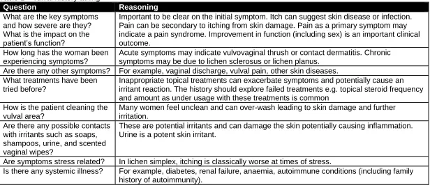
Table 1 – vulval history taking
Question Reasoning
What are the key symptoms and how severe are they? What is the impact on the patient’s function?
Important to be clear on the initial symptom. Itch can suggest skin disease or infection. Pain can be secondary to itching from skin damage. Pain as a primary symptom may indicate a pain syndrome. Improvement in function (including sex) is an important clinical outcome.
How long has the woman been experiencing symptoms?
Acute symptoms may indicate vulvovaginal thrush or contact dermatitis. Chronic symptoms may be due to lichen sclerosus or lichen planus.
Are there any other symptoms? For example, vaginal discharge, vulval pain, other skin diseases. What treatments have been
tried before?
Inappropriate topical treatments can exacerbate symptoms and potentially cause an irritant reaction. The history should explore failed treatments e.g. topical steroid frequency and amount as under usage with these treatments is common
How is the patient cleaning the vulval area?
Many women feel unclean and can over-wash leading to skin damage and further irritation.
Are there any possible contacts with irritants such as soaps, shampoos, urine, and scented vaginal wipes?
These are potential irritants and can damage the skin potentially causing inflammation. Urine is a potent skin irritant.
Are symptoms stress related? In lichen simplex, itching is classically worse at times of stress.
Are there any other skin conditions present?
For example, eczema or psoriasis (sometimes hidden as cracking behind the ears, a scaly scalp or umbilical erythema)
Vulval examination
A full vulval examination requires good lighting and each part of the vulva should be examined systematically including the mons pubis, inguinal folds, outer and inner labia (majora and minora), clitoris (body and hood), perineum, vestibule and anus (Fig 1). Hart’s line is the junction
between the vestibule and the inner labia and marks change in epithelium type from mucosal type to stratified squamous. Ideally, digital and speculum examinations are important to rule out erosions, mucosal thickening, adhesions, and scarring as can be seen in conditions such as erosive lichen planus and lichen sclerosus (2).
Vulval pathology may be a manifestation of a general skin condition and therefore a complete examination including hidden sites such as the umbilicus and natal cleft needs to be considered. Furthermore, examination of other non-keratinised or mucosal surfaces, including the oral cavity, eyes and mouth should be performed. This allows a more complete assessment of disease extent and diagnosis especially for diseases that are not solely restricted to the vulval region such as psoriasis, eczema, lichen sclerosus, pemphigus vulgaris, pemphigoid and erosive lichen planus.
Table 2: Terminology of lesions that may be seen in the vulval area Lesion
terminology
Description Example
Fissure A thin ‘hairline’ crack in the skin surface due to excessive dryness Psoriasis and lichen sclerosus Excoriation Scratch mark, may be single or multiple As seen in any itchy skin conditions
e.g. atopic eczema, lichen sclerosus
Erosion A shallow denuded area due to loss of the epidermis (surface layer of skin)
Erosive lichen planus
Ulcer Full thickness loss of the epidermis (top layer of skin) +/- dermis Aphthous ulceration,
Macule Flat area of colour change Vulval melanosis
Ecchymosis (subcutaneous purpura) seen in lichen sclerosus Nodule Large palpable lesion greater than 0.5 cm in diameter Squamous cell carcinoma, Scabies Papule Small palpable lesion less than 0.5cm in diameter Genital warts
Molluscum contagiosum Seborrhoeic keratosis Plaque A palpable flat lesion greater than 0.5 cm diameter. It may be elevated
or may be a thickened area without being visibly raised above the skin surface
Vulval Intraepithelial Neoplasia Squamous cell carcinoma
Vesicle Small fluid filled blister less than 0.5cm diameter Bullous pemphigoid Lichenification An accentuation of skin markings commonly associated with
thickening of epidermis usually caused by scratching or rubbing
Lichen simplex
Investigations
Vaginal swabs
These are indicated when the history suggests possible primary or secondary infection with bacterial, candida and viral infections. Infection is a common cause for loss of symptom control in inflammatory dermatoses. This may be a reason why lichen sclerosus appears initially well
controlled with potent steroids and then flares. Occasionally, genital herpes can be a cause of vulval symptoms and a viral culture swab may be necessary.
Vulval biopsy
areas, 4) when there is poor response to treatment following the initial diagnosis. The site selected for biopsy should be tissue-representative of the lesion or area of abnormality. This is usually at the edge of the lesion and should also include some normal tissue. The most central area may be inflamed or necrotic, which may give minimal tissue diagnosis because of the inflammation present. An excisional biopsy can be
problematic if the diagnosis is subsequently found to be cancer or VIN in that re-excision is often required. This can lead to further skin loss that might compromise function. Multiple mapping biopsies are indicated in cases of suspected multifocal disease. A 4mm Keyes punch biopsy is adequate and should ideally be carried out under local anaesthetic at the initial visit if possible. It can provide adequate tissue for histology but is not adequate if immunofluorescence is also required to exclude immunobullous disease when two biopsies may be needed. Inflammatory vulval lesions often have indistinct inflammatory pathology and a diagnosis should always be a clinic pathological correlation.
Patch testing
Patch testing by a dermatologist is indicated when allergic contact dermatitis is considered (see section on ‘Contact Dermatitis’). Common allergens include topical anaesthetics, fragrances, sodium lauryl sulphate and topical neomycin (4,5). Allergic contact dermatitis to the adhesive used in sanitary pads is relatively common and should be considered particularly if the fully keratinised epithelium is affected [6]. Since
sensitisation can occur at any time it is a further reason why symptoms initially well controlled with topical therapies can flare. Clinical history is very important to determine potential sensitising agents which can then be applied to the skin during the patch testing process.
The multidisciplinary team
Many chronic, rare and difficult vulval problems require a multidisciplinary input. A local vulval clinic might be of help in dealing with difficult patients and those with unusual skin conditions when expert help is needed. Other members of vulval service might include dermatology, genito-urinary medicine, physiotherapy, pain management, psychosexual therapy, pathology and urogynaecology.
Specific vulval conditions
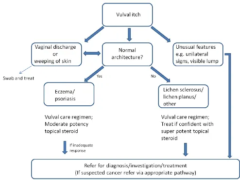
Vulval itching is often the presenting complaint of a vulval skin condition. Itch is a symptom and not a diagnosis. The causes can be separated into skin disease (vulval dermatoses), infection and premalignant/malignant disease. The algorithm outlined in figure 4 is a useful when
assessing patients. Many of the conditions outlined below require treatment with frequent topical steroids and emollients with a focus on patient education and compliance. The ‘Treatment principles’ section below outlines the general principles of these treatments.
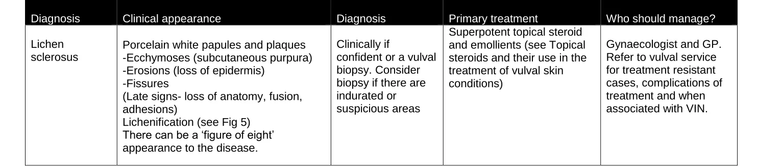
Table 3 - summary of the clinical features and treatment of specific skin diseases.
Diagnosis Clinical appearance Diagnosis Primary treatment Who should manage?
Lichen sclerosus
Porcelain white papules and plaques -Ecchymoses (subcutaneous purpura) -Erosions (loss of epidermis)
-Fissures
(Late signs- loss of anatomy, fusion, adhesions)
Lichenification (see Fig 5) There can be a ‘figure of eight’ appearance to the disease.
Clinically if
confident or a vulval biopsy. Consider biopsy if there are indurated or suspicious areas
Superpotent topical steroid and emollients (see Topical steroids and their use in the treatment of vulval skin conditions)
Lichen planus ‘Classical’ lichen planus - violaceous, well demarcated plaques with overlying lacy white lines which usually affect the labia majora and surrounding skin
Erosive lichen planus - ‘glazed’ erythema or erosions symmetrically distributed at vaginal introitus. White slightly raised edge to lesions. Lacy white lines (Wickham’s striae) in
surrounding skin. (see Fig 6). May have loss of anatomy
Clinical assessment and biopsy from the edge of an erosion.
Lichen planus may be seen in the mouth or normal skin
Superpotent topical steroid and emollients (see Topical steroids and their use in the treatment of vulval skin conditions)
.
Dermatologist or gynaecologist. Refer to vulval service for treatment resistant cases, complications of treatment and when associated with VIN. Erosive lichen planus is difficult to treat so refer to a vulval service.
Atopic eczema Symmetrically inflamed, erythematous, weepy skin. No loss of anatomy. May be satellite lesions and poorly defined edges
Clinical history and examination to include other skin sites for other signs of eczema.
Moderate (e.g clobetasone butyrate 0.05%) or potent (e.g. Mometasone furoate 0.1%) topical steroid plus emollients to gain control of inflammation.
Dermatologist and GP
Contact dermatitis
Irritant form - Poorly defined erythema present where irritant has been applied Allergic form – Erythema extends
outside of area where allergen has been applied
Clinical history and examination.
Patch testing if allergic contact dermatitis suspected
Moderate (e.g clobetasone butyrate 0.05%) or potent (e.g. Mometasone furoate 0.1%) topical steroid plus emollients to gain control of inflammation.
Strict avoidance of irritants/allergens.
Seborrhoeic eczema
Glazed skin in the intralabial sulci Clinical examination of other sites – scalp, eyebrows, nasolabial folds for erythema and fine scaling
Moderate (e.g clobetasone butyrate 0.05%) or potent (e.g. Mometasone furoate 0.1%) topical steroid plus emollients to gain control of inflammation.
Dermatologist and GP
Psoriasis Classically, well demarcated, scaly erythematous plaques, but vulval psoriatic plaques are smooth, glossy and often salmon-pink in colour. Often no scale in vulval creases but
surrounding skin may have typical scaly lesions of psoriasis. There is no scarring or loss of anatomy (Fig 3).
Clinical assessment to include
examination of ‘hidden sites’ for other signs of psoriasis eg . knees, elbows, umbilicus, scalp, ears, lower back and nails Biopsy if unsure.
Moderate potency topical steroid plus emollients as recommended by NICE guidance (7) As the skin folds can become particularly macerated, there is a chance of secondary bacterial or fungal infection. A combination topical
preparation (e.g clobetasone butyrate
0.05%/oxytetracycline 3%/nystatin) may be helpful.
Dermatologist
Lichen simplex Lichenification of the skin with erosions from chronic scratching.
Usually no loss of anatomy but can give thick ‘leathery’ skin This is often
superimposed on other itchy skin disorders such as eczema and lichen sclerosus.
Clinical history and examination.
Superpotent topical steroid and emollient. Once control of symptoms is achieved, moderate potency topical steroid may be required intermittently. Secondary infection with candida or bacteria is common and may need treatment (2).
Dermatologist and GP.
Intertrigo
(See also
Flexural rash which may involve the groin, natal cleft, submammary region and abdominal ‘apron fold’. Common in
Infection of the flexural areas with thrush (Candida
Treatment is usual using both oral antibiotics and antifungal agents together
vulvovaginal candidiasis )
overweight patients. albicans),
erythrasma, (Corynebacterium minutissmum) and tinea species
with the regular use of an emollient to improve the skin barrier function at this site.
Lichen sclerosus
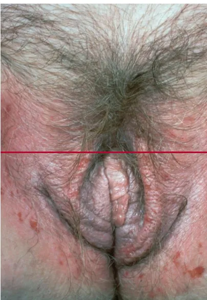
Lichen sclerosus is an auto-immmune, chronic, inflammatory skin condition that has a predilection for the genital skin in both sexes(Figure 5). Early stage disease may be subtle and masked especially if topical steroids have been used prior to referral.
The general management of lichen sclerosus is with topical steroids and emollients and is detailed below (8). There is a small risk of cancer (less than 5% risk)(9) so patients should be encouraged to self exam on a regular (suggested monthly) basis to detect skin cancers. Changes may include raised lesions irregular lesions, ulceration and persistent eroded areas. Skin fissuring at the posterior fourchette and the initial treatment should focus on digital massage of the steroid into the fissure on a daily basis, the encouragement in the use of vaginal dilators and a good lubricant. If these measures do not help then consideration should be given to the use of surgical division or refashioning of the fissure or skin bridge.
Lichen planus
Lichen planus is another autoimmune chronic condition (10). Two main forms of lichen planus may affect the vulval area. ‘Classical’ lichen planus outlined in table 2 is usually very successfully treated with topical steroids and emollients. In contrast, patients with erosive lichen planus usually present with pain and burning as erosions occur at the entrance to the vagina and may affect the vaginal mucosal surface. This variant leads to considerable scarring and loss of anatomy (Figure 6). In contrast to the ‘classical’ plaque form of lichen planus this disorder is painful rather than itchy and is responds less well to therapy. Referral to a vulval service is important to optimise disease management. Although the evidence is not as clear cut as with lichen sclerosus, there is probably a small chance of malignant potential in lesions of lichen planus (11).
Vulval dermatitis
The terms dermatitis and eczema are often used interchangeably. There are different types of dermatitis that can affect the vulval area. It is important to note that vulval anatomy is normal with eczematous conditions and scarring does not occur.
Contact dermatitis may be either ‘irritant’ or ‘allergic contact’. Irritant contact dermatitis is particularly common and may be triggered by soaps, perfumes, medicaments, urine, faeces and sweat. The barrier function of the skin becomes impaired by local irritants and can subsequently be made worse by continued application of the product. Clinical signs of vulval irritant dermatitis include poorly defined intralabial erythema that is occurs anywhere where the irritant has been present Small fissures and erosions may be present. Lichenification occurs in longstanding disease (12). All of these clinical features are demonstrated in Fig. 2. It is common that fissures become secondarily infected with skin pathogens or candida. It is important to realise this when treatment strategies are implemented. Allergic contact dermatitis is less common; around one fifth of patients with vulval skin conditions having a relevant positive patch test result (13). It can be difficult to distinguish from irritant contact dermatitis clinically, but affected skin usually extends outside of the genital area in ‘non-contact’ areas. This is because it is an immune mediated hypersensitivity reaction. The only way to confidently diagnose allergic contact dermatitis is through patch testing (see section on ‘Investigations’) .
Vulval seborrhoeic eczema is difficult to distinguish from psoriasis. It will often manifest as glazed skin in the interlabial sulci bilaterally. Fine scale and erythema at other affected body sites such as the nasolabial folds, scalp and eyebrows can aid the diagnosis (14).
Vulvovaginal candidiasis. Acute vulval candidiasis is likely to resolve quickly with treatment and patients are unlikely to present to a
gynaecologist and be managed in primary healthcare. However recurrent candidiasis (more than 6 attacks a year) or chronic infection can be more subtle and more recalcitrant to treatment. Patients with an existing vulval condition may develop candidiasis as a secondary problem which can be overlooked e.g. a patient with lichen sclerosus treated with topical steroids. There should be a low threshold for taking swabs for infection especially those patients who fail to respond to treatment. A suggested treatment regime for recurrent candidiasis includes initial treatment followed by a maintenance regime for 6 months (eg Fluconazole 100mg weekly for 6 months). Cessation of therapy may result in relapse in at least 50% of women (15)
Treatment principles for all vulval disease
There are no core clinical outcome measures for vulval skin disease (16).Suggested clinical outcomes include:
1) A reduction in symptoms (eg less itch, fewer flare-ups) 2) An improvement in function (eg sexual function, mobility)
General principles Initial principles of management are the same for all vulval skin conditions and a holistic approach is required. Good
education, support and counselling are required with extra time given to address the disease process, discussing general vulval care measures and managing patient expectation (2). It is useful to provide information leaflets, direct patients to relevant patient-oriented websites and write down instructions for applying topical agents (see ‘Additional resources’ for sources of patient information). The use of a mirror or model in the clinic setting is helpful to show patients where to apply their topical treatments.
Correct barrier function The goal of therapy is to correct barrier function and reduce inflammation. For washing, soap and other routine cleaning agents (e.g. wipes) should be avoided, as they are likely to act as irritants and sensitising allergens. Irritation from urinary and faecal
incontinence need to be addressed as these will be a common cause of irritant vulvitis and make underlying skin pathology worse. ‘Soap substitution’ with a bland cream or ointment based emollient is best for cleansing. The same agent can then be used as an emollient to both provide a barrier to the site and sooth inflamed skin. There is no preferred emollient to use and some can cause irritation. Emollient creams (not ointments) can be placed in the fridge. Patients find this soothing and lowering skin temperature is thought to reduce itch through central
inhibitory pathways (17).
Topical steroids and their use in the treatment of vulval skin conditions
Inflammation reduction associated with skin disease (such as in lichen planus, lichen sclerosus and eczema) is achieved by the use of topical steroids. Topical steroids are often ineffectively used in the vulval area due to concerns from patients and non-specialists about side-effects, particularly skin or mucosal atrophy. It is important therefore to use the correct strength of steroid for the necessary length of time on the appropriate body site. Mucosal surfaces such as the vulval vestibule are remarkably resistant to steroid atrophy.
In contrast, keratinised surfaces such as the labiocrural folds, perineum, perianal area and thighs can develop skin thinning and striae (stretch marks) with inappropriate use with potent topical steroids (2). Overusage of topical steroids appears as thinned skin which appears redder and is reversible in the early stages. In the later stages permanent telangiectasia and striae can develop. Topical calcineurin inhibitors in the vulval region reduce inflammation and do not cause skin atrophy. However their role is not fully understood and there is a theoretical risk of long term localised immunosuppression from these agents causing skin cancers (18).
In lichen sclerosus and lichen planus, the use of superpotent topical steroids is recommended as first-line therapy. Cochrane systematic reviews of interventions for both lichen sclerosus and lichen planus (19,20) have been published. There is reasonable randomised controlled trial evidence for the use of topical steroids in lichen sclerosus and The British Association of Dermatologists suggest the use of the
In general topical steroids should be used once-daily. There is no evidence to suggest that daily application is superior, although twice-daily has greater potential to cause side effects (23). Ointments are preferable to creams as they contain fewer constituents and therefore have a lower chance of causing irritation/contact allergy. Once control of inflammation and symptoms has been achieved, topical steroids should be reduced to the minimum frequency required to maintain remission. The concept of ‘weekend therapy’, that is, applying topical steroids on two consecutive days per week, is effective in atopic eczema patients (24) and can extrapolated to chronic vulval diseases such as lichen sclerosus and lichen planus where long-term maintenance therapy is required. A patient with these conditions will use approximately 30-60 g of topical steroid per year as maintenance therapy (8). Topical steroids should only be used on affected areas to prevent side-effects in adjacent skin..
Failure to respond to treatment If a patient fails to respond to appropriate treatment the following should be considered:
1. Poor adherence to prescribed treatment regimen – ‘steroid phobia’ is a well recognised problem when treating skin conditions. Many healthcare professionals, including pharmacists, will compound the issue by advising the patient to use sparingly and recommending not to use on ‘sensitive areas’. This cautious approach can be detrimental to the patient’s treatment plan. The patient should be advised to apply the topical steroid in terms of the finger tip unit (A finger tip is from the very end of the finger to the first crease in the finger. It does NOT mean a blob on the fingertip). The number of fingertip units required is usually one to two but is specifically tailored to the patient depending upon surface area affected by the condition (25).
2. Inaccurate placement of topical steroid – the patient may be applying the topical treatment to an unaffected area. Especially common if the patient is elderly and unable to use a mirror to see what they are doing. In clinic the exact location of application should be
explained, diagrams/photographs or models can be used as an aid.
3. Continued exposure to irritants – urine or faeces, external products such as wipes or non prescribed topical treatments and over washing with water can all contribute towards irritation and ongoing symptoms.
4. Incorrect diagnosis – if adherence and skin care practices are assessed as adequate, it may be that the diagnosis given is incorrect. An allergic contact dermatitis to topical treatments may have occurred (see section on Patch Testing) or there may be pre-malignant or malignant change in the affected area. If there is any concern a biopsy should be taken.
Conclusion
Vulval skin conditions are common and generally easy to diagnose by an accurate history and examination. Sometimes further investigations such as vulval swabs, patch-tests and biopsies are needed. For patients with unusual clinical features, or who fail to respond to adequate therapy, pre-malignancy or malignancy should be considered as an alternative diagnosis. Complex, rare and treatment resistant patients should be referred to a vulval service.
Further information is available on line from the following resources; Dermnetnz: The Dermatology Resource www.dermnetnz.org
FIGURES
Figure 1: Schematic representation of the normal adult vulva. Copyright “Dawn Danby and Paul Waggoner”, c/o ISSVD
Figure 2: Contact dermatitis affecting the female genitalia. Note poorly defined erythema of the genitocrural skin, erosions due to excoriation in the inflamed skin and white thickening (lichenification) of the labia majora due to scratching.
Figure 3: Psoriasis affecting the female genitalia. Well demarcated erythema surrounding the anogenital area with typical psoriatic plaques in surrounding skin. The genital plaque has typical scale in the perianal area, but lack of scale in the perineal and vulval areas.
Figure 4– algorithm for the management of vulval itching
References
1. Coulson C, Crowley T. Current thoughts on psychosexual disorders in women. THE OBSTETRICIAN & GYNAECOLOGIST Volume 9, Issue 4, October 2007, Pages: 217–222,
2. Schlosser BJ, Mirowski GW. Approach to the patient with vulvovaginal complaints. Dermatol Ther. 2010;23(5):438-48. doi: 10.1111/j.529-8019.2010.01348.x.
3. Margesson LJ. Overview of treatment of vulvovaginal disease. Skin Therapy Letter. 2011;16(3):5-7.
4. Bhate K1, Landeck L, Gonzalez E, Neumann K, Schalock PC. Genital contact dermatitis: a retrospective analysis. Dermatitis.2010 Nov-Dec;21(6):317-20.
McPherson T1Cooper S. Vulval lichen sclerosus and lichen planus..Dermatol Ther. 2010 Sep-Oct;23(5):523-32 5. O'Gorman SM1, Torgerson RR Allergic contact dermatitis of the vulva. .Dermatitis. 2013 Mar-Apr;24(2):64-72.
6. Wujanto L, Wakelin S. Allergic contact dermatitis to colophonium in a sanitary pad-an overlooked allergen? Contact Dermatitis. 2012;66(3):161-2. doi:10.1111/j.1600-0536.2011.02006.x.
7. NICE. The assessment and management of psoriasis. 2012. guidance.nice.org.uk/cg153.
8. Neill SM, Lewis FM, Tatnall FM, Cox NH. British Association of Dermatologists' guidelines for the management of lichen sclerosus 2010. The British journal of dermatology. 2010;163(4):672-82. doi:10.1111/j.1365-2133.2010.09997.x.
9. Kirtschig G, Becker K, Günthert A, Jasaitiene D, Cooper S, Chi CC, Kreuter A, Rall KK, Aberer W, Riechardt S, Casabona F, Powell J, Brackenbury F, Erdmann R, Lazzeri M, Barbagli G, Wojnarowska F. Evidence-based (S3) Guideline on (anogenital) Lichen sclerosus.J Eur Acad Dermatol Venereol. 2015 Oct;29(10):e1-43. doi: 10.1111/jdv.13136. Epub 2015 Jul 22
10. Simpson RC, Murphy R Is vulval erosive lichen planus a premalignant condition? Arch Dermatol. 2012 Nov;148(11):1314-6 11. McPherson T, Cooper S. Vulval lichen sclerosus and lichen planus. Dermatologic Therapy. 2010;23(5):523-32
12. Welsh B, Howard A, Cook K. Vulval itch. Australian family physician. 2004;33(7):505-10.
13. Goldsmith PC, Rycroft RJ, White IR, Ridley CM, Neill SM, McFadden JP. Contact sensitivity in women with anogenital dermatoses. Contact Dermatitis. 1997;36(3):174-5.
14. Farage MA, Miller KW, Ledger WJ. Determining the cause of vulvovaginal symptoms. Obstetrical & gynecological survey. 2008;63(7):445-64. doi:10.1097/OGX.0b013e318172ee25.
15. http://cks.nice.org.uk/candida-female-genital
16. Simpson RC, Thomas KS, Murphy R. Outcome measures for vulval skin conditions: a systematic review of randomized controlled trials.Br J Dermatol. 2013 Sep;169(3):494-501
17. Carstens E, Jinks SL. Skin cooling attenuates rat dorsal horn neuronal responses to intracutaneous histamine. Neuroreport. 1998;9(18):4145-9.
18. Thaçi D1, Salgo R.Malignancy concerns of topical calcineurin inhibitors for atopic dermatitis: facts and controversies. Clin Dermatol. 2010 Jan-Feb;28(1):52-6.
20. Chi CC, Kirtschig G, Baldo M, Brackenbury F, Lewis F, Wojnarowska F. Topical interventions for genital lichen sclerosus. Cochrane Database Syst Rev. 2011(12):CD008240. doi:10.1002/14651858.CD008240.pub2.
21. Simpson RC, Littlewood SM, Cooper SM, Cruickshank ME, Green CM, Derrick E et al. Real-life experience of managing vulval erosive lichen planus: a case-based review and U.K. multicentre case note audit. The British journal of dermatology. 2012;167(1):85-91.
doi:10.1111/j.1365-2133.2012.10919.x.
22. Cooper SM, Wojnarowska F. Influence of treatment of erosive lichen planus of the vulva on its prognosis. Archives of Dermatology. 2006;142(3):289-94.
23. Green C, Colquitt JL, Kirby J, Davidson P. Topical corticosteroids for atopic eczema: clinical and cost effectiveness of once-daily vs. more frequent use. The British journal of dermatology. 2005;152(1):130-41. doi:10.1111/j.1365-2133.2005.06410.x.
24. Berth-Jones J, Damstra RJ, Golsch S, Livden JK, Van Hooteghem O, Allegra F et al. Twice weekly fluticasone propionate added to emollient maintenance treatment to reduce risk of relapse in atopic dermatitis: randomised, double blind, parallel group study. BMJ. 2003;326(7403):1367. doi:10.1136/bmj.326.7403.1367.
25. Lawton S1, Littlewood S.Vulval skin disease: clinical features, assessment and management. Nurs Stand. 2006 Jun 28-Jul 4;20(42):57-63
Contribution of authorship
David Nunns conceived the idea, wrote the outline, supervised the writing. DN, RS, RM and AW edited and approved the final version.
Disclosure of interests/disclaimer
DN is a Trustee of the Vulval Pain Society, Education and Training Group Chair, BSSVD
25 CPD questions True False
The following conditions should ideally be managed within specialist care (vulval clinic).
Lichen sclerosus follow-up
F – follow-up can be with GPs or general gynaecology/dermatology clinics
Unifocal vulval intraepithelial neoplasia
F – can be managed by general gynaecologists
Vulvodynia
T – treatment resistant vulvodynia should be referred to the vulval service but all general gynaecolgogists should be able to start basic treatment
Bullous disease
T – a rare, complex condition
With regards vulval disease management
Creams have better moisturizing properties than ointments F
With an ulcerated vulval lesion, a punch biopsy should be taken from the central area of the lesion as this represents the most useful site for diagnosis.
F – Biopsies should be taken from the edge of the lesion at the junction of the normal skin and the lesion. Areas of inflammation are often histologically non specific.
In the absence of a diagnosis, mapping vulval biopsies should be taken in symptomatic patients even if the skin looks normal.
F – biopsying clinically normal skin should be discouraged as there is little additional diagnostic benefit. Normal skin biopsy reports are often reported as showing ‘inflammation’ which can lead to inappropriate prescription of topical steroids.
First line treatment with topical steroids for vulval lichen sclerosus and erosive lichen planus should be reducing potency regime for three months
F – Superpotent topical steroids should be used in with reducing frequency
Vulval psoriasis should be treated with a superpotent toptical steroid
F – NICE guidance recommends that moderate potency topical steroids are used
Side effects from topical steroid use are uncommon
T – If an appropriate strength and frequency of topical steroid are used, side effects such as atrophy and striae are uncommon.
Dermatologist patch testing should be considered for all patients with recurrent vulval itching F – only for those patients where allergic contact dermatitis require exclusion
Biopsy is always required to diagnose lichen sclerosus
F – Biopsy is required if the diagnosis is unclear, there has been poor response to standard treatment, or if there are indurated or suspicious areas on clinical examination
Erosive lichen planus presents with pain rather than itch
T – Erosive lichen planus is usually painful due to mucosal erosion. Classical lichen planus is itchy
The risk of squamous cell carcinoma development in lichen sclerosus is 10% F – less than 5%
With regards skin lesion morphology
A papule is a small palpable lesion less than 0.5cm in diameter T
An ulcer is a shallow denuded area due to loss of the epidermis (surface layer of skin) F – this is an erosion
Patients with vulval skin lichenification usually have painful, thickened skin in the area scratching
A vesicle is a small fluid filled blister less than 0.5cm diameter T
Vulval intraepithelial neoplasia is commonly plaque-like T
With regards topical steroids in the management of vulval lichen sclerosus
Follow-up treatment is usually with daily treatment of superpotent topical steroid ointments in patients who are in remission F – usually treatment is twice a week, but this can be increased depending on symptoms
Twice a day treatment is more likely than once a day treatment to produce remission of symptoms F – once a day treatment frequently produces good symptom control
May predispose a patient to secondary candida infection T
Will reverse anatomical change in advanced disease
F – skin texture will improve but the anatomical loss will not reverse