AbstrAct
Stromal cells like synovial fibroblasts gained great interest over the years, since it has become clear that they strongly influence their environment and neighbouring cells. The current review describes the role of synovial fibroblasts as cells of the innate immune system and expands on their involvement in inflammation and cartilage destruction in rheumatoid arthritis (RA). Furthermore, epigenetic changes in RA synovial fibroblasts and studies that focused on the identification of different subsets of synovial fibroblasts are discussed.
In most autoimmune diseases, including rheumatic diseases, research focused on the analysis of immune cells and their role in initiation and perpetuation of disease. Since a couple of years, however, it has been acknowl-edged that immune cells and local stroma have a significant role in disease pathogen-esis. Stromal cells reveal an unexpected complexity in phenotype and function in different organs and in different diseases.
The stroma builds the structural framework of an organ or tissue. Studies analysing stromal cells in different settings revealed interac-tions between stromal cells and immune cells, which substantially contribute to shaping the immune response. Furthermore, stromal cells were recognised to have immunological func-tions and to be able to recognise pathogens and elicit an immune response. Intestinal, dermal, gingival and synovial fibroblasts are only some examples of stromal cells that have been shown to express innate immune recep-tors, in particular toll-like receptors (TLR). They react with the production of cytokines and chemokines to pathogen sensing and are able to process and to present antigens via major histocompatibility complex (MHC) II receptors.1–5 Therefore, these cells are part of the innate immune system and play an important role as local pathogen and damage sensors and activators of the immune system.
Synovial fibroblasts are the main stromal cells of the joint synovium. They are found in the synovial sublining and lining layer, which, in a healthy joint, is one to two cell layers thick and is interspersed with tissue-resident macrophages. Synovial fibroblasts produce
the extracellular matrix (ECM) components of the synovial fluid and thus are important for cartilage integrity and lubrication of the joint. In rheumatoid arthritis (RA), the lining layer thickens and immune cells infiltrate the sublining layer of the synovium (figure 1). Synovial fibroblasts proliferate, become acti-vated and invade and destroy the adjacent cartilage.
The analysis of synovial fibroblast in RA reaches back to the late 60s and early 70s with first studies done in particular by HG Fass-bender and CW Castor and colleagues. Already in 1972, Castor and Buckingham noted that ‘extracts of Gram-negative bacteria, applied to fibroblast cultures, markedly increased hyaluronic acid production, glucose utilisa-tion, and lactate output’,6 anticipating studies on TLR signalling and metabolic changes in synovial fibroblasts that were published decades later. Since then, numerous studies have characterised many different aspects of synovial fibroblast biology. Usually, cultured synovial fibroblasts from patients undergoing joint replacement surgery or synovial biop-sies were used for these studies. Thereby, the synovium is enzymatically digested or fragmented, and adherent cells are used for analysis after three to four passages.7 These cultures are highly homogeneous for the expression of fibroblasts markers such as CD90 or proly-4-hydroxylase. However, it should be kept in mind that long-term culture of synovial fibroblasts can have an influence
Review
Synovial fibroblasts in 2017
Caroline Ospelt
To cite: Ospelt C. Synovial fibroblasts in 2017. RMD Open 2017;3:e000471. doi:10.1136/ rmdopen-2017-000471
Received 20 July 2017 Revised 14 September 2017 Accepted 28 September 2017
Department of Rheumatology, Center of experimental Rheumatology, University Hospital and University of Zurich, Zurich, Switzerland
Correspondence to Dr Caroline Ospelt; Caroline. Ospelt@ usz. ch
Key messages
► Synovial fibroblasts are carry a variety of innate
immune receptors and are able to present antigen.
► in rheumatoid arthritis (RA), synovial fibroblasts
promote joint destruction via their attachment to cartilage and thus are key cells in RA pathogenesis.
► RA synovial fibroblasts are stably activated and
show alterations in their epigenetic landscape.
► Synovial fibroblasts of different joints and of the
lining and sublining layer differ in their phenotype and thus represent distinct subtypes of the synovial fibroblast population.
on May 7, 2020 by guest. Protected by copyright.
http://rmdopen.bmj.com/
Figure 1 Osteoarthritis (A) and rheumatoid arthritis (RA) synovium (B). Synovial fibroblasts are found in the lining as well as in the sublining layer of the synovium. Thickening of the lining layer, lymphocytic infiltration and increased formation of blood vessels can be seen in RA synovium.
on their phenotype and that some characteristics of syno-vial fibroblasts, for example, the proliferative potential change over several passages.8 9 Furthermore, it has not yet been clarified how homogeneous this cultured cell population is, considering that different subtypes of synovial fibroblasts have been described as mentioned in more detail below.
Due to the vast amount of studies conducted with synovial fibroblasts in various rheumatic diseases, not all aspects of synovial fibroblast function in health and disease can be covered by the current review article. Instead, it focuses on the role of synovial fibroblasts as innate immune cells, their interaction with other immune cells and how this influences the pathogenesis of RA. Furthermore, epigenetic changes and their potential consequences in RA synovial fibroblasts are mentioned, and studies that subcategorise synovial fibroblasts into different subsets based on functional and embryonic differences are discussed.
Synovial fibroblaSTS aS innaTe immune CellS
By the expression of various pattern recognition recep-tors, synovial fibroblasts are well equipped to sense invading pathogens as well as joint damage. Synovial fibroblasts express TLR1–7, but very low or not-detect-able basal transcript levels of TLR8–10.2 10
TLR3 and TLR7 bind double-stranded and single-stranded RNA, respectively. In addition, the cytosolic pattern recognition receptors (PRR) RIG-I and MDA5, which both bind double-stranded RNA are expressed and active in synovial fibroblasts.10 Necrotic cells in the syno-vial fluid were suggested to be a possible source of endog-enous ligands leading to the activation of TLR3-mediated pathways in synovial fibroblasts in RA.11 In a large scale comparison of cytokine profiles between RA synovial fluid and stimulated synovial fibroblasts, the cytokine profiles obtained by stimulation of synovial fibroblasts with tumour necrosis factor (TNF) α, interleukin (IL) 1 or the TLR3 ligand Poly(I:C) reached the highest simi-larity with RA synovial fluids among 10 different stimuli
used.12 The seven cytokines most strongly induced by these stimuli in synovial fibroblasts were also among the 12 cytokines with the highest levels in RA synovial fluids, corroborating the strong contribution of synovial fibro-blasts to the inflammatory environment in RA joints. Unfortunately, other TLR ligands were not included in the stimulation experiments in this study.
TLR2, which forms heterodimers with TLR1 or TLR6, and TLR4 are both sensors of bacterial lipoproteins. A variety of endogenous ligands have been described to bind and activate TLR2 and TLR4, and many of them have been found at increased levels in RA joints.13–17 Some like heat-shock proteins, low-molecular-weight hyaluronan or high-mobility group protein 1 were initially described as direct ligands, but highly purified preparations were later not confirmed to stimulate TLR activation.18–20 Instead, it was suggested that these molecules facilitate binding of TLR ligands and thus amplify the cellular response to TLR activation. However, it is not clear in how far recom-binant or highly purified preparations resemble their counterparts in vivo. Post-translational modifications and the formation of immune complexes can influence binding and activity of TLR ligands in vivo. Fibrinogen, which was described as endogenous ligand of TLR4,21 was shown to be more potent to activate macrophages and synovial fibroblasts in its citrullinated form.22 23 Also free histones that can activate TLR2 and TLR424 were found to be more potent stimulators if citrullinated and even more if incorporated in an immune complex.25 Interestingly, activation of monocytes by synovial fluids was significantly better blocked with an TLR4 body if the patients had citrullinated protein anti-bodies (ACPA) directed against citrullinated fibrinogen and citrullinated histone 2A, pointing toward a strong contribution of TLR4-mediated disease pathways in these patients.26 Thus, in the RA joint environment, the high levels of protein citrullination and the formation of immune complexes comprising citrullinated proteins and ACPA might further promote the binding of endog-enous ligands to TLRs and cellular activation.
Interestingly, high content of citrullinated peptides was found in exosome preparations from synovial fluids of patients with RA, osteoarthritis (OA) and reactive arthritis, suggesting that extracellular vesicles might be of importance in the distribution and presentation of citrullinated peptides to various cell populations.27 Even though the amount of exosomes was similar between the patient’s groups, the content of proteins differed in that α2-macroglobulin, IgG1 γ-chain and fibronectin were only present in RA exosomes.27 Plasma-derived extra-cellular vesicles from RA patients but not from healthy individuals were shown to activate TLR4 on human embryonic kidney cells, on human monocytic cell lines and on murine bone-marrow derived monocytes.28 The authors could show that oxidised lipids in the membrane of RA exosomes were responsible for the activation of TLR4 and that the subsequent response resulted in the upregulation of genes that are important in resolving
on May 7, 2020 by guest. Protected by copyright.
http://rmdopen.bmj.com/
rather than promoting inflammation and substantially differed from a lipopolysaccharide-induced response. Synovial fibroblasts were found to produce proinflam-matory mediators on incubation with extracellular vesi-cles.29–31 Even though it remains to be analysed whether extracellular vesicles from RA synovial fluid can elicit a specific TLR4 response in synovial fibroblasts, these studies support the hypothesis that extracellular vesicles with RA-specific content and/or modifications might activate synovial fibroblasts via TLR4 and thus modulate inflammation and joint destruction.
There are not many published studies investigating the expression and function of TLR9 and other DNA sensors such as AIM2 in synovial fibroblasts, and thus, it is not yet clarified in how much synovial fibroblasts can contribute to an immunoresponse induced by DNA sensing.32 After 24-hour stimulation with unmethylated CpG-rich DNA, the ligand of TLR9, no increase in protein levels of IL 6 or IL-8 could be measured,33 and only a small increase in transcript levels of IL-6 and IL-8 was measured after 4 hours of stimulation.34 An interesting study published in 2017 could show that neutrophil extracellular traps (NETs) can be internalised by synovial fibroblasts via a pathway involving the receptor for advanced glycation end-products and TLR9.35 Internalisation of NETs led to upregulation of MHC class II by synovial fibroblasts via IL-17B and presentation of citrullinated peptides to anti-gen-specific T cells. Injection of synovial fibroblasts that had internalised NETs into humanised HLA-DRB*04:01 transgenic mice led to formation of auto-antibodies against citrullinated histones H3 and H4 and citrulli-nated myeloperoxidase, which are all constituents of NETs,35 proving the effectiveness of antigen presentation by synovial fibroblasts. Earlier studies also showed that synovial fibroblasts in vivo and in vitro,9 mainly after stim-ulation with interferon γ (IFNγ),36 37 express MHC class II and that they can present antigens to T cells.38 It would be interesting to see whether also TLR stimulation can influence the expression and function of MHC class II in synovial fibroblasts as it was described in other cell types.
Once activated, TLRs were shown to induce the produc-tion of cytokines, chemokines and matrix metalloprotein-ases (MMPs) in synovial fibroblasts.2 12 39 High levels of the B cell survival factors B cell activating factor (BAFF) and a proliferation-inducing ligand (APRIL) produced by synovial fibroblasts after TLR3 stimulation promote antibody production and class switching of B cells.40 Interestingly, stimulation of one PRR also influences the expression of others. Stimulation of TLR3 in synovial fibroblasts leads to a robust upregulation of TLR3 itself, TLR2 and the cytoplasmic PRRs NOD1 and NOD2, which both sense bacterial products.2 41 42 Activation of NOD1 in turn synergises with TLR2 and TLR4 pathways.42 Therefore, activation of one PRR increases the reactivity of synovial fibroblasts to various endogenous and exog-enous PRR ligands, which in an environment with high levels of endogenous ligands such as the RA synovium might lead to a constant build-up of the proinflammatory
response. Furthermore, in contrast to macrophages, the production of proinflammatory cytokines is not damp-ened after repeated stimulation of TLRs in synovial fibro-blasts.43 This lack of tolerisation might further contribute to sustaining the inflammatory response in RA synovium.
For the sake of completeness, it should be mentioned that several other stimuli, for example, a variety of cyto-kines, growth factors44 and adipokines,45–47 were found to activate synovial fibroblasts and promote their proin-flammatory and joint destructive behaviour. However, it would go beyond the scope of this review to discuss all of them.
inTeraCTionS of Synovial fibroblaSTS wiTh T CellS Based on findings that various types of stromal cells such as bone marrow-derived mesenchymal stem cells and dermal and synovial fibroblasts inhibit T cell prolifera-tion, it has been proposed that stromal cells in general possess immunosuppressive properties.48 49 For the suppression of T cell proliferation by synovial fibroblasts, soluble factors are needed that are only induced in syno-vial fibroblasts via direct cell-cell contact between synosyno-vial fibroblasts and T cells.49 50 A recent study could show that the inhibition of T cell proliferation is at least partly medi-ated via depletion of tryptophan by synovial fibroblasts.50 This study also showed that OA synovial fibroblasts are more efficient in suppressing T cell proliferation than RA synovial fibroblasts.
Whereas some studies showed that co-culture of T cells with synovial fibroblast reduced the production of IFNγ and IL-10, but not tumour necrosis factor (TNF) or IL-17A by T cells,49 50 other studies showed increased production of IFNγ, TNF and IL-17 by T cells co-cultured with synovial fibroblasts.51 52 This effect was mediated via membrane-bound IL-1551 and the CD40 pathway.52 The discrepancies between these studies probably stem from differences in isolation, culturing and stimulation tech-niques. It is hard to estimate which in vitro system most closely reflects the situation in vivo.
Stromal cells, including synovial fibroblasts, maintain T cells in a resting G0/G1 state and extend their survival.49 53 Thus, synovial fibroblasts keep T cells in the tissue in a non-proliferative state and protect them from under-going apoptosis. In an inflammatory state, this might have profound effects on the persistence of inflammation and might actively counteract resolution of inflammation. Indeed, in the synovium of RA patients, differentiated T cells are protected from apoptotic cell death.54 Together, these data point to a modulation of the T cell phenotype and function by synovial fibroblasts, rather than general immunosuppressive properties. Whether this interac-tion between stroma and invading T cells is promoting or counteracting disease development remains, however, elusive up to now.
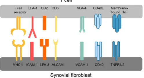
Various different direct adhesive and signalling inter-actions have been described between synovial fibroblasts and T cells as summarised in figure 2. These interactions
on May 7, 2020 by guest. Protected by copyright.
http://rmdopen.bmj.com/
Figure 2 Overview of key interactions between T cells and synovial fibroblasts. ALCAM, activated leukocyte cell adhesion molecule; CD40L, CD40 ligand; ICAM-1,
intercellular adhesion molecule 1; LFA, lymphocyte function-associated antigen; MHC II, major histocompatibility complex II; TNFR1/2, tumour necrosis factor receptor 1/2; VCAM-1, vascular cell adhesion protein 1; VLA-4, very late antigen-4.
first promote infiltration and retention of T cells in the synovium, for example, very late antigen-4 (VLA-4) and vascular cell adhesion protein 1 (VCAM-1) interactions, and second activate intracellular signalling pathways in both T cells and synovial fibroblasts. The connec-tion between lymphocyte funcconnec-tion-associated antigen 1 (LFA-1/CD11a) on T cells and intercellular adhe-sion molecule 1 (ICAM-1/CD54) on synovial fibroblasts is of particular interest because of its ability to enhance antigen presentation and T cell activation.55 LFA-1/ ICAM-1 binding lowers the threshold for presented antigens to induce T cell activation, thereby allowing T cell activation by antigens that without this co-stimula-tion would not elicit a T cell response.56 LFA-1 binding by ICAM-1 also amplifies signalling pathways activated by T cell receptor binding.56 Interestingly, presentation of superantigens in conjunction with ICAM-1 strongly inhibits the production of IL-10 by T cells and thus coun-teracts self-limitation of the immune response.57 Whether this ICAM-1-dependent modulation of the T cell cytokine profile occurs in RA synovium is, however, not known. Similar to ICAM-1/LFA-1, binding of LFA-3 to CD2 and of activated leukocyte cell adhesion molecule (ALCAM/ CD166) to CD6 optimises antigen-induced T cell responses.58 59
Also, synovial fibroblast functions are modulated by T cells, and synovial fibroblasts produce higher levels of cytokines such as IL-6 and IL-8 in co-culture with T cells.51 52 60 61 Blockade of TNF could inhibit this response of synovial fibroblasts, even though no soluble TNF was found in the co-cultures.61 The authors therefore concluded that membrane-bound TNF on T cells must be responsible for this effect.
In summary, there is clear evidence that synovial fibro-blasts can present antigens to T cells with concurrent activation of an immune response and that synovial fibro-blasts themselves are activated by this cell-cell contact. The effect on the phenotype and function of such cell-cell
contacts on T cells is, however, not clarified in detail, and studies are complicated by the complexity of the in vivo situation in the RA synovium, which is hard to mimic in an artificial in vitro environment.
The role of Synovial fibroblaSTS in The formaTion of eCTopiC lymphoid STruCTureS and b Cell Survival Based on the histology of the synovium, patients with RA can be grouped into three subtypes. The fibroid patho-type is characterised by little infiltration of immune cells; in the myeloid pathotype, immune cells invade diffusely and monocytes/macrophages are the predom-inant immune cell type, and in the lymphoid pathotype, ectopic lymphoid structures (ELS) that are formed by B and T cell aggregates are found.62 63 Whether and how ELS contribute to disease pathogenesis has not been clar-ified in detail, but affinity maturation of B cells in ELS and local production of auto-antibodies in ELS could be shown in RA synovium.64 65 Based on their cytokine expression profile, synovial fibroblasts are believed to promote ELS formation and function. Synovial fibro-blasts of patients with RA and psoriatic arthritis were shown to express higher levels of CCL19 and CCL21 than synovial fibroblasts from patients with OA.66 Both factors are being considered important for the development and maintenance of ELS (for review, see Bombardieri et al67). Similarly, receptor activator of NF-κB ligand (RANKL)68 and IL-769 are expressed at higher levels by synovial fibroblasts from RA patients compared with OA and are crucial factors in ELS development. Synovial fibroblasts furthermore promote retention and survival of plasma cells that matured in ELS or infiltrated through the circu-lation in the synovium by the production of CXCL12, IL-6, BAFF and APRIL.40 70 71 It is, however, still unclear to which extent synovial fibroblasts are actually involved in the appearance of ELS and why they only develop in a subgroup of RA patients.
inTeraCTionS of Synovial fibroblaSTS wiTh monoCyTeS and maCrophageS
There is an intimate relationship between synovial fibro-blasts and tissue resident synovial macrophages (type A synoviocytes) in the synovial tissue. The origin of these tissue resident macrophages in the synovium is not known to date. Apart from the long-lived, self-renewing tissue macrophages, monocytes are recruited from the circu-lation and mature into monocyte-derived macrophages
(MDM), in particular during inflammation.72 MDM
show remarkable plasticity, and in K/BxN serum trans-fer-induced arthritis in mice, they were shown to switch from a proinflammatory phenotype promoting arthritis to an anti-inflammatory phenotype necessary to resolve arthritis at a later stage of disease.73 The real extent of variability of MDM in inflamed synovium can probably only be estimated by applying singe cell based technolo-gies in the future.
on May 7, 2020 by guest. Protected by copyright.
http://rmdopen.bmj.com/
Surprisingly, little data are available addressing the role of synovial fibroblasts in macrophage differenti-ation and phenotypes, even though in other organs, it has been shown that the local stroma is crucial for the formation of tissue-specialised phenotypes of macro-phages.74 Synovial fibroblasts can support the survival of monocytes in two-dimensional and three-dimen-sional co-cultures.75 76 Co-culture experiments with syno-vial fibroblasts and MDM showed that synosyno-vial fibroblasts suppressed the TNF-induced IFN response of MDM.77 This effect was found to be based on yet unidentified, soluble factors produced by synovial fibroblasts after stimulation with TNF. Strikingly, co-culture of synovial fibroblasts regulated almost one-third of TNF-induced genes in MDM, and there was substantial overlap of syno-vial fibroblast-regulated genes with genes that were found to be specific for synovial macrophages isolated from RA patients.77 These data strongly support a role of synovial fibroblasts in modulating the phenotype of macrophages in the RA synovium, and more studies focusing on this issue would be of high interest.
how do Synovial fibroblaSTS invade CarTilage
One of the first described and most characteristic features of RA synovial fibroblasts is their capability of invading and destroying cartilage. There is evidence from mouse models that cartilage structure has to be impaired before synovial fibroblasts can attach to carti-lage. In TNF transgenic (hTNFtg) mice, loss of cartilage proteoglycan is a prerequisite for the attachment of and thus cartilage damage by synovial fibroblasts.78 Further-more, collagenase or IL-1 injection into mouse joints attracts intravenously injected human RA synovial fibro-blasts to the joint and promotes attachment and degra-dation.79 80 These data support the interesting hypothesis that synovial fibroblasts that leave the inflamed synovium can migrate through the body and spread RA to addi-tional joints. Which factors are responsible for this attrac-tion and whether the same mechanisms are operative in human RA joints are, however, not known to date.
Integrins and syndecans are adhesion molecules that have been implicated in the attachment of synovial fibro-blasts to cartilage and ECM proteins. In hTNFtg mice, attachment of synovial fibroblasts to cartilage was substan-tially diminished when α2β1 integrin or syndecan 4 expres-sion was knocked out.78 81 The functions of syndecan 4 are, however, manifold, and apart from promoting binding to ECM proteins, it binds a variety of growth factors (eg, basic fibroblast growth factor) induces cytoskeletal rear-rangement and activates intracellular signalling path-ways (for review, see Afratis et al82). Therefore, also other properties than ECM binding could be responsible for the positive effect of syndecan 4 knockout in the hTNFtg arthritis model. In RA synovium, the expression of the integrin subunits α5, αv and β1 was specifically increased at the site of synovial invasion into cartilage.83 Cultured RA synovial fibroblasts adhered significantly more to
collagen type IV, fibronectin, laminin and tenascin than normal synovial fibroblasts and expressed increased levels of α5β1 integrins.84 Interestingly, syndecan 4 functions as co-receptor of α5β1 integrins in the formation of focal adhesions and cell migration as shown in a melanoma cell line.85 They also might cooperate in the RA synovium to promote cartilage destruction.
α5β1 integrins bind to fibronectin, and levels of fibronectin are increased in RA synovium and synovial fluids.86 Shiozawa et al found fibronectin at the surface of RA, but not OA or healthy cartilage, and staining increased inversely to proteoglycan content.87 It was suggested that cartilage impairment in RA exposes carti-lage fibronectin or stimulates fibronectin deposition, which might promote binding by synovial fibroblasts.88 However, additional factors are necessary to promote attachment of synovial fibroblasts to cartilage because the content of fibronectin is also greatly increased in OA cartilage,89 but cartilage damage in OA patients does not lead to attachment and invasion of synovial fibroblasts.
Synovial fibroblasts damage cartilage by the produc-tion of MMPs, a disintegrin and metalloproteinase with thrombospondin motifs (ADAMTs) and cathepsins. In particular, MMP1, 3, 9, 13, 14 and 15; ADAMTS4 and 5; and cathepsins B, L and K are highly expressed by synovial fibroblasts in RA joints (for detailed review, see Rengel et al90). The production of these protein-ases is induced by the proinflammatory environment, for example, TNF and IL-1 in the inflamed joint. Also, epigenetic changes have been found that maintain the expression of MMPs at a high level in RA synovial fibro-blasts.91 Even though many attempts have been made to influence cartilage destruction by pharmacological targeting of MMPs, none of the trials up to now was successful, partly because of lack of effect and partly because of serious side effects. An overview of factors that are implicated in the attachment of RA synovial fibro-blasts to cartilage and cartilage destruction is given in figure 3.
inTeraCTion of Synovial fibroblaSTS wiTh endoThelial CellS and oSTeoClaSTS
Blood vessel growth is increased in RA synovium, and synovial fibroblasts have been shown to produce a variety of factors that promote angiogenesis such as vascular endothelial growth factor and CXCL12 (for detailed review, see Elshabrawy et al92). Nevertheless, oxygen concentrations are strongly reduced in RA synovium.93 This hypoxic environment has severe consequences for the function of synovial fibroblasts by promoting muta-tions of mitochondrial DNA leading to mitochondrial dysfunction, changes in metabolic activity and increased migratory and invasive properties.94 In addition, hypoxia was recently shown to induce the release of extracel-lular RNA from synovial fibroblasts, which might influ-ence the attachment of synovial fibroblasts to cartilage and subsequent invasion.95 The increase in the amount
on May 7, 2020 by guest. Protected by copyright.
http://rmdopen.bmj.com/
Figure 3 Attachment of rheumatoid arthritis (RA) synovial fibroblasts to cartilage and cartilage destruction. Impairment of cartilage structure and loss of proteoglycan were
described as initiating events in the attachment of synovial fibroblasts to cartilage. Increased expression of α5β1 and its co-receptor syndecan 4 in RA synovial fibroblasts promotes binding of fibronectin, which can be deposited or exposed in damaged cartilage. High levels of proinflammatory factors such as tumour necrosis factor (TNF), interleukin (IL) 1 and toll-like receptor (TLR) ligands stimulate synovial fibroblasts to produce matrix-degrading molecules such as matrix metalloproteinases (MMPs), a disintegrin and metalloproteinase with thrombospondin motifs (ADAMTS) and cathepsins.
of blood vessels supports the recruitment of leukocytes in the inflamed synovium. Interactions between syno-vial, but not dermal fibroblasts with endothelial cells, were shown to increase the adhesion and migration of lymphocytes and neutrophils.96 97 Interestingly, under inflammatory conditions (in vitro stimulation with TNF and IFNγ), synovial fibroblasts from non-inflamed and resolving arthritis inhibited lymphocyte recruitment, while RA synovial fibroblasts did not.98 This was also true for synovial fibroblasts isolated from RA patients before they fulfilled RA classification criteria, showing that this change in fibroblast behaviour occurs at a very early stage of disease.98
Bone erosion is another classical feature of RA that is influenced by synovial fibroblasts. Production of proin-flammatory cytokines and RANKL by synovial fibroblasts directly promotes osteoclastogenesis (for detailed review, see Wehmeyer et al99). Myostatin, a member of the trans-forming growth factor-β family, was shown to be strongly expressed by synovial fibroblasts in RA synovium and to enhance RANKL-mediated osteoclastogenesis.100
epigeneTiC imprinTing of Synovial fibroblaSTS
Epigenetic mechanisms can induce stable changes in gene expression without altering the DNA sequence. They are essential during development and define tissue- and cell-specific gene expression. Later in life, the epigenetic landscape can still be modulated by environ-mental influences, which is a possibility for cells to adapt to an altered environment. However, shifts in the epige-netic landscape can also lead to the stable imprinting of pathological changes in gene expression. In RA synovial fibroblasts, a stably activated, aggressive phenotype has been described more than 20 years ago.101 102 Since then, a variety of epigenetic modifications, mostly DNA meth-ylation, but also histone modifications and changes in non-coding RNA have been described to be altered in RA synovial fibroblasts compared with OA or healthy syno-vial fibroblasts and have been made responsible for the development of this disease-specific phenotype. The find-ings of the various studies analysing epigenetic changes in RA synovial fibroblasts will only briefly discussed here, since several review articles were recently published that focused in more detail on this particular topic.103–105
Studies analysing changes in DNA methylation in RA synovial fibroblasts could show that specific genes and pathways connected to the pathogenesis of RA are affected by these changes, which suggests that these alter-ations do not occur arbitrarily but are introduced by directed processes.106–108 Which processes might cause the changes in DNA methylation in RA synovial fibro-blasts is largely unknown. Besides hypomethylation and hypermethylation connected to coding regions and gene expression in RA, a globally hypomethylated state, which presumably stems from a loss of DNA methylation in sequences harbouring retrotransposons, were found.109 Global hypomethylation often occurs in high prolifer-ative cells and is connected to an increased recycling of polyamines, competing with the DNA methylating enzyme for the same methyl donor.110 How the subse-quent expression of retrotransposable elements such as long interspersed element-1 (LINE-1) contributes to the aggressive phenotype of synovial fibroblasts is, however, not known.
The study of histone modifications is technically much more challenging than the analysis of DNA methylation. Accordingly, few studies analysing histone modifications were conducted in synovial fibroblasts. As yet, mainly histone acetylation and its modifying enzymes have been studied.111 Histone deacetylases (HDACs) have emerged as therapeutic targets in RA with HDAC inhib-itors showing promising anti-inflammatory properties in a variety of cell types and animal models. Probably, this effect is based on modulation of histone acetylation as well as of changes in acetylation of non-histone proteins, such as transcription factors.
Apart from DNA methylation and histone modifi-cations, non-coding RNAs, in particular microRNAs, are altered in RA synovial fibroblasts.112 113 MicroRNAs are small non-coding RNA molecules that can inhibit
on May 7, 2020 by guest. Protected by copyright.
http://rmdopen.bmj.com/
translation of proteins by binding of complementary messenger RNA (mRNA). The identification of the targets and pathways that are altered by differentially expressed microRNAs in RA synovial fibroblasts is, however, chal-lenging, since one microRNA can bind several hundred different target mRNAs and one mRNA can be targeted by several microRNAs.
In summary, major advances have been made in the identification of epigenetic modifications that are altered in RA synovial fibroblasts. Epigenetic research now needs to go a step further and decipher how and at which stage of the disease these changes are introduced and what functional consequences they really have.
SubTypeS of Synovial fibroblaSTS
In earlier days, it was assumed that all fibroblasts throughout the body are similar and their function is limited to providing the extracellular framework for the more specialised tissue and immune cells. Nowadays, it is well known that fibroblasts from different anatom-ical sites are distinct and fulfil specialised functions; for example, synovial fibroblasts interact differently with local cells and immune cells than dermal fibroblasts and produce different molecules on stimulation.2 96
Newer data support the notion that the population of synovial fibroblasts is also heterogeneous and that several distinct subtypes of synovial fibroblasts exist. Synovial fibroblasts isolated from different joints show distinct phenotypes.114 115 They differ in their gene expression patterns, epigenetic marks and functions, which could explain why some joints are more susceptible to develop certain kinds of arthritis than others are.114 In particular, Hox genes that are expressed in a strictly controlled, time- and location-dependent manner during limb devel-opment are differentially expressed in synovial fibroblasts from different joints and maintain the same pattern of expression that was established during embryogenesis.114 Not a lot is known about the function of Hox genes in adult cells, but their continuous expression in a joint-spe-cific manner together with some preliminary experi-ments114 suggests that they have a role in joint physiology and probably also in joint pathology.
Also within the synovium, heterogeneous populations of synovial fibroblasts are found. Synovial fibroblasts in the lining layer express the adhesion molecule VCAM-1, phosphodiesterase, cadherin-11 and gp38 (podo-planin), whereas synovial fibroblasts in the sublining layer express CD90 and CD248.116 117 This separation is even more pronounced in the hyperplastic lining layer of RA synovium and might play a role in the adhesive properties of RA synovium. Cadherin-11-deficient mice lack the typical organisation of the synovium into lining and sublining layer and develop ameliorated arthritis compared with wild-type mice as shown in the K/BxN serum transfer model.116 However, also CD248 knockout mice develop less severe collagen antibody-in-duced arthritis than wild-type mice, with less synovial
hyperplasia and cartilage destruction, suggesting that also molecules expressed primarily in the sublining layer can influence synovial hyperplasia and invasion.118 These studies underline the importance of synovial fibroblasts in arthritis development, but further studies are needed to fully characterise differences in synovial fibroblasts within the synovium and their specific roles in arthritis development.
ConCluSion and ouTlook
There is an increasing interest of the research commu-nity in analysing synovial fibroblasts and their role in arthritis development. From the many in vitro and in vivo studies, we learnt that synovial fibroblasts are critical in promoting destructive arthritis and produce a variety of factors that drive joint inflammation and joint destruc-tion. They are innate immune cells that can sense path-ogens and danger molecules in their environment and respond by the production of chemokines and cytokines. Furthermore, they are able to present antigen and elicit an adaptive immune response. In RA, they exhibit a stably altered phenotype and attach to and destroy cartilage. At the same time, they show profound changes in their epig-enome, which might be responsible for this alteration in phenotype.
Despite this wealth of knowledge gained in the last years, many questions are still open that need to be answered to fully understand the role of synovial fibro-blasts in the development of destructive arthritis. It is not clear at which stage of disease and how synovial fibro-blasts become activated and phenotypically altered in RA. Furthermore, studies that connect epigenetic changes with functional changes in synovial fibroblasts are still rare. Cell adhesion pathways are predicted to be altered by changes in DNA methylation in RA synovial fibro-blasts,107 but it is not really known which adhesion mole-cules are crucial for the attachment of synovial fibroblasts to cartilage, a process that is typical for RA synovium and a key step in disease development. Finally, the question why this process only happens in some joints, but not in others, remains unresolved. Further characterisation of the different subtypes of synovial fibroblasts might clarify the question which type of synovial fibroblasts drive destruction at which anatomical site.
The fascinating technical advances of recent years, such as single cell based transcriptome and proteome analysis, gene editing by the CRISPR/Cas9 system and high-resolution imaging techniques, will certainly drive scientific insights in the years to come. With the help of these techniques, it will be possible to get a clear picture of the biology of synovial fibroblasts in health and disease and their interactions with their neigh-bouring cells and tissues. Interdisciplinary integration of data and knowledge gained from genetics, immune and stroma cell analysis will be key to develop a comprehen-sive model of disease pathogenesis and preventive treat-ment strategies.
on May 7, 2020 by guest. Protected by copyright.
http://rmdopen.bmj.com/
Contributors CO planned and wrote the manuscript. Competing interests None declared.
provenance and peer review Commissioned; externally peer reviewed. open access This is an Open Access article distributed in accordance with the Creative Commons Attribution Non Commercial (CC BY-NC 4.0) license, which permits others to distribute, remix, adapt, build upon this work non-commercially, and license their derivative works on different terms, provided the original work is properly cited and the use is non-commercial. See: http:// creativecommons. org/ licenses/ by- nc/ 4. 0/
© Article author(s) (or their employer(s) unless otherwise stated in the text of the article) 2017. All rights reserved. No commercial use is permitted unless otherwise expressly granted.
RefeRences
1. Owens BM, Steevels TA, Dudek M, et al. CD90(+) stromal cells are non-professional innate immune effectors of the human colonic
mucosa. Front Immunol 2013;4:307.
2. Ospelt C, Brentano F, Rengel Y, et al. Overexpression of toll-like receptors 3 and 4 in synovial tissue from patients with early rheumatoid arthritis: toll-like receptor expression in early and
longstanding arthritis. Arthritis Rheum 2008;58:3684–92.
3. Umetsu DT, Katzen D, Jabara HH, et al. Antigen presentation by
human dermal fibroblasts: activation of resting T lymphocytes. J
Immunol 1986;136:440–5.
4. Wassenaar A, Snijders A, Abraham-Inpijn L, et al. Antigen-presenting properties of gingival fibroblasts in chronic adult
periodontitis. Clin Exp Immunol 1997;110:277–84.
5. Uehara A, Takada H. Functional TLRs and NODs in human gingival
fibroblasts. J Dent Res 2007;86:249–54.
6. Buckingham RB, Castor CW. The effect of bacterial products on synovial fibroblast function: hypermetabolic changes induced by
endotoxin. J Clin Invest 1972;51:1186–94.
7. Rosengren S, Boyle DL, Firestein GS. Acquisition, culture,
and phenotyping of synovial fibroblasts. Methods Mol Med
2007;135:365–75.
8. Neumann E, Riepl B, Knedla A, et al. Cell culture and passaging alters gene expression pattern and proliferation rate in rheumatoid arthritis synovial fibroblasts. Arthritis Res Ther 2010;12:R83. 9. Zimmermann T, Kunisch E, Pfeiffer R, et al. Isolation and
characterization of rheumatoid arthritis synovial fibroblasts from primary culture—primary culture cells markedly differ from
fourth-passage cells. Arthritis Res 2001;3:72–6.
10. Carrión M, Juarranz Y, Pérez-García S, et al. RNA sensors in human osteoarthritis and rheumatoid arthritis synovial fibroblasts:
immune regulation by vasoactive intestinal peptide. Arthritis Rheum
2011;63:1626–36.
11. Brentano F, Schorr O, Gay RE, et al. RNA released from necrotic synovial fluid cells activates rheumatoid arthritis synovial fibroblasts
via Toll-like receptor 3. Arthritis Rheum 2005;52:2656–65.
12. Jones DS, Jenney AP, Swantek JL, et al. Profiling drugs for rheumatoid arthritis that inhibit synovial fibroblast activation. Nat Chem Biol 2017;13:38–45.
13. Okamura Y, Watari M, Jerud ES, et al. The extra domain
A of fibronectin activates Toll-like receptor 4. J Biol Chem
2001;276:10229–33.
14. Kokkola R, Sundberg E, Ulfgren AK, et al. High mobility group box chromosomal protein 1: a novel proinflammatory mediator in
synovitis. Arthritis Rheum 2002;46:2598–603.
15. Hasegawa M, Nakoshi Y, Muraki M, et al. Expression of large tenascin-C splice variants in synovial fluid of patients with
rheumatoid arthritis. J Orthop Res 2007;25:563–8.
16. Huang QQ, Sobkoviak R, Jockheck-Clark AR, et al. Heat shock protein 96 is elevated in rheumatoid arthritis and
activates macrophages primarily via TLR2 signaling. J Immunol
2009;182:4965–73.
17. Connolly M, Rooney PR, McGarry T, et al. Acute serum amyloid A is an endogenous TLR2 ligand that mediates inflammatory and
angiogenic mechanisms. Ann Rheum Dis 2016;75:1392–8.
18. Warger T, Hilf N, Rechtsteiner G, et al. Interaction of TLR2 and TLR4 ligands with the N-terminal domain of Gp96 amplifies innate
and adaptive immune responses. J Biol Chem 2006;281:22545–53.
19. Hreggvidsdottir HS, Ostberg T, Wähämaa H, et al. The alarmin HMGB1 acts in synergy with endogenous and exogenous danger
signals to promote inflammation. J Leukoc Biol 2009;86:655–62.
20. Ebid R, Lichtnekert J, Anders H-J. Hyaluronan is not a ligand but a regulator of toll-like receptor signaling in mesangial cells:
role of extracellular matrix in innate immunity. ISRN Nephrol
2014;2014:1–11.
21. Smiley ST, King JA, Hancock WW. Fibrinogen stimulates
macrophage chemokine secretion through toll-like receptor 4. J
Immunol 2001;167:2887–94.
22. Sokolove J, Zhao X, Chandra PE, et al. Immune complexes containing citrullinated fibrinogen costimulate macrophages via Toll-like receptor 4 and Fcγ receptor. Arthritis Rheum 2011;63:53–62.
23. Sanchez-Pernaute O, Filkova M, Gabucio A, et al. Citrullination enhances the pro-inflammatory response to fibrin in rheumatoid
arthritis synovial fibroblasts. Ann Rheum Dis 2013;72:1400–6.
24. Xu J, Zhang X, Monestier M, et al. Extracellular histones are mediators of death through TLR2 and TLR4 in mouse fatal liver
injury. J Immunol 2011;187:2626–31.
25. Sohn DH, Rhodes C, Onuma K, et al. Local joint inflammation and histone citrullination in a murine model of the transition
from preclinical autoimmunity to inflammatory arthritis. Arthritis
Rheumatol 2015;67:2877–87.
26. Hatterer E, Shang L, Simonet P, et al. A specific anti-citrullinated protein antibody profile identifies a group of rheumatoid arthritis
patients with a toll-like receptor 4-mediated disease. Arthritis Res
Ther 2016;18:224.
27. Skriner K, Adolph K, Jungblut PR, et al. Association of
citrullinated proteins with synovial exosomes. Arthritis Rheum
2006;54:3809–14.
28. Manček-Keber M, Frank-Bertoncelj M, Hafner-Bratkovič I, et al.
Toll-like receptor 4 senses oxidative stress mediated by the oxidation of
phospholipids in extracellular vesicles. Sci Signal 2015;8:ra60.
29. Berckmans RJ, Nieuwland R, Kraan MC, et al. Synovial microparticles from arthritic patients modulate chemokine
and cytokine release by synoviocytes. Arthritis Res Ther
2005;7:R536–44.
30. Messer L, Alsaleh G, Freyssinet JM, et al. Microparticle-induced release of B-lymphocyte regulators by rheumatoid synoviocytes. Arthritis Res Ther 2009;11:R40.
31. Jüngel A, Distler O, Schulze-Horsel U, et al. Microparticles stimulate the synthesis of prostaglandin E(2) via induction of cyclooxygenase 2 and microsomal prostaglandin E synthase 1. Arthritis Rheum 2007;56:3564–74.
32. Paludan SR, Bowie AG. Immune sensing of DNA. Immunity
2013;38:870–80.
33. Kyburz D, Rethage J, Seibl R, et al. Bacterial peptidoglycans but not CpG oligodeoxynucleotides activate synovial fibroblasts by
toll-like receptor signaling. Arthritis Rheum 2003;48:642–50.
34. Hu F, Mu R, Zhu J, et al. Hypoxia and hypoxia-inducible
factor-1α provoke toll-like receptor signalling-induced inflammation in
rheumatoid arthritis. Ann Rheum Dis 2014;73:928–36.
35. Carmona-Rivera C, Carlucci PM, Moore E, et al. Synovial fibroblast-neutrophil interactions promote pathogenic adaptive immunity in
rheumatoid arthritis. Sci Immunol 2017;2:eaag3358.
36. Boots AM, Wimmers-Bertens AJ, Rijnders AW. Antigen-presenting
capacity of rheumatoid synovial fibroblasts. Immunology
1994;82:268–74.
37. Burmester GR, Dimitriu-Bona A, Waters SJ, et al. Identification of three major synovial lining cell populations by monoclonal antibodies directed to Ia antigens and antigens associated with
monocytes/macrophages and fibroblasts. Scand J Immunol
1983;17:69–82.
38. Tran CN, Davis MJ, Tesmer LA, et al. Presentation of arthritogenic peptide to antigen-specific T cells by fibroblast-like synoviocytes. Arthritis Rheum 2007;56:1497–506.
39. Pierer M, Rethage J, Seibl R, et al. Chemokine secretion of rheumatoid arthritis synovial fibroblasts stimulated by Toll-like
receptor 2 ligands. J Immunol 2004;172:1256–65.
40. Bombardieri M, Kam NW, Brentano F, et al. A BAFF/APRIL-dependent TLR3-stimulated pathway enhances the capacity of rheumatoid synovial fibroblasts to induce AID expression and Ig
class-switching in B cells. Ann Rheum Dis 2011;70:1857–65.
41. Ospelt C, Brentano F, Jüngel A, et al. Expression, regulation, and signaling of the pattern-recognition receptor nucleotide-binding oligomerization domain 2 in rheumatoid arthritis synovial
fibroblasts. Arthritis Rheum 2009;60:355–63.
42. Yokota K, Miyazaki T, Hemmatazad H, et al. The pattern-recognition receptor nucleotide-binding oligomerization domain—containing protein 1 promotes production of inflammatory mediators
in rheumatoid arthritis synovial fibroblasts. Arthritis Rheum
2012;64:1329–37.
43. Klein K, Frank-Bertoncelj M, Karouzakis E, et al. The epigenetic architecture at gene promoters determines cell type-specific LPS
tolerance. J Autoimmun 2017;83:122–33.
on May 7, 2020 by guest. Protected by copyright.
http://rmdopen.bmj.com/
44. Lefèvre S, Schwarz M, Meier FMP, et al. Disease-specific effects of matrix and growth factors on adhesion and migration of rheumatoid
synovial fibroblasts. J Immunol 2017;198:4588–95.
45. Ehling A, Schäffler A, Herfarth H, et al. The potential of adiponectin
in driving arthritis. J Immunol 2006;176:4468–78.
46. Boström EA, Svensson M, Andersson S, et al. Resistin and insulin/ insulin-like growth factor signaling in rheumatoid arthritis. Arthritis Rheum 2011;63:2894–904.
47. Meier FM, Frommer KW, Peters MA, et al. Visfatin/pre-B-cell colony-enhancing factor (PBEF), a proinflammatory and cell
motility-changing factor in rheumatoid arthritis. J Biol Chem
2012;287:28378–85.
48. Haniffa MA, Wang XN, Holtick U, et al. Adult human fibroblasts are potent immunoregulatory cells and functionally equivalent to
mesenchymal stem cells. J Immunol 2007;179:1595–604.
49. Jones S, Horwood N, Cope A, et al. The antiproliferative effect of mesenchymal stem cells is a fundamental property shared by all
stromal cells. J Immunol 2007;179:2824–31.
50. Tykocinski LO, Lauffer AM, Bohnen A, et al. Synovial fibroblasts selectively suppress Th1 cell responses through IDO1-mediated
tryptophan catabolism. J Immunol 2017;198:3109–17.
51. Miranda-Carús ME, Balsa A, Benito-Miguel M, et al. IL-15 and the initiation of cell contact-dependent synovial fibroblast-T lymphocyte cross-talk in rheumatoid arthritis: effect of
methotrexate. J Immunol 2004;173:1463–76.
52. Cho ML, Yoon CH, Hwang SY, et al. Effector function of type II collagen-stimulated T cells from rheumatoid arthritis patients:
cross-talk between T cells and synovial fibroblasts. Arthritis Rheum
2004;50:776–84.
53. Scott S, Pandolfi F, Kurnick JT. Fibroblasts mediate T cell survival:
a proposed mechanism for retention of primed T cells. J Exp Med
1990;172:1873–6.
54. Salmon M, Scheel-Toellner D, Huissoon AP, et al. Inhibition
of T cell apoptosis in the rheumatoid synovium. J Clin Invest
1997;99:439–46.
55. Krzesicki RF, Fleming WE, Winterrowd GE, et al. T lymphocyte adhesion to human synovial fibroblasts. role of cytokines and the interaction between intercellular adhesion molecule 1 and CD11a/
CD18. Arthritis Rheum 1991;34:1245–53.
56. Brunmark A, O'Rourke AM. Augmentation of mature CD4+ T cell responses to isolated antigenic class II proteins by fibronectin and
intercellular adhesion molecule-1. J Immunol 1997;159:1676–85.
57. Labuda T, Wendt J, Hedlund G, et al. ICAM-1 costimulation induces IL-2 but inhibits IL-10 production in superantigen-activated human
CD4+ T cells. Immunology 1998;94:496–502.
58. Moingeon P, Chang HC, Wallner BP, et al. CD2-mediated adhesion
facilitates T lymphocyte antigen recognition function. Nature
1989;339:312–4.
59. Hassan NJ, Simmonds SJ, Clarkson NG, et al. CD6 regulates T-cell responses through activation-dependent recruitment of the positive
regulator SLP-76. Mol Cell Biol 2006;26:6727–38.
60. Yamamura Y, Gupta R, Morita Y, et al. Effector function of
resting T cells: activation of synovial fibroblasts. J Immunol
2001;166:2270–5.
61. Tran CN, Lundy SK, White PT, et al. Molecular interactions between T cells and fibroblast-like synoviocytes: role of membrane tumor
necrosis factor-alpha on cytokine-activated T cells. Am J Pathol
2007;171:1588–98.
62. Dennis G, Holweg CT, Kummerfeld SK, et al. Synovial phenotypes in rheumatoid arthritis correlate with response to biologic
therapeutics. Arthritis Res Ther 2014;16:R90.
63. Pitzalis C, Kelly S, Humby F. New learnings on the pathophysiology
of RA from synovial biopsies. Curr Opin Rheumatol
2013;25:334–44.
64. Scheel T, Gursche A, Zacher J, et al. V-region gene analysis of locally defined synovial B and plasma cells reveals selected B cell expansion and accumulation of plasma cell clones in rheumatoid
arthritis. Arthritis Rheum 2011;63:63–72.
65. Humby F, Bombardieri M, Manzo A, et al. Ectopic lymphoid structures support ongoing production of class-switched
autoantibodies in rheumatoid synovium. PLoS Med 2009;6:e1.
66. Pickens SR, Chamberlain ND, Volin MV, et al. Characterization
of CCL19 and CCL21 in rheumatoid arthritis. Arthritis Rheum
2011;63:914–22.
67. Bombardieri M, Lewis M, Pitzalis C. Ectopic lymphoid neogenesis
in rheumatic autoimmune diseases. Nat Rev Rheumatol
2017;13:141–54.
68. Takayanagi H, Iizuka H, Juji T, et al. Involvement of receptor activator of nuclear factor kappaB ligand/osteoclast differentiation factor in osteoclastogenesis from synoviocytes in rheumatoid
arthritis. Arthritis Rheum 2000;43:259–69.
69. Harada S, Yamamura M, Okamoto H, et al. Production of interleukin-7 and interleukin-15 by fibroblast-like synoviocytes
from patients with rheumatoid arthritis. Arthritis Rheum
1999;42:1508–16.
70. Burger JA, Zvaifler NJ, Tsukada N, et al. Fibroblast-like synoviocytes support B-cell pseudoemperipolesis via a stromal cell-derived factor-1- and CD106 (VCAM-1)-dependent mechanism. J Clin Invest 2001;107:305–15.
71. Ohata J, Zvaifler NJ, Nishio M, et al. Fibroblast-like synoviocytes of mesenchymal origin express functional B cell-activating factor
of the TNF family in response to proinflammatory cytokines. J
Immunol 2005;174:864–70.
72. Thurlings RM, Wijbrandts CA, Bennink RJ, et al. Monocyte scintigraphy in rheumatoid arthritis: the dynamics of monocyte
migration in immune-mediated inflammatory disease. PLoS One
2009;4:e7865.
73. Misharin AV, Cuda CM, Saber R, et al. Nonclassical Ly6C(-) monocytes drive the development of inflammatory arthritis in mice. Cell Rep 2014;9:591–604.
74. Liddiard K, Taylor PR. Understanding local macrophage
phenotypes in disease: shape-shifting macrophages. Nat Med
2015;21:119–20.
75. Darrieutort-Laffite C, Boutet MA, Chatelais M, et al. IL-1β and
TNFα promote monocyte viability through the induction of GM-CSF
expression by rheumatoid arthritis synovial fibroblasts. Mediators
Inflamm 2014;2014:1–10.
76. Kiener HP, Watts GF, Cui Y, et al. Synovial fibroblasts self-direct multicellular lining architecture and synthetic function in
three-dimensional organ culture. Arthritis Rheum 2010;62:742–52.
77. Donlin LT, Jayatilleke A, Giannopoulou EG, et al. Modulation of
TNF-induced macrophage polarization by synovial fibroblasts. J
Immunol 2014;193:2373–83.
78. Korb-Pap A, Stratis A, Mühlenberg K, et al. Early structural changes in cartilage and bone are required for the attachment and invasion of inflamed synovial tissue during destructive inflammatory arthritis. Ann Rheum Dis 2012;71:1004–11.
79. Hillen J, Geyer C, Heitzmann M, et al. Structural cartilage damage attracts circulating rheumatoid arthritis synovial fibroblasts into affected joints. Arthritis Res Ther 2017;19:40.
80. Lefèvre S, Knedla A, Tennie C, et al. Synovial fibroblasts spread
rheumatoid arthritis to unaffected joints. Nat Med 2009;15:1414–20.
81. Peters MA, Wendholt D, Strietholt S, et al. The loss of α2β1 integrin suppresses joint inflammation and cartilage destruction in mouse
models of rheumatoid arthritis. Arthritis Rheum 2012;64:1359–68.
82. Afratis NA, Nikitovic D, Multhaupt HA, et al. Syndecans—key
regulators of cell signaling and biological functions. Febs J
2017;284:27–41.
83. Lowin T, Straub RH, Neumann E, et al. Glucocorticoids increase alpha5 integrin expression and adhesion of synovial fibroblasts but inhibit ERK signaling, migration, and cartilage invasion. Arthritis Rheum 2009;60:3623–32.
84. Rinaldi N, Schwarz-Eywill M, Weis D, et al. Increased expression of integrins on fibroblast-like synoviocytes from rheumatoid arthritis in vitro correlates with enhanced binding to extracellular matrix
proteins. Ann Rheum Dis 1997;56:45–51.
85. Mostafavi-Pour Z, Askari JA, Parkinson SJ, et al. Integrin-specific signaling pathways controlling focal adhesion formation and cell
migration. J Cell Biol 2003;161:155–67.
86. Vartio T, Vaheri A, Von Essen R, et al. Fibronectin in synovial fluid
and tissue in rheumatoid arthritis. Eur J Clin Invest 1981;11:207–12.
87. Shiozawa K, Shiozawa S, Shimizu S, et al. Fibronectin on the
surface of articular cartilage in rheumatoid arthritis. Arthritis Rheum
1984;27:615–22.
88. Shiozawa S, Yoshihara R, Kuroki Y, et al. Pathogenic importance of fibronectin in the superficial region of articular cartilage as a local factor for the induction of pannus extension on rheumatoid articular
cartilage. Ann Rheum Dis 1992;51:869–73.
89. Brown RA, Jones KL. The synthesis and accumulation
of fibronectin by human articular cartilage. J Rheumatol
1990;17:65–72.
90. Rengel Y, Ospelt C, Gay S. Proteinases in the joint: clinical
relevance of proteinases in joint destruction. Arthritis Res Ther
2007;9:221.
91. Araki Y, Tsuzuki Wada T, Aizaki Y, et al. Histone methylation and STAT-3 differentially regulate interleukin-6-induced matrix metalloproteinase gene activation in rheumatoid arthritis synovial
fibroblasts. Arthritis Rheumatol 2016;68:1111–23.
92. Elshabrawy HA, Chen Z, Volin MV, et al. The pathogenic role of
angiogenesis in rheumatoid arthritis. Angiogenesis 2015;18:433–48.
93. Ng CT, Biniecka M, Kennedy A, et al. Synovial tissue hypoxia and
inflammation in vivo. Ann Rheum Dis 2010;69:1389–95.
on May 7, 2020 by guest. Protected by copyright.
http://rmdopen.bmj.com/
94. Biniecka M, Canavan M, McGarry T, et al. Dysregulated
bioenergetics: a key regulator of joint inflammation. Ann Rheum Dis
2016;75:2192–200.
95. Zimmermann-Geller B, Köppert S, Fischer S, et al. Influence of extracellular RNAs, released by rheumatoid arthritis synovial
fibroblasts, on their adhesive and invasive properties. J Immunol
2016;197:2589–97.
96. McGettrick HM, Smith E, Filer A, et al. Fibroblasts from different sites may promote or inhibit recruitment of flowing lymphocytes by
endothelial cells. Eur J Immunol 2009;39:113–25.
97. Lally F, Smith E, Filer A, et al. A novel mechanism of neutrophil recruitment in a coculture model of the rheumatoid synovium. Arthritis Rheum 2005;52:3460–9.
98. Filer A, Ward LSC, Kemble S, et al. Identification of a transitional
fibroblast function in very early rheumatoid arthritis. Ann Rheum Dis
2017:annrheumdis-2017-211286.
99. Wehmeyer C, Pap T, Buckley CD, et al. The role of stromal cells in
inflammatory bone loss. Clin Exp Immunol 2017;189:1–11.
100. Dankbar B, Fennen M, Brunert D, et al. Myostatin is a direct regulator of osteoclast differentiation and its inhibition reduces
inflammatory joint destruction in mice. Nat Med 2015;21:1085–90.
101. Müller-Ladner U, Kriegsmann J, Franklin BN, et al. Synovial fibroblasts of patients with rheumatoid arthritis attach to and invade
normal human cartilage when engrafted into SCID mice. Am J
Pathol 1996;149:1607–15.
102. Müller-Ladner U, Kriegsmann J, Gay RE, et al. Oncogenes in
rheumatoid arthritis. Rheum Dis Clin North Am 1995;21:675–90.
103. Doody KM, Bottini N, Firestein GS. Epigenetic alterations in
rheumatoid arthritis fibroblast-like synoviocytes. Epigenomics
2017;9:479–92.
104. Ospelt C, Gay S, Klein K. Epigenetics in the pathogenesis of RA. Semin Immunopathol 2017;39:409–19.
105. Cribbs A, Feldmann M, Oppermann U. Towards an understanding of the role of DNA methylation in rheumatoid arthritis: therapeutic
and diagnostic implications. Ther Adv Musculoskelet Dis
2015;7:206–19.
106. Nakano K, Whitaker JW, Boyle DL, et al. DNA methylome signature
in rheumatoid arthritis. Ann Rheum Dis 2013;72:110–7.
107. Whitaker JW, Shoemaker R, Boyle DL, et al. An imprinted rheumatoid arthritis methylome signature reflects pathogenic
phenotype. Genome Med 2013;5:40.
108. de la Rica L, Urquiza JM, Gómez-Cabrero D, et al. Identification of novel markers in rheumatoid arthritis through integrated analysis
of DNA methylation and microRNA expression. J Autoimmun
2013;41:6–16.
109. Karouzakis E, Gay RE, Michel BA, et al. DNA hypomethylation
in rheumatoid arthritis synovial fibroblasts. Arthritis Rheum
2009;60:3613–22.
110. Karouzakis E, Gay RE, Gay S, et al. Increased recycling of polyamines is associated with global DNA hypomethylation
in rheumatoid arthritis synovial fibroblasts. Arthritis Rheum
2012;64:1809–17.
111. Angiolilli C, Baeten DL, Radstake TR, et al. The acetyl code in
rheumatoid arthritis and other rheumatic diseases. Epigenomics
2017;9:447–61.
112. Churov AV, Oleinik EK, Knip M. MicroRNAs in rheumatoid arthritis:
altered expression and diagnostic potential. Autoimmun Rev
2015;14:1029–37.
113. Kolarz B, Majdan M. Epigenetic aspects of rheumatoid arthritis:
contribution of non-coding RNAs. Semin Arthritis Rheum
2017;46:724–31.
114. Frank-Bertoncelj M, Trenkmann M, Klein K, et al. Epigenetically-driven anatomical diversity of synovial fibroblasts guides
joint-specific fibroblast functions. Nat Commun 2017;8:14852.
115. Ai R, Hammaker D, Boyle DL, et al. Joint-specific DNA methylation and transcriptome signatures in rheumatoid
arthritis identify distinct pathogenic processes. Nat Commun
2016;7:11849.
116. Lee DM, Kiener HP, Agarwal SK, et al. Cadherin-11 in synovial lining
formation and pathology in arthritis. Science 2007;315:1006–10.
117. Croft AP, Naylor AJ, Marshall JL, et al. Rheumatoid synovial fibroblasts differentiate into distinct subsets in the presence of
cytokines and cartilage. Arthritis Res Ther 2016;18:270.
118. Maia M, de Vriese A, Janssens T, et al. CD248 and its cytoplasmic
domain: a therapeutic target for arthritis. Arthritis Rheum
2010;62:3595–606.
on May 7, 2020 by guest. Protected by copyright.
http://rmdopen.bmj.com/