Original Article
Values of contrast-enhanced ultrasound combined
with BI-RADS in differentiating benign
and malignant breast lesions
Zhenyu Cai1, Mingkui Li2,3, Yiqing Zhang2, Rongrong Ru2, Chun Yao4
1Department of Ultrasound, Hangzhou Hospital of Zhejiang Medical and Health Group, Hangzhou 310022,
Zhejiang, China; 2Department of Ultrasound, Zhejiang Xiaoshan Hospital, Hangzhou 311200, Zhejiang, China; 3Management Section, Trying Doctor Group, Hangzhou 311200, Zhejiang, China; 4Department of Ultrasound,
Hangzhou Hospital of Traditional Chinese Medicine, Hangzhou 310007, Zhejiang, China
Received December 23, 2017; Accepted Septembery 10, 2018; Epub November 15, 2018; Published November 30, 2018
Abstract: Background: Conventional ultrasound with breast imaging reporting and data system (BI-RADS) is often used for diagnosing the breast lesions, but has not considered the blood supply of the tumor. This study aimed to investigate the values of contrast enhanced ultrasound (CEUS) combined with BI-RADS in differentiating benign and malignant breast lesions. Methods: One hundred and forty-nine patients with 149 breast lesions were enrolled. The conventional ultrasound and CEUS examinations were performed on the breast lesions. The images of conventional ultrasound were analyzed and categorized based on BI-RADS. After CEUS, the BI-RADS categorizing of CEUS was corrected. The sensitivity, specificity and accuracy of each method were calculated, and the receiver operating char-acteristic (ROC) curve was drawn to determine the diagnostic efficiency. Results: In conventional ultrasound, there were 20, 30, 89 and 10 cases with BI-RADS category 2, 3, 4 and 5, respectively. All ultrasound image features had significant difference between benign and malignant groups, including lesion shape, direction, margin, echo type, posterior echo, calcification and Adler grade of blood flow (P < 0.001). In CEUS, there were 45, 27, 44 and 33 cases with BI-RADS category 2, 3, 4 and 5, respectively. All the CEUS image features had significant difference between benign and malignant groups, including enhancement intensity, enhancement range, margin after enhancement, contrast agent distribution and tumor nourishing vessel distribution (P < 0.001). There was no significant difference of sensitivity between conventional ultrasound and CEUS (P > 0.05), but the specificity and accuracy of CEUS were significantly higher than those of conventional ultrasound, respectively (P < 0.01). The AUC of CEUS was significantly higher than that of conventional ultrasound (P < 0.001). Conclusion: CEUS combined with BI-RADS can significantly improve the differentiation ability of breast lesions, and it is worthy of application in clinic.
Keywords: Contrast-enhanced ultrasound, BI-RADS, breast lesions
Introduction
Breast cancer is a cancer in women, and it has the highest incidence worldwide [1]. China is one of the countries with fastest growing inci-dence of breast cancer [2]. The mortality rate of breast cancer has leapt to the second place in malignant tumors, of which the main reason is that the breast cancer has not obtained effec-tive early diagnosis [3]. Therefore, the early diagnosis and effective treatment has become the key to improve the survival rate of breast cancer patients. Conventional imaging meth-ods for breast lesions include ultrasound, molybdenum target examination and magnetic
consider-Their diagnostic results on benign and malig-nant breast lesions based on BI-RADS catego-ries were analyzed. The objective was to pro-vide a basis for further application of CEUS to diagnosis of breast lesions.
Subjects and methods
Subjects
One hundred and forty-nine patients with 149 breast lesions receiving breast mass surgery in Zhejiang Xiaoshan Hospital (Hanzhou, China) from June 2013 to September 2016 were enrolled in this study. Al patients were female. The age was 24-65 years, with average age of 41.5 ± 6.9 years. The patient came to visit with the breast pain and finding or touching of breast masses. All patients underwent con-ventional ultrasound and CEUS examination of breast lesions before surgery. All the excised lesions underwent the pathological examina-tion. All patients had no liver, kidney, major car-diovascular or cerebrovascular disease. The study protocol was approved by the Ethics Committee of Zhejiang Xiaoshan Hospital. In- formed consent was obtained from all partici-pants after explanation of the procedure and purpose of the study to all of them.
Examination methods
[image:2.612.90.378.82.296.2]MyLab™ ClassC ultrasonic diagnostic instru-ment (Esaote Inc., Genoa, Italy) was used for the ultrasound examination. The probe type Table 1. Pathological types of breast lesions
Pathological type n Percentage (%)
Invasive ductal carcinoma 31 20.8
Intraductal carcinoma 12 8.1
Invasive ductal carcinoma with intraductal carcinoma 7 4.7 Invasive ductal carcinoma with other cancers 3 2.0
Intraductal papillary carcinoma 3 2.0
Invasive lobular carcinoma 2 1.3
Mucous adenocarcinoma 2 1.3
Carcinoma in situ 1 0.7
Adenoma of adenoma 30 20.1
Adenosis 18 12.1
Cyst 18 12.1
Intraductal papilloma 15 10.1
Plasma cell mastitis 3 2.0
Sclerosing adenosis 3 2.0
Tuberculosis 1 0.7
[image:2.612.90.290.314.471.2]ing the blood supply of the tumor. The application of microbubble ultrasound con-trast agent can enhance the vascular signals in ultrasonic imaging, which is helpful to improve the ability of ultra-sonic evaluation of breast lesions, and differentially di- agnose the benign and ma- lignant breast lesions [6-8]. The effective combination of above two techniques can greatly increase the differen-tiation ability of breast le- sions. In this study, the con-ventional ultrasound and co- ntrast-enhanced ultrasound (CEUS) were applied to the patients with breast lesions.
Figure 1. Pathological image of invasive ductal car-cinomav (HE, 10×). There was obvious infiltration of fat, with unclear boundary with the surrounding tis-sue and crisscrossing vessels.
[image:2.612.89.289.538.685.2]respectively. Firstly, the convention-al two-dimensionconvention-al ultrasound ex- amination was performed to ob- serve the conditions of lesion in- cluding lesion size, shape, direction, margin, echo, posterior echo, calci-fication and blood flow distribu- tion. The ultrasound images were saved. Then, the largest viewing section was selected for CEUS examination. The instrument was adjusted to double contrast mode. SonoVue ultrasound contrast agent (4.8 ml; Shanghai Bolaike Xinyi Pharmaceutical Co., Ltd., Shanghai, China) was bolusly injected into the elbow vein of patients, followed by injection of 5 ml 0.9% sodium chlo-ride solution. The time was record-ed from the starting of contrast agent injection. The dynamic imag-es of the entire CEUS procimag-ess were saved.
Image analysis and diagnosis
[image:3.612.91.355.92.198.2]Conventional ultrasound images were classified by BI-RADS. The CEUS images were read, and the features of lesion were described. The observed image features in- cluded enhancement intensity, en- hancement range, enhancement margin, contrast agent distribution and tumor nourishing vessels. The malignant lesions were determin- ed based on the signs as follows: i) the lesions presented rapid and obvious high enhancement; ii) the enhancement range was larger than conventional ultrasound; iii) the enhancement margin was irreg-ular; iv) the contrast agent distribu-tion was heterogeneous; v) there were twisted nourishing vessels in or around the lesions. The BI- RADS categories were adjusted after CEUS. The BI-RADS category of cases with 3 or more malignant signs was upgraded by one catego-ry, and that with one malignant sign was degraded by one category. The BI-RADS category with 1-2 malig-nant signs was maintained at the Table 2. BI-RADS categories by conventional ultrasound and
pathological findings
BI-RADS category n Pathological finding [n (%)] Benign Malignant
2 20 20 (100) 0 (0)
3 30 29 (96.7) 1 (3.3)
4A 34 27 (79.4) 7 (20.6)
4B 30 11 (36.7) 19 (63.3)
4C 25 1 (4) 24 (26)
5 10 0 (0) 10 (100)
BI-RADS, breast imaging reporting and data system.
Table 3. Image features of conventional ultrasound and pathological findings
Image features Pathological finding (n) X2 P
Benign Malignant
Shape 44.59 < 0.001
Circular 10 3
Oval 60 12
Irregular 18 46
Direction 45.44 < 0.001
Parallel 80 24
Vertical 8 37
Margin 31.62 < 0.001
Clear 48 7
Vague 17 32
Leaflet 15 16
Burr 8 6
Echo type 17.15 0.002
No 16 0
High 3 1
Low 51 52
Equal 5 1
Mixed 13 7
Posterior echo 45.15 < 0.001
Enhanced 25 7
Attenuated 12 41
No obvious change 51 13
Calcification 16.35 < 0.001
No calcification 63 30 Coarse calcification 17 9 Micro calcification 8 22
Adler grade of blood flow 40.86 < 0.001
0 29 5
1 33 12
2 23 19
3 3 25
was LA523 and LA522, respectively, with the
[image:3.612.91.353.258.692.2]they met the degrading and upgrading condi-tion, respectively. Category 4 and above were determined as positive diagnostic values. The sensitivity, specificity and accuracy of each method were calculated, and the receiver oper-ating characteristic (ROC) curve was drawn to determine the diagnostic efficiency.
Statistical analysis
SPSS 22.0 software (SPSS Inc., Chicago, IL, USA) was used for statistical analysis. The enu-meration data were presented as number. The image features between benign and malignant breast lesions were compared using X2 test.
The sensitivity, specificity and accuracy of con-ventional ultrasound and CEUS were compared using X2 test. The diagnostic efficiencies of two
methods were analyzed using ROC curve, and the area under curve (AUC) between two meth-ods was compared using Z test. P < 0.05 was considered as statistically significant.
Results
Basic information of patients
In 149 cases of breast lesions, the pathological examination confirmed 61 cases of malignant lesions and 88 cases of benign lesions. The pathological types of breast lesions were sh- own in Table 1. The representative pathological images of invasive ductal carcinoma and ade-noma of adeade-noma were shown in Figures 1 and 2, respectively.
BI-RADS categories by conventional ultrasound and pathological findings
BI-RADS categories by conventional ultrasound and pathological findings were shown in Table
2. The image features of conventional ultra-sound and pathological findings were shown in Table 3. All the image features of conventional ultrasound had significant difference between benign and malignant groups, including lesion shape, direction, margin, echo type, posterior echo, calcification and Adler grade of blood flow (P < 0.001).
BI-RADS categories by CEUS and pathological findings
BI-RADS categories by conventional ultrasound combined with CEUS and pathological findings were shown in Table 4. The distribution to lesions with BI-RADS category corrected from conventional ultrasound to CEUS was shown in Table 5. The image features of CEUS and patho-logical findings were shown in Table 6. All the image features of CEUS had significant differ-ence between benign and malignant groups, including enhancement intensity, enhance-ment range, margin after enhanceenhance-ment, con-trast agent distribution and tumor nourishing vessel distribution (P < 0.001). The benign lesions presented homogeneous low or equal enhancement and clear margin, unexpanded enhancement range. There was no obvious nourishing vessel inside or around the lesions (Figure 3). The malignant lesions presented the irregular and burr margin. The lesion range after enhancement was expanded, and the enhancement intensity was significantly im- proved, with a mixture of high enhancement zone and low enhancement zone. The con- trast reagent was heterogeneously distributed in the lesions. There were twisted tumor nour-ishing vessels inside or around to the lesions (Figure 4).
Comparison of diagnostic efficiency between conventional ultrasound and CEUS
Diagnosis results of conventional ultrasound and CEUS were compared. Using BI-RADS cat-egory 4 and above as the positive diagnostic values, the sensitivity, specificity and accuracy of conventional ultrasound and CEUS were cal-culated (Table 7). There was no significant dif-ference of sensitivity between conventional ultrasound and CEUS (P > 0.05), but the speci-ficity and accuracy of CEUS were significantly higher than those of conventional ultrasound, respectively (P < 0.01). The ROCs of conven-tional ultrasound and CEUS were shown in Table 4. BI-RADS categories by CEUS and
pathological findings
BI-RADS category n Pathological finding [n (%)] Benign Malignant
2 45 45 (100%) 0 (0%)
3 27 27 (100%) 0 (0%)
4A 17 14 (82.4%) 3 (17.6%)
4B 8 1 (12.5%) 7 (87.5%)
4C 19 1 (5.3%) 18 (94.7%)
5 33 0 (0%) 33 (100%)
Figure 5. The AUC and the test result were shown in Table 8, which indicated that, the CEUS had higher diagnostic efficiency com-pared with conventional ultrasound.
Discussion
Breast cancer is a disease depending on the angiogenesis [9]. The vascular anatomy and hemodynamics are different between benign and malignant tumors, which has provided a
[image:5.612.90.357.99.201.2]The BI-RADS category 4 and above are used as the boundary of positive diagnosis. The conven-tional ultrasound has high sensitivity, but the specificity and accuracy are low. Improving the specificity is the key for diagnosis without reducing the sensitivity. In this study, after CEUS there were 23 lesions of BI-RADS catego-ry 4C upgrading to categocatego-ry 5 and 1 lesion of category 3 upgrading to category 4A. They were all pathologically confirmed as malignant. In these 24 cases, CEUS displayed typical high Table 5. Distribution to lesions with BI-RADS categories
cor-rected from conventional ultrasound to CEUS
BI-RADS category in conventional ultrasound BI-RADS category in CEUS n
3 2 25
3 4A 1
4A 3 23
4B 4A 5
4B 4C 17
4C 5 23
[image:5.612.91.356.258.562.2]BI-RADS, breast imaging reporting and data system. CEUS, contrast-en-hanced ultrasound.
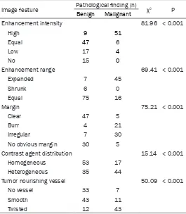
Table 6. Image features of CEUS and pathological findings Image feature Pathological finding (n) χ2 P
Benign Malignant
Enhancement intensity 81.96 < 0.001
High 9 51
Equal 47 6
Low 17 4
No 15 0
Enhancement range 69.41 < 0.001
Expanded 7 45
Shrunk 6 0
Equal 75 16
Margin 75.21 < 0.001
Clear 47 5
Burr 4 21
Irregular 7 30
No obvious margin 30 5
Contrast agent distribution 15.14 < 0.001
Homogeneous 53 17
Heterogeneous 35 44
Tumor nourishing vessel 50.09 < 0.001
No vessel 33 7
Smooth 43 11
Twisted 12 43
CEUS, contrast-enhanced ultrasound.
pathophysiological basis for Do- ppler ultrasound of breast lesions. However, as an important exami-nation method, the color Doppler ultrasound has limitation in dis-playing the intratumoral vessels [10]. As a new technique of ultra-sonic imaging, CEUS has been widely carried out in the abdomen and the heart examination. So- noVue is a pure blood-pool con-trast agent, of which the microbub-ble diameter (2.5 μm) is smaller than the capillary diameter, so it can well reflect the distribution of microvascular network in the lesion [11]. It has provided a new method for detecting micro vascu-lar perfusion status. In the present study, based on the difference in vascular microcirculation of benign and malignant tumors, the CEUS was applied to differentiating the benign and malignant breast le- sions. Results of this study has provided a basis for further appli-cation of CEUS to diagnosis of breast lesions.
enhancement, expanded enlargement range, burr margin, homogeneous contrast agent dis-tribution and tumor nourishing vessels, which were similar to the results of previous study [13]. In 23 lesions, the BI-RADS category was graded from 4A to 3, and the final pathology confirmed as benign. In these 23 cases, the conventional ultrasound presented the
[image:6.612.90.378.72.277.2]suspi-attenuated posterior echo, micro calcification in lesion and grade Adler 1 blood flow. CEUS showed the homogeneous equal enhance-ment, no obvious boundary of enhancement margin, expanded enhancement range, and no tumor vessel. In the other case, the pathologi-cal result indicated the carcinoma muciparum. The conventional ultrasound showed irregular
Figure 3. Double-image synchronous display of benign breast lesion by con-trast-enhanced ultrasound. The right contrast image showed homogeneous low enhancement and clear margin, with non-enlarged lesion area compared with the left two-dimensional image.
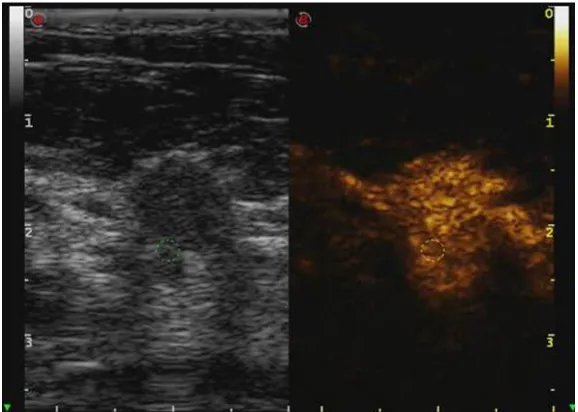
Figure 4. Double-image synchronous display of malignant breast lesion by contrast-enhanced ultrasound. The right contrast image showed heteroge-neous high enhancement and burr margin, with significantly enlarged lesion area compared with the left two-dimensional image.
cious malignant signs such as irregular shape, vertical growth, heterogeneous inter-nal echo and microcalcifica-tion. However, the CEUS sh- owed homogeneous equal or low enhancement, unexpand-ed enhancement range, clear margin and no obvious tumor vessels.
Morphological characteristics of blood vessels in breast cancer are uneven thickness of internal diameter, tortuous expansion of shape and arte-riovenous fistula formation. On the contrary, the blood vessels of the benign breast tumor present regular internal diameter and regular shape. These features are the vascu-lar anatomical basis of CEUS features for benign and ma- lignant breast lesions [14]. Results of the present study prove that, CEUS can accu-rately reflect the different per-fusion modes caused by the difference in internal vascular structure between benign and malignant tumors, thus identi-fying the benign and malig-nant breast lesions.
[image:6.612.89.378.346.552.2]shape, vertical growth, homogeneous low echo, no change of posterior echo, slight calcifica- tion, and grade Adler 1 blood flow. CEUS show- ed homogeneous high enhancement, clear enhancement margin, unexpanded enhance-ment range and access of tumor vessels. It did not contain all the typical malignant features. Above two cases illustrate that, there is the neovascularization in some benign and malig-nant borderline tumors, which is similar to the malignant tumors. In addition, in some malig-nant tumors with special pathological type,
biopsies, thus reducing the psychological and economic burden of patients. The number of lesions with category 3 and below is increased, which can increase the number of patients included in the follow-up observation. There was no significant difference of sensitivity between conventional ultrasound and CEUS, but the specificity and accuracy of CEUS were significantly higher than that of conventional ultrasound. The AUC in CEUS was significantly higher than that of conventional ultrasound. This indicates that, the CEUS has higher diag-Table 7. Sensitivity, specificity and accuracy of conventional
ultra-sound and CEUS
Index Conventional ultrasound CEUS X2 P
Sensitivity 98.4% 100% 1.613 0.204
Specificity 55.7% 81.8% 15.853 < 0.001
Accuracy 73% 89% 8.3171 0.004
[image:7.612.89.377.97.151.2]CEUS, contrast-enhanced ultrasound.
Figure 5. Receiver operating characteristic curves of conventional ultra-sound and CEUS. CEUS, contrast-enhanced ultraultra-sound.
Table 8. Area under curve and test result of conventional ultra-sound and CEUS
Method AUC SE 95% CI Z P
Conventional ultrasound 0.770 0.038 0.695-0.845 5.569 < 0.001
CEUS 0.909 0.025 0.859-0.959
CEUS, contrast-enhanced ultrasound.
[image:7.612.90.374.178.419.2] [image:7.612.92.377.494.536.2]nostic efficiency than conventional ultrasound in differentiating benign and malignant breast lesions.
This study still has some limitations. First of all, due to the insufficient sample size, this study has not investigated the angiographic charac-teristics of benign and malignant lesions, es- pecially the inflammatory lesions, benign le- sions with malignant tendency and malignant lesions with special pathological types. Se- condly, this study only observed the qualitative indexes of lesions. The quantitative indexes including enhancement peak value, time reach-ing peak, extinction time and so on are not included in the research. This needs to be fur-ther investigated. In conclusion, CEUS com-bined with BI-RADS can significantly improve the differentiation ability of breast lesions, and it is worthy of application in clinic.
Disclosure of conflict of interest
None.
Address correspondence to: Dr. Chun Yao, Depart- ment of Ultrasound, Hangzhou Hospital of Traditional Chinese Medicine, 453 Tiyuchang Road, Hangzhou 310007, Zhejiang, China. Tel: +86-571-85827841; E-mail: yaochunhz@126.com
References
[1] Siegel RL, Miller KD, Jemal A. Cancer statistics, 2016. CA Cancer J Clin 2016; 66: 7-30. [2] Chen WQ, Zheng RS. Death and survival of
fe-male breast cancer in China. Chin J Clin Oncol 2015; 42: 668-674.
[3] Siegel R, Naishadham D, Jemal A. Cancer sta-tistics, 2013. CA Cancer J Clin 2013; 63: 11-30.
[4] Zippel DB, Papa MZ. The use of MR imaging guided focused ultrasound in breast cancer patients; a preliminary phase one study and review. Breast Cancer 2005; 12: 32-38. [5] Sickles EA, D’Orsi CJ. How should screening
breast US be audited? The BI-RADS perspec-tive. Radiology 2014; 272: 316-320.
[6] Wan CF, Du J, Fang H, Li FH, Zhu JS, Liu Q. En-hancement patterns and parameters of breast cancers at contrast-enhanced US: correlation with prognostic factors. Radiology 2012; 262: 450-459.
[7] Wang X, Xu P, Wang Y, Grant EG. Contrast-en-hanced ultrasonographic findings of different histopathologic types of breast cancer. Acta Radiol 2011; 52: 248-255.
[8] Wan C, Du J, Fang H, Li F, Wang L. Evaluation of breast lesions by contrast enhanced ultra-sound: qualitative and quantitative analysis. Eur J Radiol 2012; 81: e444-450.
[9] Gasparini G. Clinical significance of the deter-mination of angiogenesis in human breast cancer: update of the biological background and overview of the Vicenza studies. Eur J Can-cer 1996; 32: 2485-2493.
[10] Shirakawa K, Kobayashi H, Sobajima J, Hashi-moto D, Shimizu A, Wakasugi H. Inflammatory breast cancer: vasculogenic mimicry and its hemodynamics of an inflammatory breast can-cer xenograft model. Breast Cancan-cer Res 2003; 5: 136-139.
[11] Dietrich CF. Comments and illustrations re-garding the guidelines and good clinical prac-tice recommendations for contrast-enhanced ultrasound (CEUS)--update 2008. Ultraschall Med 2008; 29: S188-202.
[12] Lee HJ, Kim EK, Kim MJ, Youk JH, Lee JY, Kang DR, Oh KK. Observer variability of breast imag-ing reportimag-ing and data system (BI-RADS) for breast ultrasound. Eur J Radiol 2008; 65: 293-298.
[13] Ricci P, Cantisani V, Ballesio L, Pagliara E, Sal-lusti E, Drudi FM, Trippa F, Calascibetta F, Er-turk SM, Modesti M, Passariello R. Benign and malignant breast lesions: efficacy of real time contrast-enhanced ultrasound vs. magnetic resonance imaging. Ultraschall Med 2007; 28: 57-62.
[14] Mohammed RA, Ellis IO, Mahmmod AM, Hawkes EC, Green AR, Rakha EA, Martin SG. Lymphatic and blood vessels in basal and tri-ple-negative breast cancers: characteristics and prognostic significance. Mod Pathol 2011; 24: 774-785.
[15] Lumachi F, Burelli P, Basso SM, Iacobone M, Ermani M. Usefulness of ultrasound scissors in reducing serous drainage after axillary dissec-tion for breast cancer: a prospective random-ized clinical study. Am Surg 2004; 70: 80-84. [16] Gal-Gombos EC, Esserman LE, Odzer SL,