Original Article
Efficacy of anterior cervical discectomy and fusion
versus artificial cervical disc replacement
for cervical degenerative disease
Bihua Lai, Jianbin Wu, Zhaowen Gao, Hong Ye
Department of Orthopaedics, The First Hospital of Nanping City Affiliated to Fujian Medical University, Nanping, Fujian Province, China
Received March 14, 2018; Accepted April 25, 2018; Epub July 15, 2018; Published July 30, 2018
Abstract: Objective: To compare the clinical efficacy of anterior cervical discectomy and fusion (ACDF) and artifi-cial cervical disc replacement (ACDR) in the management of cervical degenerative disease in patients. Methods: Seventy-nine patients with cervical degenerative disease admitted to The First Hospital of Nanping City Affiliated to Fujian Medical University from January 2012 to December 2014 were assigned to undergo ACDF or ACDR and followed for 3 years. In the ACDF group, 24 patients were male and 19 were female, with an age range of 43 to 55 years (mean, 48.9±1.6 years). All the patients had 36 months of follow-up. In the ACDR group, 21 patients were male and 15 were female and they varied in age from 42 to 57 years (mean, 49.1±1.4 years) and were followed up for 36 months. There were no remarkable differences between the two groups in basic data, as well as imaging data at baseline. The Japanese Orthopaedic Association (JOA) score, the neck disability index (NDI), the visual analogue scale (VAS) for pain in the neck and upper-limb, the Odom scale score, the cervical spine range of motion (ROM) and the cervical curvature index were employed to assess the clinical efficacy of the surgeries in the patients. Results: Greater improvements in the JOA, VAS, and NDI scores after surgery were noted than those before surgery in the two groups (P<0.05), but the corresponding scores were insignificantly different between the two groups (P>0.05). At the final follow-up after surgery, the excellent and good rate (83.72%) of the Odom scores in the ACDF group was insignificantly different from that (88.89%) in the ACDR group (P>0.05). The surgical segments in the ACDF group were fully fused, but the ROM of the surgical segments in the ACDR group was 7.4±3.9, which was mildly different from than before surgery. The cervical spine ROM in the ACDF group was remarkably smaller than that of the ACDR group (P<0.05), and that of the ROM group before surgery (P<0.05). However, the ROM of the cervical spine in the ACDR group at final follow-up after surgery differed insignificantly from that before surgery (P>0.05). The values for the cervical curvature index were strikingly lower in the ACDF group than in ACDR group at 3, 12, 24, and 36 months after surgery. The cervical curvature index in the ACDF group at 36 months was remarkably lower than that before surgery (P<0.05), but the cervical curvature index in the ACDR group was insignificantly different from that before surgery (P>0.05). Conclusion: Among the patients with cervical degenerative disease, the clinical efficacy of ACDR was similar to that of ACDF, but ACDR was superior to ACDF in maintaining the ROM and physiological curvature of the cervical spine.
Keywords: Anterior cervical discectomy and fusion, artificial cervical disc replacement, cervical degenerative dis-ease, efficacy comparison
Introduction
With the rapid development of society, increas-ingly people spend longer times bending over their desks to work or bowing their heads to play mobile phone games, cervical degenera-tive disease is more prevalent and has become a common and frequently-occurring clinical dis-ease [1]. The special structure and the great
recognized that degeneration of the cervical spine contributed to spinal stenosis and spinal cord compression. Since anterior cervical dis-cectomy and fusion (ACDF) was introduced in the 1950s, this technology has been extensive-ly applied in the clinical treatment of cervical degenerative disease, and has also known as the gold standard [3]. Although ACDF has great-ly improved the clinical symptoms, the long-term follow-up results demonstrate the loss of normal ROM of the cervical spine in the patients after the surgery. The consequence of adjacent segment degeneration was reported by the scholars worldwide [4, 5]. In this case, anterior cervical non-fusion, namely, artificial cervical disc replacement (ACDR) was developed and has been used in clinical practice [6]. The big-gest advantages of ACDR are maximum reten-tion of ROM of the surgical segments and little influence on intervertebral space. As a result, the postoperative kinematic characteristics of the entire cervical spine are close to the preop-erative physiological profile. Additionally, ACDR is associated with reduced postoperative sur- gical segment fusion which otherwise might result in dysfunction of the segment, central-ized stress and excessive motion of adjacent segments. ACDR does not increase the stress of adjacent segments, instead it is effective in preventing the complication of accelerated degeneration due to stress changes in adja-cent segments [7]. Nevertheless, few studies are focused on follow-ups regarding the safety and efficacy of ACDR [8].
Therefore, in the current study, we made 3 years of follow-up with the patients and com-pared the efficacy and safety of the ACDF and ACDR, in hope of providing potent evidence for the planning of the protocols in treatment of cervical degenerative disease.
Materials and methods
Patients
Between January 2012 and December 2014, a total of 79 patients with cervical degenera-tive disease admitted to the Department of Orthopedics in The First Hospital of Nanping City Affiliated to Fujian Medical University were recruited in this study. Among the 79 patients, 45 were male and 34 were female, with an age ranging from 42 to 57 years (mean, 49.2±1.7 years). Forty-two patients had cervical
lotic myelopathy while 37 had cervical spondy-lotic radiculopathy. Patients were eligible for enrollment if they had cervical spondylotic myelopathy or radiculopathy, previous 6-week ineffective standard care before enrollment, spinal stenosis, anterior neural compression suitable for anterior approach, and provided written informed consent. Patients were ineli-gible for enrollment if they had undergone sur-geries for cervical degenerative disease, had inflammation, deformity, or tumor in the cervi-cal vertebrae, were required to have other con-comitant surgeries, complicated with acute cervical trauma, severe osteoporosis, cervical instability, or associated with ossification of the posterior longitudinal ligaments. Patients were also excluded if they had severe disease involv-ing in the heart, liver, or kidney, or they were unsuitable for treatment by ACDF or ACDR. This study was approved by Ethics Committee Hospital.
Methods
The enrolled patients underwent the surgeries under general anesthesia. Placed in a supine position and with a thin pillow under the shoul-der, each patient stretched out the neck back-ward. The operative part of the patient was fully exposed by the standard Smith-Robinson method [9]. All the patients were managed by the method. After routine sterilization and drap-ing, a transverse incision was made on the right side of the neck. After the skin incision, subcu-taneous tissue and platysma were dissected layer by layer till the junction between the inter-nal jugular sheath and the carotid sheath, where the blunt dissection was used to dissect till the prevertebral fascia, allowing the lesion segment fully exposed. Subsequently, the patients in the two groups were treated with the following different procedures.
this segment was completely relieved. The pro-cessed intervertebral space was implanted with Syncage-C Cage (Synthes, Sweden) and bone fragments, and then compressed to real-ize a chimeric fixation. After the completion of the surgery, the surgical site was visualized under radiography (Toshiba TOSHIBA, Japan). When the surgery was confirmed as satisfacto-ry, appropriate titanium end-plates and screws were used for fixation. After that, suturing was performed. The patients were required to wear neck support for a month after surgery and allowed to ambulate 2 days after surgery.
ACDR procedure: Under the guidance of radiog-raphy, the diseased intervertebral disc and its surrounding diseased tissues were removed. After distraction of the involved intervertebral space, the midline was found out for channel grinding. The center was ground after localiza-tion. A disc-shaped grinding drill was utilized to grind and dissect the posterior border of the vertebral level, whereas a column-shaped grinding drill was used to grind and dissect the superior and inferior end-plates. The residual osteophytes and ligaments were completely scraped with a curette, and the bilateral nerve root canals were open wider. The osteophytes at the Luschka’s joint were completely scraped off with the curette. After full relief of the com-pression, hemostasis and washing were con-ducted, followed by implantation of artificial intervertebral discs of the right size. After com-pletion of the surgery, the surgical site was visualized under the radiography. When the sur-gery was confirmed as satisfactory, suturing was performed. The patients were required to wear neck support for 2 weeks after surgery and allowed to ambulate 2 days after the surgery.
Clinical efficacy assessment
The clinical efficacy of the surgery in the patients was evaluated using the Japanese Orthopaedic Association (JOA) score, the neck disability index (NDI), the visual analogue scale (VAS) score for pain in the neck and upper-limb, as well as the Odom scale score [10-13]. The JOA scores were assessed with the 17-point scoring criteria [14].
The NDI score consists of 10 items (each hav-ing six 0-5-point scorhav-ing criteria) and ranges
from 0 to 50, with higher scores indicating more severe dysfunction or disability of perfor-mance status in patients.
On the VAS scale, 0 point indicates no pain; 1-3 indicates mild pain; 4-6 indicates tolerable pain affecting sleep; 7-10 indicates unbearable pain.
On the Odom scale, excellent efficacy indica- tes the clinical symptoms disappear, and the patient can work normally. Good efficacy indi-cates most of the clinical symptoms disappear, and the patient can go on working. Fair efficacy indicates the symptoms are relieved, but the patient cannot work normally, whereas poor efficacy indicates the symptoms are not relieved, and they affect the patient’s normal work. The formula for calculation of the excel-lent and good rate of the Odom score in pati- ents was as follows: Excellent and good rate = (Excellent + Good)/Total number of patients * 100%.
The values for the Cobb angles of the patients were measured according to the data from the imaging system in The First Hospital of Nanping City Affiliated to Fujian Medical University. Over the whole range of overextension to over-flex-ion, the Cobb angle values of the C2-C7 seg-ments on the radiographs were measured to evaluate the ROM of the entire cervical spine. The overall curvature of the cervical spine was defined as the intersection angle formed by the inferior border of C2 segment and the inferior border of C7 segment, whereas the ROM of the entire cervical spine was defined as the chang-es in the overall cervical curvature in the preop-erative and postoppreop-erative overextension to over-flexion of cervical spine on the radiographs [15, 16]. The measurements were utilized to evaluate the changes in the normal physiologi-cal functions of the cerviphysiologi-cal spine.
Statistical analysis
Fisher exact probability tests. Rank sum tests were employed for intergroup comparisons of class variables and the paired rank sum tests for intragroup comparisons. P<0.05 was deemed as significantly different.
[image:4.612.92.339.85.388.2]insignificantly different between the two groups at the same time points (P>0.05), but the scores at 36 months in the two groups differed remarkably from those before surgery (P<0.05,
Table 2).
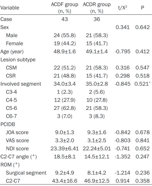
Table 1. Basic and clinical characteristics of the patients Variable ACDF group (n, %) ACDR group (n, %) t/X2 P
Case 43 36
Sex 0.341 0.642
Male 24 (55.8) 21 (58.3) Female 19 (44.2) 15 (41.7)
Age (year) 48.9±1.6 49.1±1.4 -0.795 0.412 Lesion subtype
CSM 22 (51.2) 21 (58.3) 0.316 0.547 CSR 21 (48.8) 15 (41.7) 0.298 0.518 Involved segment 34.0±3.4 35.0±2.8 -0.845 0.521*
C3-4 1 (2.3) 2 (5.6)
C4-5 12 (27.9) 10 (27.8) C5-6 27 (62.8) 21 (58.3)
C6-7 3 (7.0) 3 (8.3)
PCIDB
JOA score 9.0±1.3 9.3±1.6 -0.842 0.678 VAS score 3.3±2.0 3.1±2.5 -0.803 0.841 NDI score 23.39±6.41 22.24±5.01 -0.741 0.652 C2-C7 angle (°) 18.5±8.1 14.5±12.1 -1.352 0.247 ROM (°)
Surgical segment 9.2±4.9 8.1±4.2 -1.214 0.236 C2-C7 43.4±16.6 46.9±12.5 0.914 0.358
[image:4.612.91.338.495.613.2]Note: *Fisher precision probability test. ACDF denotes anterior cervical discectomy and fusion, ACDR artificial cervical disc replacement, CSM cervical spondylotic myelopathy, CSR cervical spondylotic radiculopathy, JOA Japanese Orthopaedic Association, VAS visual analogue scale, NDI neck disability index, PCIDB preoperative clinical and imaging data at baseline and ROM range of motion.
Table 2. JOA scores of the patients before and after sur-gery
Time ACDF group (n=43) ACDR group (n=36) t P Pre-surgery 9.3±1.6 9.0±1.3 -0.842 0.678 3 mon after surgery 14.9±1.1 15.6±1.7 0.985 0.741
12 mon 14.9±1.8 14.8±1.4 0.841 0.514
24 mon 15.3±1.2 14.4±1.7 0.548 0.301 36 mon 15.4±1.4 14.3±2.0 0.558 0.324
t 2.354 2.187
P 0.024* 0.013*
Note: *Comparison of the JOA scores before and 36 mon after surgery. ACDF denotes anterior cervical discectomy and fusion, ACDR artificial cervical disc replacement, and JOA Japanese Orthopaedic Association.
Results
Basic and clinical characteristics of the patients
A total of 79 patients were recruited in this study. Among them, 43 patients were assigned to receive ACDF (ACDF group). There were 24 males and 19 females with an age of 43-55 years (mean, 48.9±1.6 years). Cervical spon-dylotic myelopathy occurred in 22 patients and cervical spondylotic ra- diculopathy in 21 patients. One patient had the surgical segment C3-4, 12 had the surgical segment C4-5, 27 had the surgical segment C5-6, 3 had the surgical segment C6-7; all the patients had 36 months of follow-up. The remaining 36 patients were as- signed to undergo ACDR (ACDR group); 21 patients were male and 15 were female, with an age of 42-57 years (mean, 49.1±1.4 years). Cervical spon-dylotic myelopathy occurred in 20 patients and cervical spondylotic ra- diculopathy in 16 patients whereas 2 patients had the surgical segment C3-4, 10 had the surgical segment C4-5, 21 had the surgical segment C5-6, and 3 had the surgical segment C6-7. All the patients had 36 months of follow-up. The patients in the two groups were generally well-balanced in basic characteristics, as well as preop-erative clinical and imaging data (P>0.05, Table 1).
JOA scores of the patients
VAS scores of the patients
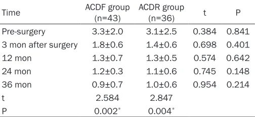
The preoperative VAS scores of the patients were high in both groups, and the results of the 3 years of follow-up after treatment indicated that the VAS scores at 3, 12, 24, and 36 months after surgery were 1.8±0.6, 1.3±0.7, 1.2±0.3, and 0.9±0.7 in the ACDF group, and 1.4±0.6, 1.3±0.5, 1.1±0.6, and 1.0±0.6 in the ACDR group. The VAS scores were insignificantly
dif-segments differed insignificantly from that before surgery in the ACDR group (P>0.05). There were insignificant differences in the ROM of cervical spine between the two groups before surgery (P>0.05). The ROM of cervical spine declined substantially in the ACDF group at the final follow-up, and was remarkably dif-ferent from that in the ACDR group at the final follow-up, and that before surgery (P<0.05,
[image:5.612.91.343.97.213.2]Table 6).
Table 3. VAS scores of the patients before and after sur-gery
Time ACDF group (n=43) ACDR group (n=36) t P Pre-surgery 3.3±2.0 3.1±2.5 0.384 0.841 3 mon after surgery 1.8±0.6 1.4±0.6 0.698 0.401
12 mon 1.3±0.7 1.3±0.5 0.574 0.642
24 mon 1.2±0.3 1.1±0.6 0.745 0.148
36 mon 0.9±0.7 1.0±0.6 0.954 0.214
t 2.584 2.847
P 0.002* 0.004*
[image:5.612.91.341.294.412.2]Note: *Comparison of the VAS scores before and 36 mon after surgery. ACDF denotes anterior cervical discectomy and fusion, ACDR artificial cervical disc replacement, and VAS visual analogue scale.
Table 4. NDI scores of the patients before and after sur-gery
Time ACDF group (n=43) ACDR group (n=36) t P Pre-surgery 23.39±6.41 22.24±5.01 0.598 0.652 3 mon after surgery 6.81±2.71 5.51±1.72 0.987 0.124 12 mon 5.01±2.03 4.51±1.76 0.687 0.541 24 mon 3.54±1.14 3.12±1.24 0.574 0.648 36 mon 3.12±1.12 2.71±0.91 0.848 0.224
t 3.187 3.014
P <0.001* <0.001*
Note: *Comparison of the NDI scores before and 36 mon after surgery. ACDF denotes anterior cervical discectomy and fusion, ACDR artificial cervical disc replacement, and NDI neck disability index.
ferent between the two groups at the same time points (P>0.05), but the scores at 36 month in both groups dif-fered strikingly from those before sur-gery (P<0.05, Table 3).
NDI scores of the patients
The preoperative NDI scores of the patients were higher than 20 points in both groups, and the results of the 3 years of follow-up after surgery showed that the NDI scores at 3, 12, 24, and 36 months after surgery we- re 6.81±2.71, 5.01±2.03, 3.54±1.14, and 3.12±1.12 in the ACDF group, and 5.51±1.72, 4.51±1.76, 3.12± 1.24, and 2.71±0.91 in the ACDR group. The JOA scores differed insig- nificantly between the two groups at the same time points (P>0.05), but the scores at 36 months in the two groups differed significantly from th- ose before surgery (P<0.05, Table 4).
Odom scores of the patients
[image:5.612.91.342.490.558.2]According to the Odom scores, insig-nificant disparities were noted be- tween the excellent and good rate (83.72%) of the ACDF group and that (88.89%) of the ACDR group at the final follow-up of surgery (P>0.05,
Table 5).
ROM of surgical segments and cervi-cal spine of the patients
The ROM of surgical segments be- fore surgery was different insignifi-cantly between the two groups (P> 0.05). The surgical segments of the patients in the ACDF group achieved full fusion at the final follow-up after surgery. The ROM (7.4±3.9) of surgical
Table 5. Odom scores of the patients at the final follow-up after surgery (n, %)
Odom score Excellent Good Fair Poor
ACDF group (n=43) 27 (62.79) 9 (20.93) 7 (16.28) 0 (0.00) ACDR group (n=36) 21 (58.33) 11 (30.56) 4 (11.11) 0 (0.00)
Z -0.158
P 0.873
Cervical curvature index of the patients
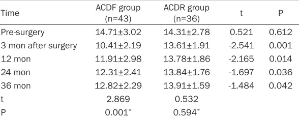
The patients in the two groups differed insigni-ficantly in the values for the cervical curvature index before surgery (P>0.05), whereas the val-ues of the cervical curvature index at 3, 12, 24, and 36 months after surgery were remarkably lower in the ACDF group than in the ACDR group (all P<0.05). Moreover, the value at 36 months in the ACDF group was considerably different from that before surgery (P<0.05, Table 7).
Discussion
It has been half a century since ACDF was applied to treat cervical degenerative disease. Multiple clinical studies demonstrate that ACDF is effective in relief of the clinical symptoms, improvement of the neurologic functions, and enhancement of the stability of the cervical spine. ACDF has been extensively used in clini-cal practice, but it has the disadvantages of constraining the ROM of the surgical segment and contributing to the degeneration of adja-cent segments [4, 5, 17]. Given the drawbacks of the conventional technique, ACDR came into
[image:6.612.91.387.86.179.2]cantly between the two groups. The VAS and NDI scores in both groups decreased consider-ably after surgery. The scores at 36 months after surgery were different from those before surgery in the same group, but differed insigni-ficantly between the two groups. The excellent and good rate of the Odom scores at the final follow-up was 83.72% in the ACDF group and 77.78% in the ACDR group, so they were differ-ent insignificantly. As a result, the patidiffer-ents in the two groups were largely similar in relief of clinical symptoms. Earlier studies revealed that ACDR was superior to ACDF in the JOA scores, VAS scores, NDI scores, the ROM of the cervical spine, as well as adverse events [18, 19]. In a study with a long-term follow-up, the VAS and NDI scores of patients increased strikingly after ACDR and the rate of reoperation in adjacent segments was 21%, so the researchers argued that ACDR was safe and effective [20]. In our current study, the VAS and NDI scores were improved remarkably at 3 months after surgery in the two groups. Though greater improve-ments were observed in the ACDR group, the difference was insignificant (P>0.05). The VAS
Table 6. ROM of surgical segments and cervical spine of the patients
Variable Case
ROM (°)
Surgical segment Cervical spine Pre-surgery Final follow-up Pre-surgery Final follow-up ACDF group 43 9.2±4.9 0 43.4±16.6 29.1±9.2* ACDR group 36 8.1±4.2 7.4±3.9 46.9±12.5 39.1±14.9
t 0.941 2.957 0.863 2.456
P 0.236 0.001 0.358 0.032
Note: *Compared with that before surgery (P<0.01). ACDF denotes anterior cervical discectomy and fusion, ACDR artificial cervical disc replacement, and ROM range of motion.
Table 7. Cervical curvature index of the patients
Time ACDF group (n=43) ACDR group (n=36) t P Pre-surgery 14.71±3.02 14.31±2.78 0.521 0.612 3 mon after surgery 10.41±2.19 13.61±1.91 -2.541 0.001
12 mon 11.91±2.98 13.78±1.86 -2.165 0.014
24 mon 12.31±2.41 13.84±1.76 -1.697 0.036
36 mon 12.82±2.29 13.91±1.59 -1.484 0.042
t 2.869 0.532
P 0.001* 0.594*
Note: *Comparison of the values for cervical curvature index before and 36 mon after surgery. ACDF denotes anterior cervical discectomy and fusion, ACDR artificial cervical disc replacement.
being accordingly. ACDR is characterized by mainte-nance of the surgical seg-ment mobility and normal-ization of the kinematics and mechanics of adjacent segments. Hence, it can not only guarantee the sta-bility of surgical segments and recovery of the cervical curvature, but also enable the surgical segments to recover the normal ROM after surgery. In this man-ner, ACDR broke out the status quo of static fixation and decompression after ACDF in patients [6].
[image:6.612.92.386.246.362.2]insignifi-and NDI scores of both groups were improved substantially over time as compared with those before surgery. Such non-synchronization was also reflected in the excellent and good rates of the Odom scores. The postoperative excellent and good rate in the ACDR group was higher than that of the ACDF group, though insignifi-cantly (P>0.05). This suggests that the fact that the patients in the ACDR group could alleviate the clinical symptoms in the early stage and continue to treat for 3 years might be related to the shorter duration of neck support use and earlier exercise of cervical spine among the patients.
When it comes to the imaging improvements, fusion and smaller ROM of the surgical seg-ments, and greater ROM of adjacent segments were found among the patients after ACDF [21]. In long-term follow-up, degeneration of adja-cent segments in different degrees, disappear-ance of physiological curvature, and backward extrusion of cervical spine were noted in the patients with ACDF [22]. By contrast, in numer-ous follow-ups, fewer events of adjacent seg-ment degeneration were observed in the pa- tients with ACDR, and the clinical efficacy of ACDR was basically similar to that of ACDF [23-27]. In our current study, we found that at the final follow-up, the surgical segments were completely fused in patients of the ACDF group, whereas the ROM (7.4±3.9) of the surgical seg-ments in the ACDR group differed insignificant-ly from that of the same group before surgery (P>0.05). Additionally, the ROM of the surgical segments in the ACDF group at the final follow-up dropped more strikingly when compared with those of the ACDR group both at the final follow-up and before surgery. The cervical cur-vature index after surgery was remarkably lower in the ACDF group than in the ACDR group, which was consistent with the results reported by Kim [28]. Moreover, according to a previous report of long-term follow-up, the improvements in the surgical segment, the adjacent segments, and the cervical curvature index in the ACDR group were greater than those in the ACDF group.
There are the following limitations in this stu- dy: no randomization was conducted to the patients in the two groups, and the sample size was small. As for the selection of surgeries, the patients should be explained in details accord-ing to their conditions. The final choice was at
the discretion of the patients, hence there was a bias of selection.
In summary, for the patients with cervical degenerative disease, ACDR was similar to ACDF in clinical efficacy but superior to ACDF in maintaining the ROM and physiological curva-ture of the cervical spine. It was safe and reli-able during follow-up.
Disclosure of conflict of interest
None.
Address correspondence to: Jianbin Wu, Depart- ment of Orthopaedics, The First Hospital of Nanping City Affiliated to Fujian Medical University, No. 317 Zhongshan Road, Yanping District, Nanping 353001, Fujian Province, China. Tel: +86-0599-8612625; E-mail: wujianbin281h@163.com
References
[1] Webb R, Brammah T, Lunt M, Urwin M, Allison T and Symmons D. Prevalence and predictors of intense, chronic, and disabling neck and back pain in the UK general population. Spine (Phila Pa 1976) 2003; 28: 1195-1202. [2] Karadimas SK, Gatzounis G and Fehlings MG.
Pathobiology of cervical spondylotic myelopa-thy. Eur Spine J 2015; 24 Suppl 2: 132-138. [3] Hibbs RA. An operation for progressive spinal
deformities: a preliminary report of three cas-es from the service of the orthopaedic hospi-tal. 1911. Clin Orthop Relat Res 2007; 460: 17-20.
[4] Luo J, Gong M, Huang S, Yu T and Zou X. Incidence of adjacent segment degeneration in cervical disc arthroplasty versus anterior cervical decompression and fusion meta-anal-ysis of prospective studies. Arch Orthop Trauma Surg 2015; 135: 155-160.
[5] Burkus JK, Traynelis VC, Haid RW Jr and Mummaneni PV. Clinical and radiographic analysis of an artificial cervical disc: 7-year follow-up from the Prestige prospective ran-domized controlled clinical trial: clinical article. J Neurosurg Spine 2014; 21: 516-528. [6] Hyun Oh C and Hwan Yoon S. Past, present,
and future of cervical arthroplasty. Keio J Med 2013; 62: 47-52.
[7] Ghogawala Z, Benzel EC, Riew KD, Bisson EF and Heary RF. Surgery vs conservative care for cervical spondylotic myelopathy: surgery is ap-propriate for progressive myelopathy. Neuro- surgery 2015; 62 Suppl 1: 56-61.
cervical spondylosis: more heterotopic ossifi-cation at 3 years of follow-up. Spine (Phila Pa 1976) 2012; 37: E1251-1259.
[9] Robinson RA and Smith GW. Anterolateral vical disc removal and interbody fusion for cer-vical disc syndrome. Sas Journal 2010; 4: 34-35.
[10] Fukui M, Chiba K, Kawakami M, Kikuchi SI, Konno SI, Miyamoto M, Seichi A, Shimamura T, Shirado O and Taguchi T. An outcome measure for patients with cervical myelopathy: Japanese Orthopaedic Association Cervical Myelopathy Evaluation Questionnaire (JOACMEQ): Part 1. J Orthop Sci 2007; 12: 227-240.
[11] Wu S, Ma C, Mai M and Li G. Translation and validation study of Chinese versions of the neck disability index and the neck pain and disability scale. Spine (Phila Pa 1976) 2010; 35: 1575-1579.
[12] Samartzis D, Shen FH, Matthews DK, Yoon ST, Goldberg EJ and An HS. Comparison of al-lograft to autograft in multilevelanterior cervi-cal discectomy and fusion with rigid plate fixa-tion. Spine J 2003; 3: 451-459.
[13] Robertson JT, Papadopoulos SM and Traynelis VC. Assessment of adjacent-segment disease in patients treated with cervical fusion or arthroplasty: a prospective 2-year study. J Neurosurg Spine 2005; 3: 417.
[14] Hirabayashi K, Miyakawa J, Satomi K, Maruyama T and Wakano K. Operative results and postoperative progression of ossification among patients with ossification of cervical posterior longitudinal ligament. Spine (Phila Pa 1976) 1981; 6: 354-364.
[15] Park MS, Kelly MP, Min WK, Rahman RK and Riew KD. Surgical treatment of C3 and C4 cer-vical radiculopathies. Spine (Phila Pa 1976) 2013; 38: 112-118.
[16] Kimura H, Shikata J, Odate S and Soeda T. Anterior corpectomy and fusion to C2 for cervi-cal myelopathy: clinicervi-cal results and complica-tions. Eur Spine J 2014; 23: 1491-1501. [17] Fernandez-Fairen M, Sala P, Dufoo M, Jr.,
Ballester J, Murcia A and Merzthal L. Anterior cervical fusion with tantalum implant: a pro-spective randomized controlled study. Spine (Phila Pa 1976) 2008; 33: 465-472.
[18] Cheng L, Nie L, Zhang L and Hou Y. Fusion ver-sus Bryan cervical disc in two-level cervical disc disease: a prospective, randomised study. Int Orthop 2009; 33: 1347-1351.
[19] Zhang Y, Liang C, Tao Y, Zhou X, Li H, Li F and Chen Q. Cervical total disc replacement is su-perior to anterior cervical decompression and fusion: a meta-analysis of prospective random-ized controlled trials. PLoS One 2015; 10: e0117826.
[20] Malham GM, Parker RM, Ellis NJ, Chan PG and Varma D. Cervical artificial disc replacement with ProDisc-C: clinical and radiographic out-comes with long-term follow-up. J Clin Neurosci 2014; 21: 949-953.
[21] Delamarter RB and Zigler J. Five-year reopera-tion rates, cervical total disc replacement ver-sus fusion, results of a prospective random-ized clinical trial. Spine (Phila Pa 1976) 2013; 38: 711-717.
[22] Goffin J, Geusens E, Vantomme N, Quintens E, Waerzeggers Y, Depreitere B, Van Calenbergh F and van Loon J. Long-term follow-up after in-terbody fusion of the cervical spine. J Spinal Disord Tech 2004; 17: 79-85.
[23] Blumenthal SL, Ohnmeiss DD, Guyer RD and Zigler JE. Reoperations in cervical total disc re-placement compared with anterior cervical fu-sion: results compiled from multiple prospec-tive food and drug administration investiga-tional device exemption trials conducted at a single site. Spine (Phila Pa 1976) 2013; 38: 1177-1182.
[24] Luo J, Gong M, Huang S, Yu T and Zou X. Incidence of adjacent segment degeneration in cervical disc arthroplasty versus anterior cervical decompression and fusion meta- analysis of prospective studies. Arch Orthop Trauma Surg 2015; 135: 155-160.
[25] Rao MJ, Nie SP, Xiao BW, Zhang GH, Gan XR and Cao SS. Cervical disc arthroplasty versus anterior cervical discectomy and fusion for treatment of symptomatic cervical disc dis-ease: a meta-analysis of randomized con-trolled trials. Arch Orthop Trauma Surg 2015; 135: 19-28.
[26] Burkus JK, Traynelis VC, Haid RW, Jr. and Mummaneni PV. Clinical and radiographic analysis of an artificial cervical disc: 7-year follow-up from the Prestige prospective ran-domized controlled clinical trial: clinical article. J Neurosurg Spine 2014; 21: 516-528. [27] Hisey MS, Bae HW, Davis RJ, Gaede S, Hoffman
G, Kim KD, Nunley PD, Peterson D, Rashbaum RF, Stokes J and Ohnmeiss DD. Prospective, randomized comparison of cervical total disk replacement versus anterior cervical fusion: results at 48 months follow-up. J Spinal Disord Tech 2015; 28: E237-243.