Original Article
Correlations of
SP-D
genetic polymorphisms and protein
expression with the pathogenesis and severity of
respiratory distress syndrome in preterm infants
Li-Qun Sun1, Gong-Liang Guo2, Lei Zhang2, Jin-Yi Wu2, Hai-Bo Li1, Chun-Juan Zhang3, Hui Tang4
1Outpatient Department of Pediatrics, The First Hospital of Jilin University, Changchun 130021, P. R. China; 2Department of Cardiology, China Japan Union Hospital, Jilin University, Changchun 130033, P. R. China; 3Department of Pediatrics, Linyi People’s Hospital, Linyi 276000, P. R. China; 4Department of Pediatrics,
Traditional Chinese Medicine Hospital of Pingyi County, Linyi 273300, P. R. China
Received April 8, 2016; Accepted September 29, 2016; Epub January 15, 2017; Published January 30, 2017
Abstract: Objective: This study is designed to explore the correlations of SP-D genetic polymorphisms and protein expression with the pathogenesis and severity of respiratory distress syndrome (RDS) in preterm infants. Methods: From January 2013 to January 2015, a total of 170 RDS preterm infants were chosen as the case group, and 204 healthy infants were selected as the control group. Polymerase chain reaction restriction fragment length polymorphism (PCR-RFLP) was used to detect the frequency distributions of rs721917 C>T and rs2243639 A>G polymorphisms in the SP-D gene. Enzyme-linked immune sorbent assay (ELASA) was used to measure the serum expressions of SP-D. Results: The frequency distributions of CC, CT and TT genotypes of rs721917 were significantly different in the case and control groups. Compared with the control group, the risk of RDS for the patients carrying the CT genotype was 2.071 times higher than that for the patients carrying the CC genotype. However, there were no statistical differences in genotype frequencies of rs2243639 between the case and control groups. Genotypes and frequency distributions of rs721917 in SP-D were different in the grading of RDS in the case group. Compared with the control group, the expression of SP-D was higher in the case group. As for rs721917 the expression of SP-D
with TT genotypes was higher than that with CC and CT genotypes. The logistic regression analysis indicated that the TT genotype in rs721917, pregnancy-induced hypertension, abnormal umbilical cord and placental abnormali-ties were the risk factors of death in preterm infants with RDS (all P<0.05). Conclusions: These findings reveal that
SP-D genetic polymorphisms and protein expression may be associated with the pathogenesis and severity of RDS in preterm infants.
Keywords: Respiratory distress syndrome, SP-D, polymorphism, preterm infants, pathogenesis, severity
Introduction
Neonatal respiratory distress syndrome (NRDS)
is a condition of pulmonary insufficiency that
initiates at or shortly after birth and increases
in severity over the first 2 days of life in its natu -ral course [1]. Evidence demonstrated that NRDS was the most common cause of
neona-tal morneona-tality in the first year after birth in the
United States [2]. It was estimated that 32% of neonatal respiratory failure was ascribed to
NRDS in China, ranking first among relevant
causes [3]. Clinically, RDS has early respiratory distress comprising cyanosis, retractions, grunting and tachypnea [1]. RDS occurs in near-ly a half of preterm infants born at less than 30
weeks of gestation [4]. Preterm birth is defined
as birth at less than 37 week of gestation and considered the most prominent cause of neo-natal morbidity and mortality in developed countries [5]. EuroNeoStat data reported an incidence of 52% at 30~31 weeks’ gestation, 74% at 28~29 weeks’, 88% at 26~27 weeks’, and 91% at 23-25 weeks’ [6]. Also, it has been demonstrated that NRDS was associated with
a deficiency in pulmonary surfactant (PS) [7, 8].
alveo-lar collapse at the end of expiration [9, 10]. SP-D, an important member of the collectin family in regulating innate immunity of the lung, is composed of monomers (43 kDa) and syn-thesized in type II pneumocytes and Clara cells [11, 12]. Human SP-D protein is encoded by the SFTPD gene and located on chromosome 10q22.2-23.1, which is 43 kDa in length and contains eight exons and seven introns [13]. It has been reported that SP-D plays an
impor-tant role in mitigating pulmonary inflammation
and infection and is also involved in the regula-tion of pulmonary surface proteins [14]. In addi-tion, SP-D gene is related to many pulmonary diseases, such as chronic obstructive pulmo-nary disease and bronchopulmopulmo-nary dysplasia [15, 16]. In view of the role of SP-D in maintain-ing the function of lung tissue, it can be a can-didate gene for the study of neonatal lung dis-ease. In this study, we explored the correlations of SP-D genetic polymorphisms and protein expression with the pathogenesis and severity of RDS in preterm infants.
Materials and methods
Study subjects
From January 2013 to January 2015, a total of 170 hospitalized RDS preterm infants were chosen as the case group. Among them, 110 were male, 60 were female, the mean gesta-tional age was (32.04 + 2.36) W and the mean weight was (1.83 + 0.48) k. The diagnosis was in accordance with European consensus guide-lines on the management of neonatal respira-tory distress syndrome (NRDS) in preterm
infants [1], specifically, (1) the cases began pro -gressive dyspnea within 12 h after birth; (2) PaO2<50 mmHg in room air, central cyanosis in room air, a requirement for supplemental oxy-gen to maintain PaO2>50 mmHg; (3) chest X-ray
indicated the specific performances like
ground-glass opacity, air bronchogram and “white lung”. The RDS related risk factors like maternal age, history of pregnancy (pregnancy induced hypertension or diabetes, etc.); deliv-ery mode; premature rupture of membranes; time of birth; placental abnormalities (placental abruption, placental previa) and abnormal umbilical cord (abnormality of umbilical cord or around neck). Another 204 preterm infants born during the same period with similar gesta-tional age and birth weight and with no obvious
symptoms of infection were recruited as the control group. Inclusion criteria for the control group: (1) chest X-ray indicated no signs of NRDS or pulmonary infection; blood routine and C-reaction protein (CRP) indicated no clear signs of infection. Exclusion criteria for both the case and control group: (1) the patients with severe congenital disease, such as complex congenital heart disease, diaphragmatic her-nia, cerebral dysplasia; (2) the patients with genetic metabolic diseases, such as phenylke-tonuria, congenital hypothyroidism and diabe-tes; (3) the patients with a clear infection in the late pregnancy and the IgM of the neonatal cases increased. The research was in accor-dance with the ethical standards and was approved by the Ethics Committee of The First Hospital of Jilin University; all of the parents or guardians of the study objects were informed consent.
Classification of frontal chest X-ray
According to the normal chest X-ray classifica -tion, the case group fell into four grades. (1) RDS grade I (n = 54): the cardiac silhouette was normal; the transparency of the lung decreased or there was diffuse granular or reticular
shad-ow in the field of bilateral pulmonary. (2) RDS
grade II (n = 58): the transparency of the lung decreased; diffuse granular shadow appeared; patchy shadow in high density was seen in
par-tial lung field; texture of double lung was fuzzy
and undistinguishable; heart border and part of the surface of diaphragm were fuzzy; air bron-chogram was obvious. (3) RDS grade III (n = 32): the transparency of the lung decreased
significantly; large particle shadow overlapped
appeared in the lung; air bronchogram was widely observed; lung texture completely disap-peared; heart and diaphragmatic surface were not clear. (4) RDS grade IV (n = 26): uniform and
dense shadow was observed in the lung field;
characteristics of air bronchogram were not clear or partially clear; heart and diaphragmatic surface were blurred, with all white images. DNA preparation and genotyping
by centrifugation. An evenly mixed blood
sam-ple (200 μl) in a 1.5 ml centrifuge tube was
placed on ice and labelled for further use, and the remaining were reserved at -80°C. Genomic DNA extraction was performed using the TIANamp Blood DNA Extraction Kit (Tiangen Biotech Co., Ltd., Beijing, China; centrifugal col-umn type: DP-318). The information on SP-D gene SNPs was searched at NCBI (http://www. ncbi.nlm.nih.gov./snp/). We selected the SNPs rs721917 (Met11Thr) and rs2243639 (Ala- 160Thr) for our study. Using Primer Premier 5.0
software, we designed and verified the poly -merase chain reactor (PCR) primers for each SNP, which were synthesized by Shanghai Sangon Biological Engineering Technology & Services Co., Ltd. (Shanghai, China); the primer sequences were shown in Table 1. The 9700 PCR instrument (Applied Biosystems, Inc., CA,
amplified product mentioned above was sent to BGI for sequencing verification.
ELISA
The peripheral blood was centrifuged for 3 min at 5000 r/min using an 80-2 type low-speed
centrifuge (Shanghai Anting Scientific
Instru-ment Factory); the supernatant was reserved in a -80°C refrigerator. The protein level of SP-D was detected using enzyme-linked immunosor-bent assay (ELISA) kit (Shanghai Bioleaf Bio- tech Co., Ltd., Shanghai, China); the operation was performed completely according to the kit requirements.
Prognostic evaluation
[image:3.612.92.372.95.190.2]According to the prognosis, the preterm infants were divided into two types. (1) The cases got Table 1. Primer sequence of rs721917 C>T and rs2243639 A>G
polymorphisms in the SP-D gene SNP Primer sequence (5’-3’)
rs721917 (C>T) Forward: TCACCTCTCAGGCCATGCTGCTCTTCCTCC Reverse: GAGCTACACATGACCAGGGTGCAAGCACTGCGC Sequencing: ATGCTGCTCTTCCTCCGA
rs2243639 (A>G) Forward: GTTCCTGTGTGTTCCTTCTTCAGGAGAAGTAGG Reverse: CCAGCTCTTTCCACTGCTCACCTGCTCACCCTG Sequencing: TCCTGTGTGTTCCTTCTTCA
Note: SNP, single nucleotide polymorphism.
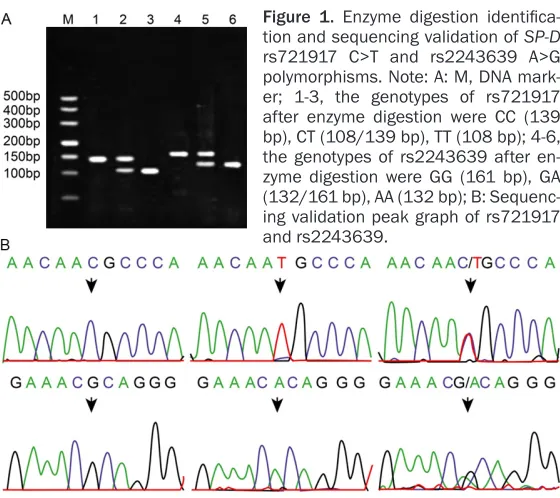
Figure 1. Enzyme digestion identifica-tion and sequencing validaidentifica-tion of SP-D
rs721917 C>T and rs2243639 A>G polymorphisms. Note: A: M, DNA mark-er; 1-3, the genotypes of rs721917 after enzyme digestion were CC (139 bp), CT (108/139 bp), TT (108 bp); 4-6, the genotypes of rs2243639 after en-zyme digestion were GG (161 bp), GA (132/161 bp), AA (132 bp); B: Sequenc-ing validation peak graph of rs721917 and rs2243639.
USA) was applied for PCR
amplification. The PCR reaction system (20 μl) included 2×PCR PLUS MIX (10 μl; DBI), DNA template (1.2 μl), forward and reverse primer (each 0.5 μl) and ultrapure water to fill the
residual volume. PCR reacti- on conditions: predegeneration for 5 min at 95°C, degenera-tion for 30 s at 94°C, anneal for 1 min at 60°C, extension for 1 min at 72°C, a total of 45 cycles, and at the end, cycle for 10 min at 72°C. The PCR
ampli-fied product (6 μl) was digested
respectively with FspI (Toyobo) and Dra III (NEB) enzymes at 37°C overnight; the reaction
system (15 μl) contained Fsp
Ienzyme (4 U) and Dra III (5 U). The enzyme-digested product
(5 μl) and 6× sample buffer (3 μl) was separated by
[image:3.612.91.371.219.468.2]improved or cured. Specifically, after standard
treatment, symptoms and signs of respiratory distress disappeared gradually; characteristic changes of RDS observed from chest X-ray dis-appeared; the cases could be independent on oxygen therapy and ventilator-supporting treat-ment (not including the cases dependent on oxygen caused by complications); or vital signs got relatively stable and the disease was con-trolled. (2) The cases got worsened and gave up treatment or died. The cases got worsened and gave up treatment: the treatment result is unsatisfactory. Within 72 hours after birth, the
vital signs were unstable; respiratory difficul -ties persisted; chest X-ray showed characteris-tic changes of RDS; out of economic consider-ation or prognosis, the family refused to con-tinue treatment. The cases died. The treatment showed no effectiveness; the disease got pro-gressively severe; vital signs disappeared and clinical death arrived.
Statistical analysis
The data was analyzed with SPSS 21.0 (SPSS Inc., Chicago, IL, USA) statistical software. Enumeration data were expressed in percent-age or rate; the differences in genotype fre-quencies were validated using chi square test. Logistic regression analysis was used to cor-rect baseline differences and calculate the risks of RDS in various genotypes which were
expressed with odds ratio (OR) and 95% confi
-dence interval (CI). Normal measurement data were expressed as mean ± standard deviation;
comparison between two groups was verified
with t test. Single factor variance analysis (one-way ANOVA) was used in comparison among three groups. Non normal distribution variables of non-normal distribution measurement data were expressed with quantile. If the data were normally distributed by logarithm transforma-tion, the means of the transformed variables in each group were compared by independent sample t test or variance analysis. Shesis anal-ysis software was applied to analyze the fre-quency of the haplotype of SP-D; Logistic regression model was used to analyze the effect of gene polymorphism and other factors on the prognosis of RDS patients. All the tests were two-sided tests. A P<0.05 was considered
statistically significant.
Results
Identification of genotyping of rs721917 C>T and rs2243639 A>G polymorphisms in the
SP-D gene
The rs721917 of SP-D gene was 139 bp in size
after PCR amplification, and generated 3 geno -types after Fsp-1 enzyme digestion, namely, CC (139 bp), TC (108 bp, 139 bp) and TT (108 bp). The rs2243639 of SP-D gene was 161 bp in
size after PCR amplification, and generated 3
[image:4.612.89.518.96.280.2]genotypes after Dra III enzyme digestion, na- Table 2. The frequency distributions of rs721917 and rs2243639 of SP-D gene in the case and con-trol groups
The case group
(n = 170) group (n = 204)The control OR (95% CI)a Pa OR (95% CI)Adjusted b Pb rs721917 (C>T)
CC 52 (30.6) 98 (48.0) ref ref
CT 70 (41.2) 78 (38.2) 1.691 (1.061-2.696) 0.026 1.750 (1.065-2.877) 0.027 TT 48 (28.2) 28 (13.8) 3.231 (1.818-5.741) <0.001 4.499 (2.251-8.990) <0.001
C 174 (51.2) 274 (67.2) ref ref
T 166 (48.8) 134 (32.8) 1.951 (1.450-2.624) <0.001 2.071 (1.490-2.878) <0.001 rs2243639 (A>G)
AA 108 (63.5) 122 (59.4) ref ref
AG 46 (27.1) 72 (35.2) 0.722 (0.459-1.134) 0.156 0.697 (0.409-1.190) 0.186 GG 16 (9.4) 10 (5.5) 1.807 (0.787-4.152) 0.158 1.854 (0.758-4.537) 0.176
A 262 (77.1) 316 (77.5) ref ref
G 78 (22.9) 92 (22.5) 1.023 (0.725 t-1.441) 0.899 1.062 (0.728-1.549) 0.755
Note: athe uncorrected OR (95% CI) and P value; bthe corrected OR (95% CI) and P value after Logistic regression model was
mely, GG (161 bp), GA (132 bp, 161 bp) and AA (132 bp) (Figure 1A). The DNA sequencing results of rs721917 and rs2243639 of SP-D gene were in full agreement (Figure 1B). The frequency distributions of genotypes and
alleles of rs721917 C>T and rs2243639 A>G
polymorphisms in the SP-D gene between the case and control groups
The genotypic distributions of SP-D gene were in accordance with the Hardy-Weinberg equilib-rium (P>0.05), which can be considered repre-sentative of the equilibrium population.
Frequency distributions of the three genotypes CC, CT and TT of SP-D rs721917 were signifi -cantly different in the case and control group
infants with RDS with different genotypes and
alleles of rs721917 C>T and rs2243639 A>G
polymorphisms
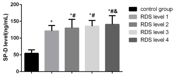
The expressions of serum SP-D in the case group and the control group were 130.04 ± 22.58 (ng/ml) and 54.62 ± 10.25 (ng/ml); the
difference was statistically significant (P< 0.001). The expression of SP-D of the patients
in the case group was significantly higher than
that of the patients in the control group (P<0.05); the expression of SP-D of the patients
with RDS level 2/3/4 was significantly higher
[image:5.612.90.366.96.277.2]than that of the patients with RDS level 1 (P<0.05); the expression of SP-D of the patients with RDS level 4 was higher than that of the patients with RDS level 2 (P<0.05) (Figure 2).
[image:5.612.87.365.312.433.2]Figure 2. Comparison of the protein expression of SP-D between the con-trol and case groups. Note: *, compared with the concon-trol group, P<0.05; #, compared with preterm infants with RDS level 1, P<0.05; &, compared with preterm infants with RDS level 2, P<0.05; RDS: respiratory distress syndrome.
Table 3. The frequency distribution of rs721917 and rs2243639 of SP-D and preterm infants with different RDS grades
SNP grade IRDS grade IIRDS grade IIIRDS grade IVRDS P
rs721917 (C>T)
CC 20 (37.0) 17 (29.3) 9 (28.1) 6 (23.1) 0.002 CT 25 (46.3) 30 (51.7) 10 (31.3) 5 (19.2) TT 9 (16.7) 11 (19.0) 13 (40.6) 15 (57.7) C 65 (60.2) 64 (55.2) 28 (43.8) 17 (32.7) 0.005 T 43 (39.8) 52 (44.8) 36 (56.2) 35 (67.3) rs2243639 (A>G)
AA 32 (59.3) 40 (69.0) 20 (62.5) 16 (61.5) 0.812 AG 18 (33.3) 12 (20.7) 8 (25.0) 8 (30.8) GG 4 (7.4) 6 (10.3) 4 (12.5) 2 (7.7) 0.904 A 82 (75.9) 92 (79.3) 48 (75.0) 40 (76.9) G 26 (20.1) 24 (20.7) 16 (25.0) 12 (23.1)
Note: RDS, respiratory distress syndrome; SNP, single nucleotide polymorphism.
(P<0.05), the same as the fre-quency distributions of C and T alleles (both P<0.05). The risk of RDS for the patients carrying the CT genotype was 1.750 times higher than that for the patients carrying the CC genotype (OR = 1.750; 95% CI = 1.065-2.877); the risk of RDS for the patients carrying the TT genotype was 4.499 times higher than that for the patients carrying the CC gen-otype (OR = 4.499; 95% CI = 2.251-8.990); the risk of RDS for the patients carrying the T allele was 2.071 times higher than that for the patients carrying the C allele (OR = 2.071; 95% CI = 1.490-2.878). Frequency distri-butions of the genotypes and alleles of SP-D rs2243639 sh-
owed no significant differences
in the case and control group (all P>0.05) (Table 2). Frequency distributions of the genotypes and alleles of SP-D rs721917
were significantly different in the
grading of RDS (P = 0.002; P = 0.005); while the frequency dis-tributions of the genotypes and alleles of SP-D rs2243639 sho-
wed no significant differences in
the grading of RDS (all P>0.05) (Table 3).
The expressions of SP-D of the genotypes CC, CT and TT of rs721917 in the case group were 123.25 ± 23.42 ng/ml, 127.81 ± 20.76 ng/ml and 140.64 ± 20.79 ng/ml, respectively; the
difference was statistically significant (F =
8.710, P<0.001). The expression of SP-D of the
genotype TT was significantly higher than that
of CC and CT genotypes (P<0.05). The expres-sions of SP-D of the genotypes CC, CT and TT of rs2243639 in the case group were 130.73 ± 23.33 ng/ml, 125.44 ± 20.46 ng/ml and 138.58 ± 21.41 ng/ml, respectively; the
differ-ence was not statistically significant (F = 2.200,
P = 0.113) (Figure 3).
Haplotype analysis of rs721917 C>T and rs2243639 A>G polymorphisms in the SP-D
gene
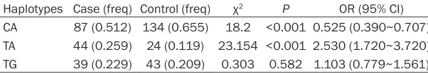
Shesis analysis software was applied for link-age disequilibrium (LD) analysis of the SNPs of SP-D and the haplotype analysis. The haplo-types with the frequency less than 3% were excluded. The results showed that the
frequen-cy of the haplotype CA was significantly differ -ent in the case and control group (P<0.001) and CA was possibly a protective haplotype for
= 1.720~3.720); and that the frequency of the
haplotype TG was not significantly different in
the case and control group (P = 0.582) (Table 4).
Logistic regression analysis for the risk factors of the prognosis of preterm infants with RDS
Of all the 170 preterm infants with RDS treated with standard treatment, 144 cases were
sig-nificantly improved and cured, and 26 died.
[image:6.612.91.522.72.203.2]Whether patients died was set as the depen-dent variable, infant sex, gestational age, body weight, maternal age, gestational hyperten-sion, gestational diabetes, premature rupture of membranes, time of birth, placental abnor-malities and umbilical cord abnormality, the expression of SPD, rs721917 and rs2243639 were all set as independent variables into Logistic regression model. The results suggest-ed that the TT genotype in rs721917, pregnan-cy-induced hypertension, abnormal umbilical cord and placental abnormalities were the risk factors of death in preterm infants with RDS (all P<0.05). However, the infant sex, gestational age, body weight, maternal age, gestational diabetes, premature rupture of membranes,
Figure 3. Comparison of the protein expression of SP-D in RDS preterm infants with various genotypes and alleles of rs721917 C>T and rs2243639 A>G polymorphisms. Note: A: Comparison of the protein expression of SP-Din RDS preterm infants with different genotypes of rs721917 C>T polymorphism; *, compared with the CC genotype,
P<0.05; #, compared with the CT genotype; B: Comparison of the protein expression of SP-D in RDS preterm infants with various genotypes in rs2243639 A>G polymorphism; RDS: respiratory distress syndrome.
Table 4. The haplotype analysis of rs721917 C>T and rs2243639 A>G polymorphisms in the SP-D gene between the case and control groups Haplotypes Case (freq) Control (freq) χ2 P OR (95% CI) CA 87 (0.512) 134 (0.655) 18.2 <0.001 0.525 (0.390~0.707) TA 44 (0.259) 24 (0.119) 23.154 <0.001 2.530 (1.720~3.720) TG 39 (0.229) 43 (0.209) 0.303 0.582 1.103 (0.779~1.561)
Note: freq, frequency; OR, odds ratio; CI, confidence interval.
RDS (OR = 0.525, 95% CI = 0.390~0.707); that the frequency of the
haplo-type TA was significantly
[image:6.612.90.391.307.359.2]time of birth, the expression of SPD and rs2243639 were not correlated with the death risk of preterm infants in RDS (Table 5). Discussion
NRDS, a disease caused by deficiency of PS, is
subject to concurrent infection, chronic lung disease, pulmonary hemorrhage, intracranial hemorrhage, premature retinopathy etc.,
great-ly influencing the survival and treatment of the
neonates [17]. The present study is aimed at exploring the correlation of SP-D gene polymor-phism and protein expression with the patho-genesis and severity of RDS in preterm infants. The research results indicated that the expres-sion of SP-D was higher in the preterm infants with RDS than the healthy infants. Studies demonstrated that high levels of plasma SP-Das a marker of pulmonary injury were associated with increased mortality [18, 19]. It was assumed that systemic circulating SP-D levels resulted from alveolar leakage into the bloodstream [20, 21]. In the lungs, SP-D has
anti-inflammatory- and anti-oxidant capacities
and protects against respiratory infections [19]. Increased SP-D levels were also detected in serum in acute or chronic lung injury and the
finding that a quick up-regulation of SP-D in
serum responding to acute airway inflamma -tion might substantiate the no-tion that SP-D translocates from the airways into the vascular system, in favor of being synthesized systemi-cally [22, 23].
[image:7.612.95.523.97.333.2]From the present study, we also found that SP-D rs721917 polymorphism was correlated with the expression of SP-D, that the patients carrying the genotypes CT and TT had higher risk of incidence of RDS than those carrying the CC genotype, the same with the cases carrying the allele T to the ones with the C allele. According to the prognosis, the cases with the genotype TT had lower likelihood of recovery than the ones with the genotypes CC and CT. The rs721917 major allele (methionine 11) was associated with the risk of severe respiratory syncytial virus bronchiolitis in infants [18]. Individuals homozygous for the rs721917 minor allele (threonine 11) were reported to have lower levels of serum SP-D and display a pre-dominance of the trimeric form of SP-D and the low level of higher oligomers affected the immu-nological ability of SP-D to bind microbes [24]. It was also demonstrated that SP-D could inhib-it the growth of mycobacterium tuberculosis and can accelerate the decomposition of the bacteria [25]. Experiment done by Hartshor et al. reported that SP-D was put together with Table 5. Logistic regression analysis for the risk factors of the prognosis of preterm infants with respi-ratory distress syndrome
Factor B S.E, Wald df Sig. Exp(B)
Exp(B) 95% CI Lower
limit Upper limit Infant sex (male vs female) 0.458 0.475 0.931 1 0.335 1.581 0.623 4.008
Gestational age -0.073 0.091 0.649 1 0.420 0.929 0.778 1.11
Body weight 0.179 0.448 0.159 1 0.690 1.196 0.497 2.879
albicans Saccharomyces, proved that SP-D may modulate host defense against bacteria and mycotic infection [26]. By binding to glyco-conjugates and lipid moietis, SP-D can facili-tate pulmonary clearance of bacterial and viral pathogens through multiple mechanisms (ag- gregation, opsonization, phagozytosis or lysis
of pathogens) confirmed by several studies
[27].
Furthermore, the SP-D expression analysis demonstrated that the higher the grade of RDS was, the higher level of SP-D was detected. In different grades of NRDS, alveolar type II cells were an indicator (AEC II) and damage,
apopto-sis and inflammatory mediators of AEC II, alveo -lar type II cells can decrease or increase the PS, resulting in severe loss of phospholipids and related proteins in the alveolar surface, which is one of the important mechanisms of NRDS [28]. SP-D is suggested to regulate sur-factant uptake and catabolism by alveolar type
II cells and influence the ultrastructure of sur -factant in the alveolus [29]. SP-D is secreted by AEC II, which can identify and remove foreign bodies and apoptotic cells in the lung and when AEC II is severely damaged or disrupted, SP-D will further release into the alveolar cavity, resulting in a marked increase in SP-D levels in
BAL fluid [22]. Todd et al. reported that the
damage and proliferation of AEC II, the increase of the alveolar epithelium and capillary perme-ability may also lead to a pronounced increase in the level of SP-D in infants with NRDS [30]. As was shown in the grading criteria, RDS grade
IV was defined as diffuse granularity or hazi -ness of the lung, air bronchogram, obscured cardiac and diaphragm borders plus white lung [31]. When these signs were detected, it indi-cated increased pulmonary foreign bodies or decreased SP-D within the lung [32]. Previous
studies also found that the deficiency or insuf
-ficiency of SP-D will lead to the susceptibility of body to pathogen infection, resulting in a more
serious inflammatory reaction [32, 33].
In summary, we have demonstrated SP-D rs721917 polymorphism may be associated with the pathogenesis and severity of RDS in preterm infants. Therefore, SP-D gene might be used as a target for the diagnosis of RDS in pre-term infants. However, further studies with a large sample size are required in order to vali-date our research results.
Acknowledgements
We would like to give our sincere appreciation to the reviewers for their helpful comments on this article.
Disclosure of conflict of interest
None.
Address correspondence to: Dr. Hai-Bo Li, Out- patient Department of Pediatrics, The First Hospital of Jilin University, No. 71, Xinmin Street, Chaoyang District, Changchun 130021, Jilin Province, P. R. China. Tel: 88782222; Fax: +86-0431-88782222; E-mail: [email protected]
References
[1] Sweet DG, Carnielli V, Greisen G, Hallman M, Ozek E, Plavka R, Saugstad OD, Simeoni U, Speer CP, Halliday HL; European Association of Perinatal Medicine. European consensus guidelines on the management of neonatal re-spiratory distress syndrome in preterm in-fants-2010 update. Neonatology 2010; 97: 402-417.
[2] Marinov B, Jekova N, Andreeva A and Hitrova S. [Antenatal ambroxol administration for pre-vention of respiratopry distress syndrome in preterm infants: preliminary report]. Akush Ginekol (Sofiia) 2011; 50: 17-22.
[3] Qian L, Liu C, Zhuang W, Guo Y, Yu J, Chen H, Wang S, Lin Z, Xia S, Ni L, Liu X, Chen C, Sun B; Chinese Collaborative Study Group for Neona-tal Respiratory Diseases. NeonaNeona-tal respiratory failure: a 12-month clinical epidemiologic study from 2004 to 2005 in China. Pediatrics 2008; 121: e1115-1124.
[4] Ramanathan R, Bhatia JJ, Sekar K and Ernst FR. Mortality in preterm infants with respirato-ry distress syndrome treated with poractant alfa, calfactant or beractant: a retrospective study. J Perinatol 2013; 33: 119-125.
[5] Blencowe H, Cousens S, Oestergaard MZ, Chou D, Moller AB, Narwal R, Adler A, Vera Gar-cia C, Rohde S, Say L and Lawn JE. National, regional, and worldwide estimates of preterm birth rates in the year 2010 with time trends since 1990 for selected countries: a system-atic analysis and implications. Lancet 2012; 379: 2162-2172.
infants--2013 update. Neonatology 2013; 103: 353-368.
[7] Whitsett JA, Wert SE and Weaver TE. Diseases of pulmonary surfactant homeostasis. Annu Rev Pathol 2015; 10: 371-393.
[8] Polin RA, Carlo WA; Committee on Fetus and Newborn; American Academy of Pediatrics. Surfactant replacement therapy for preterm and term neonates with respiratory distress. Pediatrics 2014; 133: 156-163.
[9] Whitsett JA, Wert SE and Weaver TE. Alveolar surfactant homeostasis and the pathogenesis of pulmonary disease. Annu Rev Med 2010; 61: 105-119.
[10] Chroneos ZC, Sever-Chroneos Z and Shepherd VL. Pulmonary surfactant: an immunological perspective. Cell Physiol Biochem 2010; 25: 13-26.
[11] Yamazoe M, Nishitani C, Takahashi M, Katoh T, Ariki S, Shimizu T, Mitsuzawa H, Sawada K, Voelker DR, Takahashi H and Kuroki Y. Pulmo-nary surfactant protein D inhibits lipopolysac-charide (LPS)-induced inflammatory cell re-sponses by altering LPS binding to its receptors. J Biol Chem 2008; 283: 35878-35888.
[12] Winkler C, Atochina-Vasserman EN, Holz O, Beers MF, Erpenbeck VJ, Krug N, Roepcke S, Lauer G, Elmlinger M and Hohlfeld JM. Com-prehensive characterisation of pulmonary and serum surfactant protein D in COPD. Respir Res 2011; 12: 29.
[13] Crouch E, Rust K, Veile R, Donis-Keller H and Grosso L. Genomic organization of human sur-factant protein D (SP-D). SP-D is encoded on chromosome 10q22.2-23.1. J Biol Chem 1993; 268: 2976-2983.
[14] Montalbano AP, Hawgood S and Mendelson CR. Mice deficient in surfactant protein A (SP-A) and SP-D or in TLR2 manifest delayed partu-rition and decreased expression of inflamma-tory and contractile genes. Endocrinology 2013; 154: 483-498.
[15] Ou CY, Chen CZ, Hsiue TR, Lin SH and Wang JY. Genetic variants of pulmonary SP-D predict disease outcome of COPD in a Chinese popula-tion. Respirology 2015; 20: 296-303.
[16] Pavlovic J, Papagaroufalis C, Xanthou M, Liu W, Fan R, Thomas NJ, Apostolidou I, Papathoma E, Megaloyianni E, DiAngelo S and Floros J. Ge-netic variants of surfactant proteins A, B, C, and D in bronchopulmonary dysplasia. Dis Markers 2006; 22: 277-291.
[17] Zhang JP, Wang YL, Wang YH, Zhang R, Chen H and Su HB. Prophylaxis of neonatal respiratory distress syndrome by intra-amniotic adminis-tration of pulmonary surfactant. Chin Med J (Engl) 2004; 117: 120-124.
[18] Sorensen GL, Dahl M, Tan Q, Bendixen C, Hol-mskov U and Husby S. Surfactant protein-D-encoding gene variant polymorphisms are linked to respiratory outcome in premature in-fants. J Pediatr 2014; 165: 683-689.
[19] Wulf-Johansson H, Thinggaard M, Tan Q, Jo-hansson SL, Schlosser A, Christensen K, Holm-skov U and Sorensen GL. Circulating surfac-tant protein D is associated to mortality in elderly women: a twin study. Immunobiology 2013; 218: 712-717.
[20] Sorensen GL, Husby S and Holmskov U. Sur-factant protein A and surSur-factant protein D vari-ation in pulmonary disease. Immunobiology 2007; 212: 381-416.
[21] Hartl D and Griese M. Surfactant protein D in human lung diseases. Eur J Clin Invest 2006; 36: 423-435.
[22] Gaunsbaek MQ, Rasmussen KJ, Beers MF, Atochina-Vasserman EN and Hansen S. Lung surfactant protein D (SP-D) response and regu-lation during acute and chronic lung injury. Lung 2013; 191: 295-303.
[23] Fujita M, Shannon JM, Ouchi H, Voelker DR, Nakanishi Y and Mason RJ. Serum surfactant protein D is increased in acute and chronic in-flammation in mice. Cytokine 2005; 31: 25-33. [24] Leth-Larsen R, Garred P, Jensenius H, Meschi
J, Hartshorn K, Madsen J, Tornoe I, Madsen HO, Sorensen G, Crouch E and Holmskov U. A common polymorphism in the SFTPD gene in-fluences assembly, function, and concentra-tion of surfactant protein D. J Immunol 2005; 174: 1532-1538.
[25] Shrivastava R, Yasir M, Tripathi M and Singh P. In silico interaction of methyl isocyanate with immune protein responsible for Mycobacteri-um tuberculosis infection using molecular docking. Toxicol Ind Health 2016; 32: 162-167. [26] Hartshorn KL, White MR, Tecle T, Tornoe I, So-rensen GL, Crouch EC and Holmskov U. Re-duced influenza viral neutralizing activity of natural human trimers of surfactant protein D. Respir Res 2007; 8: 9.
[27] Hilgendorff A, Heidinger K, Bohnert A, Klein-steiber A, Konig IR, Ziegler A, Lindner U, Frey G, Merz C, Lettgen B, Chakraborty T, Gortner L and Bein G. Association of polymorphisms in the human surfactant protein-D (SFTPD) gene and postnatal pulmonary adaptation in the preterm infant. Acta Paediatr 2009; 98: 112-117.
[28] Goto H, Ledford JG, Mukherjee S, Noble PW, Williams KL and Wright JR. The role of surfac-tant protein A in bleomycin-induced acute lung injury. Am J Respir Crit Care Med 2010; 181: 1336-1344.
ultrastructure and uptake by alveolar type II cells. Am J Physiol Lung Cell Mol Physiol 2005; 288: L552-561.
[30] Todd DA, Marsh MJ, George A, Henderson NG, Barr H, Sebastian S, Clark GT, Koster G, Clark HW and Postle AD. Surfactant phospholipids, surfactant proteins, and inflammatory markers during acute lung injury in children. Pediatr Crit Care Med 2010; 11: 82-91.
[31] Makri V, Hospes B, Stoll-Becker S, Borkhardt A and Gortner L. Polymorphisms of surfactant protein B encoding gene: modifiers of the course of neonatal respiratory distress syn-drome? Eur J Pediatr 2002; 161: 604-608.
[32] More JM, Voelker DR, Silveira LJ, Edwards MG, Chan ED and Bowler RP. Smoking reduces sur-factant protein D and phospholipids in patients with and without chronic obstructive pulmo-nary disease. BMC Pulm Med 2010; 10: 53. [33] Barlo NP, van Moorsel CH, Ruven HJ, Zanen P,