Original Article
Roles of heat shock protein 70 toward hypoxia-inducible
factor 1α (HIF-1α) blockade in newborn rats with
hypoxia-induced pulmonary hypertension
Le Wang, Mingxia Li
Department of Neonatology, The First Affiliated Hospital of Xinjiang Medical University, Urumqi 830011, Xinjiang,
PR China
Received March 1, 2018; Accepted July 13, 2018; Epub December 15, 2018; Published December 30, 2018
Abstract: The aim of the present study was to investigate the roles of adenovirus-mediated heat shock protein 70 (HSP70) in newborn rats with hypoxia-induced pulmonary hypertension (HPH). A total of 128 newborn rats were randomly divided into an HPH group (H) and control group (C), with 8 rats in each group. According to transfection solutions received, group H was subgrouped into three different groups: Group H1, which received saline, group H2, which received transfection of an empty virus, and group H3, which received transfection of HSP70 virus. Sub-sequently, pulmonary artery pressure (mPAP) was measured in all groups and levels of HSP70, hypoxia-inducible factor 1α (HIF-1α), endothelin-1 (ET-1), and inducible nitric oxide synthase (iNOS) were measured at different time points. mPAP in groups H1 and H2 at 3 days, 7 days, 10 days, and 14 days was significantly higher than in group C (P<0.05). Furthermore, immunohistochemical expression intensity of HSP70 in group H3 on days 3, 7, and 10 was significantly enhanced (P<0.05), compared to groups H1 and H2. However, expression of HIF-1α, ET-1, and iNOS in group H3 was reduced (P<0.01). Intergroup comparisons of HSP70 expression determined that expression of HSP70 in group H3 was enhanced (P<0.05), however expression of HIF-1α, ET-1, and iNOS was reduced (P<0.05), compared to groups H1 and H2. Results of current study demonstrate that expression of adenovirus-mediated HSP70 in newborn HPH rats may downregulate expression of HIF-1α, ET-1, and iNOS, reducing mPAP. Therefore, adenovirus-mediated HSP70 may be developed as a novel therapeutic method of treating neonatal HPH.
Keywords: Heat shock protein 70, adenovirus, hypoxia-inducible factor 1α, hypoxia induced pulmonary hyperten -sion, newborn rats
Introduction
Hypoxia-induced pulmonary hypertension (HPH) of newborns is a common acute critical illness. Incidence of HPH in newborns is 2-6/1000 live births. Early pulmonary vascular spasms are reversible with timely treatment. In advanced pulmonary vascular remodeling, persistent pul-monary hypertension of the in newborn (PPHN) [1] is often developed. It is difficult to correct the hypoxic respiratory failure and it is difficult to treat the mortality rate of newborns when PPH is high, reaching to 5~30%. It has been demonstrated that hypoxia inducible factor 1α (HIF-1α) is a key transcription factor of hypoxia [2], serving important roles in the pathogenesis of HPH [3-5]. HIF-1α regulates expression of its downstream target genes endothelin 1 (ET-1; a
novel therapeutic strategies for treatment of neonatal HPH. Heat shock protein 70 (HSP70) [12] promotes the degradation of HIF-1α under hypoxic conditions [13]. Therefore, the current study used gene transfection with adenovirus as the vector to increase HSP70 expression in lung tissues. The aim of the current study was to identify whether HSP70 expression down-regulates expression of HIF-1α, ET-1, and iNOS, while reducing mPAP, thus inhibiting the devel-opment of HPH.
Materials and methods
Animals
A total of 128 healthy Wistar neonatal rats [7-10 days old; weighing 30-40 g; animal li- cense number, SCXK (Xin2011-0004)] and 16 mother rats [60-70 days old; weighing 230-250 g] were provided by the Animal Experiment Center, the Medical Research Center of the First Affiliated Hospital of Xinjiang Medical University (Xinjiang, China). Rats were randomly divided into two groups (n=8 per subgroup): HPH group (H) and control group (C). According to transfection solutions received, rats in group H were subgrouped into 3 groups: Group H1 received a saline, group H2 received an empty virus, and group H3 received Ad-HSP70 virus. Rats in group H were injected with 5 μl 1010 PFU/ml Ad-HSP70 virus and Ad-GFP (Hanbio Biotechnology, Shanghai, China) viral transfec-tion solutransfec-tion or sterile saline, via the tail veins. Rats were not anesthetized prior to this proce-dure. The HPH model was established immedi-ately following injections [14]. Transfected neo-natal rats were placed, together with their mothers, in a normal-pressure hypoxic cabin infused with 8% nitrogen-oxygen mixture gas. An oxygen concentration monitor (CY-100B, Lihua Science & Technology Co. Ltd., Hangzhou, China) was used for 8 hours of monitoring of oxygen concentrations, maintaining it at 8-10%. All rats were subjected to a 12-hour light-dark cycle and, apart from the absence of hypoxic conditions in group C, rats in all groups were kept under the same conditions (temperature was 21-25°C, relative humidity was 55%-60%, rats were fed with basic feed, sterilized sterile water, and free feeding water). The current study was performed in strict accordance with recommendations in the Guide for the Care and Use of Laboratory Animals of the National
Institutes of Health [15]. The animal use proto-col was reviewed and approved by the Ins- titutional Animal Care and Use Committee (IACUC) of Xinjiang Medical University.
Measurement of mPAP
After being kept in hypoxic conditions for 3, 7, 10, and 14 days, neonatal rats in each group (n=8 per subgroup) were conventionally anes-thetized (intraperitoneal injection of ketamine (75 mg/kg), atropine (0.375 mg/kg), and diaz-epam (7.5 mg/kg) mixture by tail veins). They were fixed, sterilized, and underwent endotra-cheal intubation. One small animal ventilator (HX-200, Taimeng Technology Co. LTd., Cheng- du, China) was then connected. Respiratory rate was kept at 120 beats/minute and tidal volume at 4 mL/minute. It was ensured that the thorax remained in symmetric fluctuation and in sync with the ventilator. Subsequently, one U-shaped incision from the right sternal margin of the chest was performed to expose the heart to the pulmonary root. A 4.5 needle was then used to pierce towards the pulmonary arterial root inverse to the direction of blood flow. The pressure sensor was then quickly connected to record mPAP.
Immunohistochemical assay
of staining intensity and positive area ratio was used as the final score: 0 point, negative (-); 1-2 points, weak positive (+); 3-4 points, positive (++); >4 points, strongly positive (+++) with 400-fold optical microscope (PIPS-2020 patho-logical image analysis software: Polaroid, USA).
Western blotting
Following sacrifice, ~100 mg of right lung tissue was rapidly sampled from each rat. This was followed by extraction of total protein using a nucleus and cytoplasm protein extraction kit (78835; Thermo Fisher Scientific, Inc., Wal- tham, MA, USA), following manufacturer instr- uctions. Lamin A/C was selected as an inter- nal standard of nuclear proteins and β-actin was selected as the internal standard of cyto-plasmic proteins. Bicinchoninic acid method (BCA) was used to determine protein concen-trations. Proteins were then quantified and degraded and underwent SDS-polyacrylamide gel electrophoresis (30 mA constant current). Proteins were then transferred to PVDF mem-branes for 1-2 hours at 100 V (wet method, 283BR11735 electrophoresis, electroporation instrument, Bio-Rad Laboratories, Inc., Hercul-
analyzer (76S/07865 gel imaging analyzer: BIO-RAD, USA) was used to acquire images and Quantity One software (BIO-RAD, USA) was used to analyze gray values of the target strips.
Statistical analysis
SPSS 20.0 software (IBM Corp., Armonk, NY, USA) was used for statistical analysis. Normally distributed measurement data are expressed as mean ± standard deviation. One-way analy-sis of variance was used for multi-group com-parisons and Student-Newman-Keul’s test was used to perform paired comparisons between two groups. Non-normally distributed measure-ment data are expressed using the median and interquartile range. P<0.05 indicates a statisti-cally significant difference.
Results
Transfection of HSP70 genes
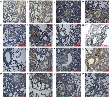
[image:3.612.93.524.69.159.2]Lung tissue slices, obtained from groups H3 and C, were observed using an immunofluores-cence microscope (Figure 1). Lung tissue from rats in group H3, that underwent hypoxia for 3,
Figure 1. Localization of heat shock protein 70 in lung tissue from rats, as determined using an immunofluorescent microscope (magnification, ×200). A. Control group; B. H3 group, day 3; C. H3 group, day 7; D. H3 group, day 10; E. H3 group, day 14.
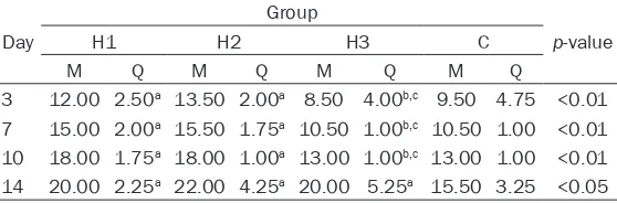
Table 1. Changes in mean pulmonary artery pressure in the four groups of newborn rats (n=8 in each group)
Day
Group
p-value
H1 H2 H3 C
M Q M Q M Q M Q
3 12.00 2.50a 13.50 2.00a 8.50 4.00b,c 9.50 4.75 <0.01
7 15.00 2.00a 15.50 1.75a 10.50 1.00b,c 10.50 1.00 <0.01
10 18.00 1.75a 18.00 1.00a 13.00 1.00b,c 13.00 1.00 <0.01
14 20.00 2.25a 22.00 4.25a 20.00 5.25a 15.50 3.25 <0.05
aP<0.05 vs. group C; bP<0.05 vs. group H1, cP<0.05 vs. group H2. Group H1, HPH group receiving saline group; group H2, an empty virus-transfected HPH group; group H3, a virus heat shock protein 70-transfected group; C, control group. M, median; Q, interquartile range. HPH, hypoxia-induced pulmonary hypertension.
[image:3.612.92.371.247.339.2]7, and 10 days (Figure 1B-D), exhibited immu-nofluorescent signal marker protein. However, those in group H3 that experienced hypoxia for 14 days (Figure 1E) and lung tissue of rats in group C (Figure 1A) exhibited no markers. mPAP changes
mPAP in groups H1 and H2 on days 3, 7, 10, and 14 were significantly greater than in rats in group C at the same time points (P<0.05). However, mPAP in group H3 at days 3, 7, and 10 was significantly lower than in groups H1 and
H2 (P<0.05; Table 1), indicating that adenovi-rus mediated HSP70 can reduce pulmonary arterial pressure.
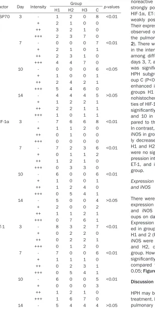
Immunohistochemical expression of HSP70, HIF-1α, ET-1, and iNOS
[image:4.612.92.525.70.454.2]Expression of HSP70, HIF-1α, ET-1, and iNOS in lung tissue taken from rats in Group C, as deter-mined by immunohistochemistry (Figure 2), was mostly negative. For groups H1 and H2, the immunoreactive intensities of HSP70 ranged from weakly positive (+) to positive (++), while
Figure 2. Immunohistochemical staining of HSP70, HIF-1α, ET-1, and iNOS in the lung tissue of newborn rats (mag
Table 2. Immunohistochemical reaction intensities of HSP70, HIF-1α, ET-1, and iNOS in lung tissues of newborn rats
Factor Day Intensity Group p-values
H1 H2 H3 C
HSP70 3 - 1 2 0 8 <0.01
+ 2 1 0 0
++ 3 2 1 0
+++ 2 3 7 0
7 - 0 0 0 7 <0.01
+ 2 1 0 1
++ 2 3 1 0
+++ 4 4 7 0
10 - 0 0 0 6 <0.05
+ 1 0 0 1
++ 2 4 2 1
+++ 5 4 6 0
14 - 4 4 4 5 >0.05
+ 1 2 2 1
++ 2 2 1 1
+++ 1 0 1 1
HIF-1α 3 - 7 6 6 8 <0.01
+ 1 1 2 0
++ 0 1 0 0
+++ 0 0 0 0
7 - 7 2 3 6 <0.01
+ 0 1 1 2
++ 1 2 1 0
+++ 0 3 3 0
10 - 6 0 0 6 <0.01
+ 1 0 0 1
++ 1 2 4 0
+++ 0 5 4 1
14 - 5 0 0 4 >0.05
+ 2 0 0 2
++ 1 1 2 1
+++ 0 7 6 1
ET-1 3 - 8 3 2 7 <0.01
+ 0 2 2 0
++ 0 2 2 1
+++ 0 1 2 0
7 - 7 0 0 6 <0.01
+ 1 1 1 0
++ 0 2 3 1
+++ 0 5 4 1
10 - 6 0 0 5 <0.01
+ 0 0 0 3
++ 1 2 1 0
+++ 1 6 7 0
14 - 5 4 4 4 >0.05
those of HIF-1α, ET-1, and iNOS ranged from positive (++) to strongly positive (+++). In group H3, the immu-noreactive intensity of HSP70 was strongly positive (+++) and those of HIF-1α, ET-1, and iNOS ranged from weakly positive (+) to positive (++). Their expression was more commonly observed on the endothelial cells of the pulmonary vascular wall (Figure 2). There were significant differences in the intensity of HSP70 expression among different HPH subgroups on days 3, 7, and 10. HSP70 expression was significantly upregulated in all HPH subgroups, compared with gr- oup C (P<0.01), and was significantly enhanced in group H3, compared to groups H1 and H2 (P<0.01). Immu- nohistochemical expression intensi-ties of HIF-1α, ET-1, and iNOS were all significantly upregulated on days 3, 7, and 10 in all HPH subgroups, com-pared to the control group (P<0.01). In contrast, levels of HIF-1α, ET-1, and iNOS in group H3 were all significant- ly decreased compared with groups H1 and H2 (P<0.01; Table 2). There were no significant differences in ex- pression intensities of HSP70, HIF-1α, ET-1, and iNOS at 14 days in each group.
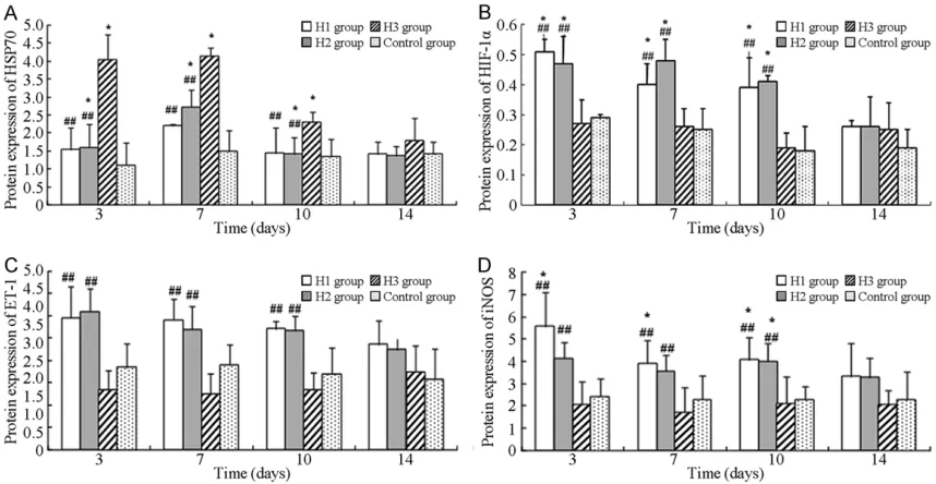
Expression of HSP70, HIF-1α, ET-1, and iNOS
There were significant differences in expression of HSP70, HIF-1α, ET-1, and iNOS among the different gr- oups on days 3, 7, and 10 (P<0.05). Expression of HSP70 was upregulat-ed in group H3, comparupregulat-ed to groups H1 and 2 (P<0.05). HIF-1α, ET-1, and iNOS were upregulated in groups H1 and H2, compared to the control group. However, their expression was significantly reduced in group H3, compared to groups H1 and 2 (P< 0.05; Figure 3).
Discussion
16], even persistent fetal circulation, may appear. It has been demonstrated that HIF-1α promotes expression of pro-vasodilators, caus-ing pulmonary vascular spasms and remodel-ing, as well as leading to an increase in mPAP [5]. Downregulating expression of HIF-1α in the early stages of hypoxia may, therefore, be a novel effective method of treating HPHN.
HSP70 promote the degradation of HIF-1α [17]. They are a group of highly conserved non-spe-cific intracellular protective proteins [18-20] that can protect cells from damage, promote the degradation and clearance of abnormal proteins, and maintain the normal physiological function of cells. The present study demon-strated that adenovirus-mediated HSP70 can be successfully targeted to pulmonary endo-thelial cells in neonatal rats, indicating that in vivo metabolism was reduced on day 14 of hypoxia. Low expression of HSP70 was detect-ed in the control group, but its expression increased in HPH groups. Hypoxia may activate HSP70, leading to an increase in expression. Expression of HSP70 was highest in Group H3 at all time points, indicating that adenovirus-mediated HSP70 transfection may induce high expression of exogenous HSP70. Positive ex- pression of HSP70 in the HPH groups was
groups H1 and H2. HPH was not evident in group H3 on days 3, 7, and 10 of hypoxia, sug-gesting that high expression of adenovirus-mediated HSP70 may block the development of HPH and induce degradation of HIF-1α. mPAP in group H3 was increased on day 14. HSP70 was downregulated in lung tissue, indicating that when the in vivo metabolic expression of adenovirus-mediated HSP70 is weakened, its ability to block the formation of HPH may be reduced. However, persistence of hypoxic con-ditions may cause an increase in mPAP.
HSP70 may mediate the degradation of HIF-1α proteins via non-VHL-dependent pathways [17]. During hypoxic conditions, HIF-1α may induce expression of ET-1 and iNOS, thus serving a damaging role in HPH [21, 22]. In the current study, immunohistochemical results indicated that HIF-1α was primarily expressed on pulmo-nary vascular endothelial cells. Immunoreactive intensities of HIF-1α in group H3 ranged from weakly positive (+) to positive (++), while those of ET-1 and iNOS ranged from weakly positive (+) to positive (++). Levels of HIF-1α were upreg-ulated in all HPH subgroups on days 3, 7, and 10. Compared to groups H1 and H2, expression of HIF-1α was downregulated in group H3, indi-cating that adenovirus-mediated HSP70 in-
+ 1 1 2 2
++ 1 2 2 1
+++ 1 1 0 1
iNOS 3 - 8 3 2 7 <0.01
+ 0 1 1 0
++ 0 2 2 1
+++ 0 2 3 0
7 - 7 0 0 6 <0.01
+ 1 1 1 1
++ 0 1 2 1
+++ 0 6 5 0
10 - 5 1 0 4 <0.01
+ 1 1 1 2
++ 1 2 2 1
+++ 1 4 5 1
14 - 5 0 0 6 >0.05
+ 2 0 0 0
++ 1 3 2 2
+++ 0 5 6 0
HSP70, heat shock protein 70; HIF-1α, hypoxia-inducible factor 1α; ET-1, endothelin-1; iNOS, inducible nitric oxide synthase; (-), negative; (+),
weakly positive; (++), positive; (+++), strongly positive.
detected 3 days following transfec-tion, peaking on day 10 and then declining. On day 14, HSP70 expres-sion in all HPH subgroups decreased. Thus, it may be hypothesized that dur-ing prolonged periods of hypoxia, cell structures and functions of lung tis-sue may be damaged, reducing ex- pression of HSP70. The present study demonstrates that the protective ef- fects of endogenous HSP70 on HPH-induced damage in the lungs are weak and cannot reduce lung injuri- es. Severe long-term oxidative stress may block the synthesis of proteins required to maintain cell growth. Mo- reover, the overgeneration of dena-tured proteins may attenuate the pro-tective abilities of HSP70. The current study demonstrates that high expres-sion of recombinant adenovirus-medi-ated HSP70 serves a protective role in HPH.
duces high expression of HSP70 and subse-quently inhibits expression of HIF-1α. Further- more, compared to the control group, expres-sion of ET-1 and iNOS in all HPH subgroups on days 3, 7, and 10 was increased, indicating that hypoxia promotes expression of these proteins. Expression of ET-1 and iNOS in group H3 was reduced, compared to groups H1 and H2, indi-cating that adenoviral-mediated HSP70 induc-es high exprinduc-ession of HSP70, subsequently inhibiting expression of HIF-1α and downregu-lating expression of its downstream target genes ET-1 and iNOS.
In conclusion, the present study demonstrates that adenovirus-mediated HSP70 may induce high expression of HSP70 in lung tissues of newborn HPH rats. Furthermore, it was demon-strated that HSP70 exhibits protective effects in pulmonary tissue in HPH by promoting the degradation of HIF-1α and downregulating ex- pression of its downstream target genes ET-1 and iNOS.
Acknowledgements
The study was supported by a grant from the National Natural Science Foundation of China (No.81360104).
Disclosure of conflict of interest
None.
Address correspondence to: Dr. Mingxia Li, Depart- ment of Neonatology, The First Affiliated Hospital of Xinjiang Medical University, 137 Liyushan Road, Urumqi 830011, Xinjiang, PR China. Tel: +86 13999129641; E-mail: mingxialidoc@163.com
References
[1] Storme L, Aubry E, Rakza T, Houeijeh A, De-barge V, Tourneux P, Deruelle P, Pennaforte T; French Congenital Diaphragmatic Hernia Study Group. Pathophysiology of persistent pulmonary hypertension of the newborn: Im-pact of the perinatal environment. Arch Cardio-vasc Dis 2013; 106: 169-177.
[2] Semenza GL. HIF-1 mediates metabolic re -sponses to intratumoral hypoxia and oncogen-ic mutations. J Clin Invest 2013; 123: 3664-3671.
[3] Kim GH, Ryan JJ, Marsboom G and Archer SL. Epigenetic mechanisms of pulmonary hyper-tension. Pulm Circ 2011; 1: 347-356.
[image:7.612.92.521.73.295.2][4] Wang L, Zhou Y, Li M and Zhu Y. Expression of hypoxia-inducible factor-1α, endothelin-1 and adrenomedullin in newborn rats with hypoxia-induced pulmonary hypertension. Exp Ther Med 2014; 8: 335-339.
[5] Wang Y, Huang Y, Guan F, Xiao Y, Deng J, Chen H, Chen X, Li J, Huang H and Shi C. Hypoxia-in-ducible factor-1alpha and MAPK co-regulate activation of hepatic stellate cells upon hypox-ia stimulation. PLoS One 2013; 8: e74051. [6] Ambalavanan N, Bulger A, Murphy-Ullrich J,
Oparil S and Chen YF. Endothelin-A receptor blockade prevents and partially reverses neo-natal hypoxic pulmonary vascular remodeling. Pediatr Res 2005; 57: 631-636.
[7] Cortese-Krott MM and Kelm M. Endothelial ni-tric oxide synthase in red blood cells: key to a new erythrocrine function? Redox Biol 2014; 2: 251-258.
[8] Enomoto M, Jain A, Pan J, Shifrin Y, Van Vliet T, McNamara PJ, Jankov RP and Belik J. Newborn rat response to single vs. combined cGMP-de-pendent pulmonary vasodilators. Am J Physiol Lung Cell Mol Physiol 2014; 306: L207-215. [9] Porta NF and Steinhorn RH. Pulmonary vasodi
-lator therapy in the NICU: inhaled nitric oxide, sildenafil, and other pulmonary vasodilating agent. Clin Perinatol 2012; 39: 149-164. [10] Kaelin WJ and Ratcliffe PJ. Oxygen sensing by
metazoans: the central role of the HIF hydroxy -lase pathway. Mol Cell 2008; 23: 393-402. [11] Porta NF and Steinhorn RH. Pulmonary vasodi
-lator therapy in the NICU: inhaled nitric oxide, sildenafil, and other pulmonary vasodilating agent. Clin Perinatol 2012; 39: 149-164. [12] Cui Y, Liu B, Xie J, Xu P, Habte-Tsion HM and
Zhang Y. Effect of heat stress and recovery on viability, oxidative damage, and heat shock protein expression inhepatic cells of grass carp (Ctenopharyngodon idellus). Fish Physiol Biochem 2014; 40: 721-729.
[13] Luo W, Zhong J, Chang R, Hu H, Pandey A and Semenza GL. Hsp70 and CHIP selectively me-diate ubiquitination and degradation of hypox-ia-inducible factor (HIF)-1alpha but Not HIF-2alpha. J Biol Chem 2010; 285: 3651-3663. [14] Belik J, Stevens D, Pan J, McIntyre BA,
Kan-tores C, Ivanovska J, Xu EZ, Ibrahim C, Panama BK, Backx PH, McNamara PJ and Jankov RP. Pulmonary vascular and cardiac effects of per-oxynitrite decomposition in newborn rats. Free Rad Biol Med 2010; 49: 1306-1314.
[15] Abud EM, Maylor J, Undem C, Punjabi A, Zaiman AL, Myers AC, Sylvester JT, Semenza GL and Shimoda LA. Digoxin inhibits develop-ment of hypoxic pulmonary hypertension in mice. Proc Natl Acad Sci U S A 2012; 109: 1239-1244.
[16] Hagner S, Welz H, Kicic A, Alrifai M, Marsh LM, Sutanto EN, Ling KM, Stick SM, Müller B, Weissmann N and Renz H. Suppression of ad-renomedullin contributes to vascular leakage and altered epithelial repair during asthma. Al-lergy 2012; 67: 998-1006.
[17] Gogate SS, Fujita N, Skubutyte R, Shapiro IM and Risbud MV. Tonicity enhancer binding pro-tein (TonEBP) and hypoxia-inducible factor (HIF) coordinate heat shock protein 70 (Hsp70) expression in hypoxic nucleus pulposus cells: role of Hsp70 in HIF-1α degradation. J Bone Miner Res 2012; 27: 1106-1117.
[18] Young JC. Mechanisms of the Hsp70 chaper-one system. Biochem Cell Biol 2010; 88: 291-300.
[19] Dong Y, Wang Y, Yu H, Liu Y, Yang N and Zuo P. Involvement of heat shock protein 70 in the DNA protective effect from estrogen. Am J Al-zheimers Dis Other Demen 2013; 28: 269-277.
[20] Sharp FR, Zhan X and Liu DZ. Heat shock pro -teins in the brain: role of Hsp70, Hsp 27, and HO-1 (Hsp32) and their therapeutic potential. Transl Stroke Res 2013; 4: 685-692.
[21] Ergorul C, Ray A, Huang W, Wang DY, Ben Y, Cantuti-Castelvetri I and Grosskreutz CL. Hy-poxia inducible factor-1alpha (HIF-1alpha) andsome HIF-1 target genes are elevated in experimental glaucoma. J Mol Neurosci 2010; 42: 183-191.