LEABHARLANN CHOLAISTE NA TRIONOIDE, BAILE ATHA CLIATH
TRINITY COLLEGE LIBRARY DUBLIN
OUscoil Atha Cliath
The University of Dublin
Terms and Conditions of Use of Digitised Theses from Trinity College Library Dublin
Copyright statement
All material supplied by Trinity College Library is protected by copyright (under the Copyright and
Related Rights Act, 2000 as amended) and other relevant Intellectual Property Rights. By accessing
and using a Digitised Thesis from Trinity College Library you acknowledge that all Intellectual Property
Rights in any Works supplied are the sole and exclusive property of the copyright and/or other I PR
holder. Specific copyright holders may not be explicitly identified. Use of materials from other sources
within a thesis should not be construed as a claim over them.
A non-exclusive, non-transferable licence is hereby granted to those using or reproducing, in whole or in
part, the material for valid purposes, providing the copyright owners are acknowledged using the normal
conventions. Where specific permission to use material is required, this is identified and such
permission must be sought from the copyright holder or agency cited.
Liability statement
By using a Digitised Thesis, I accept that Trinity College Dublin bears no legal responsibility for the
accuracy, legality or comprehensiveness of materials contained within the thesis, and that Trinity
College Dublin accepts no liability for indirect, consequential, or incidental, damages or losses arising
from use of the thesis for whatever reason. Information located in a thesis may be subject to specific
use constraints, details of which may not be explicitly described. It is the responsibility of potential and
actual users to be aware of such constraints and to abide by them. By making use of material from a
digitised thesis, you accept these copyright and disclaimer provisions. Where it is brought to the
attention of Trinity College Library that there may be a breach of copyright or other restraint, it is the
policy to withdraw or take down access to a thesis while the issue is being resolved.
Access Agreement
By using a Digitised Thesis from Trinity College Library you are bound by the following Terms &
Conditions. Please read them carefully.
INVESTIGATION OF GENE
AMPLIFICATION AND EXPRESSION IN
f ^ T R I N I T Y C O L L E G ^
n 5 OCT 2fiQ7
Declaration
a) This thesis has not been subm itted previously for a degree at this or any other
university.
b) T his thesis is m y ow n w ork how ever the contribution o f Dr. A m anda M urphy w ho
constructed the tissue m icroarrays m ust be acknow ledged.
i | - ^ . , . '• ■ ^ > ‘‘ ^»..'i^''r‘3 -- --‘ ^ -j ^ ' ' • •• r ^ > ■ » ' • > * ’ - • • - • •
L'"’-1
^-; N&^-;<^-;r^
■'
■ ••;ngL<«rf^ \ ‘ ^ ’ ^ • -5 ^ . r'C ^.r-'- ■ • • jV * .'
*-'WV-■" • , T ^ iL v t-,'iT i:- ''/^ i.i.i. ‘amS^i.^W'fcLw. -.J?
■
.y'i- ^ j ...
V r “ , "l-fVy-T’ .
''*•
, ,1 ■>.! > " • < .t f i : ? " " : • :
f t *
.
"4jSSk
' ,
Uj ;/-i-st.i.
‘ - . i ; J i i * t , ^ •• i> ' . -w -', '.-T - : -•
■ • ,;V ■ ■
; J
Summary
The purpose o f this thesis w as to identify significant genetic am plifications and protein
expression of a gene o r genes that have not been recognised as overam plified to date in
prostate cancer.
M ethods used:
1) Investigation o f possible gene am plifications in prostate cancer:
Laser capture m icrodissection w as used to identify/capture m alignant glands from three
tissue sam ples o f prostate can cer and benign glands from one case o f benign prostatic
hyperplasia. By utilising this technology we could ensure that w e w ere exam ining genetic
changes in epithelial cells only and thus avoid the background noise o f genetic changes
v/ithin the stroma.
DNA was extracted from the m icrodissected tissue and am plification o f the extracted
DNA was perform ed by D O P-PC R .
CGH m icroarray analysis was used to detect possible gene am plifications w ithin the
DNA o f the sam ples.
Major findings in the investigation o f possible gene am plifications in prostate cancer
using C G H m icroarray analysis:
The gene T opoisom erase II alp h a gene w hich is involved in cell replication was
s.gnificantly am plified in all ou r prostate cancer sam ples. T his gene was am plified to the
greatest extent in the case o f a dvanced horm one insensitive prostate cancer.
The second m ajor area o f investigation o f this thesis was to identify w hether there was
significant protein overexpression o f T opoisom erase II alpha gene in a larger cohort of
prostate cancer patients. T his w as achieved by an im m unohistochem ical study using 100
prostate cancer sam ples and 42 controls (benign prosatic hyperplasia).
The results o f this study were analysed by the SPSS statistical package using the U M ann
W hitney test.
M ajor findings o f the im m unohistochem ical study:
There w as no statistically significant difference betw een the groups benign prostatic
hyperplasia and prostate carcinom a G leason Score 6 (P=0.245). T he difference betw een
the g ro u p ’s prostate carcinom a G leason Scores 6 and 7 was statistically significant
(P<0.008). In addition there w as a highly statistically significant difference betw een the
groups prostate carcinom a G leason Score 7 and G leason scores 8,9 and 10 (not selected
for horm onal status), (P=0.000).
W hen the category o f G leason score 8,9,10 was analysed as regard to horm one treatm ent
and insensitivity (n=15) and cases w ith no horm one treatm ent (n=34) the difference in
T opoisom erase indices approached statistical significance (P=0.081).
C onclusion:
CHAPTER ONE
1.1 Introduction
T h is study investigates gene am plification and expression in prostate cancer.
In this introductory chapter w e shall review w hat is know n regarding genetic changes in
prostate cancer.
prostate cancer but not from it, therefore the survival rate has increased. The A m erican
C ancer Society recom m ends in its’ prostate cancer screening guidelines that men be
inform ed on what is know n and w hat is uncertain about the benefits and lim itations o f
early detection o f prostate cancer so that they can m ake an inform ed decision about
testing. Therefore the early diagnosis o f prostate cancer through screening creates
difficulties in predicting the outcom e o f individual patients. T he difficulty is in
distinguishing betw een clinically indolent prostate cancers, w hich will be
asym ptom atic, and aggressive prostate cancers w ith the potential to kill the patient.
G leason grading on histopathological exam ination is the best prognostic indicator to
date in prostate cancer how ever inter-observer variation can occur, grading on biopsies
m ay not correlate with the prostatectom y specim en due to sam pling problem s and cases
o f m orphologically identical prostate cancer can behave differently.
1.2 New Technologies
m ost consistently reported m olecular pathological findings in prostate cancer together
w ith new concepts and technologies.
1.3 Hereditary Prostate Cancer
Prostate cancer can be divided epidem iologically into hereditary and sporadic forms[3]
but it is not possible to distinguish these 2 groups on a m olecular level. Highly
penetrant inherited genes conferring the prostate cancer phenotype have not been
identified.
Linkage studies using genetic m arkers to search for chrom osom al regions that show
excessive sharing o f inherited alleles in cancer fam ilies have been helpful in identifying
im portant cancer susceptibility genes in o th er cancers. H ow ever sim ilar studies using
prostate cancer fam ilies have not yielded the sam e success.
A lthough possible inherited prostate cancer susceptibility genes have been identified
such as ELA C 2, RN ASEL, M S R l, N S B l and C H EK 2 genes in som e fam ilies, the
proportion o f cases o f hereditary prostate cancer attributable to germ line m utations in
these loci is small. M any studies have not supported the role o f these genes in
a susceptibility gene m ight be substantially increased only in the appropriate genetic,
dietary and environm ental background.[4] W e will briefly outline the m ost significant
hereditary prostate cancer susceptibility genes to date.
1.3.1 Possible Cancer Susceptibility Genes
ELA C 2
E L A C 2 w as the first identified possible hereditary prostate cancer gene. T he function
o f ELA C 2 is not definitively know n and it has been proposed as a m etal dependent
hydrolase. A ssociation of ELA C2 genotypes w ith fam ilial prostate cancer have been
reported. [5]
H ow ever m ultiple large subsequent studies have not provided confirm atory evidence of
this association.[6][7|
O verall it appears that if ELA C2 plays a role in prostate cancer it is a w eak role.
H ost R esponse to Infection Genes;
R N A SE L
R N A S E L is a ribonuclease that degrades viral and cellular RN A and can produce
apoptosis on viral infection. M utations in the R N A SE L gene have been identified in
fam ilial and sporadic prostate cancer in m ultiple studies.[8][9][10][11][12] O ther
studies have not supported these find in g s.[1 3 ][I4 ]
M SR l
M S R l, encodes a m acrophage scavenger receptor responsible for cellular uptake o f
m olecules including bacterial cell wall products. T he im portance o f M S R l as a prostate
cancer susceptibility gene in hereditary prostate can cer is controversial. G erm line
M S R l m utations have been linked to prostate c an cer in som e fam ilies w ith prostate
cancer and in sporadic prostate cancer.[15][16] H ow ever a recent report, w hich
investigated 163 fam ilies with fam ilial prostate cancer, did not provide confirm atory
evidence o f the role o f M S R l in fam ilial prostate cancer.[17]
Possible m echanism s o f action by which m utations o f these host response to infection
genes increase the risk o f prostate cancer is that they m ay predispose to chronic
inflam m ation due to failure o f viral RNA and bacterial degradation. There is
accum ulating know ledge supporting the role o f inflam m ation in prostate cancer, which
we will refer to again later in the article.
Cell Cycle C heckpoint Genes:
cancer o f the lym phatic system . The N B S l gene w hich is involved in this hum an
genetic disorder, codes for a protein, nibrin, involved in the processing/repair o f DNA
double strand breaks and in cell cycle checkpoints. [18] M utations in the gene for the
N ijm egen breakage syndrom e (N B S l) have been identified in both sporadic and
fam ilial cases o f prostate cancer and are associated w ith a small increased risk o f
prostate cancer. [19]
C H EK 2
The C H E K 2 gene is an upstream regulator o f p53 in the D N A -dam age-signalling
pathw ay. C H E K 2 m utations have been identified in both sporadic and fam ilial cases o f
prostate cancer and are associated w ith a sm all increased risk o f prostate
cancer.[20][211
Table 1
Hereditary Prostate Cancer Genes
Gene
C hrom osom al
locus
Putative function
Status in prostate
cancer
ELA C2
17p
M etal dependent hydrolase
Unknown
RN A SEL
iq
R ibonuclease that degrades
viral and cellular R N A and
can produce apoptosis on
viral infection
Deleted
M SR l
8p
Encodes a m acrophage
scavenger receptor
responsible for cellular
uptake o f m olecules
including bacterial cell wall
products
Deleted
NBSI
5p
Encodes for a protein,
nibrin, involved in the
processing/repair o f DNA
double strand breaks and in
cell cycle checkpoints
Deleted
C H EK 2
22q
U pstream regulator o f p53
in the D N A -dam age-
The study o f hereditary prostate cancer genes is in its ’ infancy and the challenge for the
future will be to detect genes o f small to m oderate effects. A dvances in statistical
m ethods to am plify signals from susceptibility genes in the presence o f heterogeneous
factors are required in order to decipher the genetics and m olecular pathology o f
hereditary prostate cancer.
1.4 Sporadic Prostate Cancer
T he vast m ajority o f prostate cancer is sporadic. In our discussion o f the m olecular
pathology o f sporadic prostate cancer we will discuss the evidence to date under the
follow ing categories; polym orphism s associated w ith increased prostate cancer risk,
som atic genetic changes and factors involved in the progression o f prostate cancer such
as the androgen receptor, grow th factors and invasion and m etastasis genes. W e will
discuss separately recent findings o f gene over and under-expression by m icro array
technology. The application o f the field o f proteom ics to the study o f prostate cancer
and current theories regarding the role o f inflam m ation in prostate cancer will also be
discussed.
1.4.1 Polymorphisms Associated with Increased Prostate Cancer Risk:
T L R 4
T L R 4 encodes a receptor, w hich is a central player in the signalling pathw ays o f the
innate im m une response to infection by G ram -negative bacteria. A T L R 4 sequence
polym orphism is associated w ith a sm all increased risk o f prostate cancer. [22] This is
in keeping with the current hypothesis o f inflam m ation having a role in prostate
carcinogenesis. We will discuss the current hypothesis o f the role o f inflam m ation in
prostate cancer later in the article.
C D K N IB (p27)
T he loss o f cell cycle control is believed to be an im portant m echanism in the
prom otion o f carcinogenesis. C D K N IB (p27) belongs to the C ip/K ip fam ily and
functions as an im portant cell cycle gatekeeper.
Growth of prostate cells depends on androgens. Genes that encode products, which play
a role inducing androgen stimulation of the prostate gland, are very significant. The
androgen receptor (AR) is currently a therapeutic target for the treatment o f prostate
cancer. Other genes involved in androgen stimulation o f the prostate such as SRD5A2
and CYP17 also hold potential as future therapeutic targets.
Androgen Receptor (AR)
The androgen receptor contains polymorphic polyglutam ine (CAG)n
trinucleotide
repeats. It has been reported in the past that shortening o f these repeats are associated
with increased prostate cancer risk.[25]
Short CAG length has also been correlated with high grade, high stage, metastatic and
fatal prostate cancers. A hypothesis that has been proposed for the influence o f the short
CAG repeat on prostate carcinogenesis is that due to its’ role in androgen receptor
function it causes an increase in activation o f androgen dependent genes.[26]
Other groups have not identified CAG repeats as a risk factor for prostate cancer and a
recent significant study and epidemiological review article have demonstrated that this
risk factor is less important than it has been regarded previously.[27][28]
CYP17
prostate cancer.[29] This allele is hypothesized to increase the rate of gene
transcription, increase androgen production and thereby increase the risk o f prostate
cancer. [30]
SRD5A2
[image:20.544.20.537.54.667.2]
(21)Table 2
Polymorphisms Associated With Increased Prostate Cancer Risk
Gene
Chrom osom al
locus
F unction
T LR 4
9q
E ncodes a receptor w hich is a central player in the
signalling pathw ays o f the innate im m une
response to infection by G ram -negative bacteria
C D K N IB
(p27)
12p
Belongs to the C ip/K ip fam ily and functions as an
im portant cell cycle gatekeeper
AR
Xq
M ay cause activation o f androgen dependent
genes
C Y P17
lOq
Enzym e responsible for the biosynthesis of
testosterone.
SR D 5A 2
2p
C onverts testosterone to the m ore potent
dihydrotestosterone
1.4.2 Polymorphisms associated with Advanced Sporadic Prostate Cancer:
[image:21.546.10.536.24.681.2]
(22)Physiologic levels o f vitam in D prom ote the differentiation and grow th arrest o f
prostate cancer cells in vitro.[33] The precise m echanism through w hich vitam in D
m ediates this effect is unknow n how ever it is m ost likely through its’ effect on cell
grow th proteins. A llelic differences in the vitam in D receptor (V D R ) gene result in
variation in V D R activ ity .[34] VDR alleles have been significantly associated w ith
prostate cancer and this association was stronger w ith advanced prostate c a n ce rs.[35]
As discussed previously polym orphism s in C D K N IA (p21‘^''’) and C D K N IB (p27'‘‘'’)
are associated w ith advanced prostate can cer.[24] In addition polym orphic variants o f a
num ber o f other genes have been proposed as possible contributors to the risk o f
prostate can cer.[36]
Table 3
Polymorphisms Associated With Advanced Prostate Cancer Risk
G ene
C hrom osom al locus
Function
V itam in D receptor
13q
Prom otes the differentiation
and growth arrest o f prostate
cancer cells in vitro
cycle gatekeepers
1.5 Somatic Genetic Changes
[image:23.547.11.538.20.777.2]
(24)Table 4:
Most Commonly Described Areas o f Chromosomal Loss and Gain in Prostate Cancer
Chromosome
locus
Putative
genes
Normal function of gene
Status o f gene in
prostate cancer
Ref. No.
7p
EGFR
Growth factor
Amplified
39
7q
CAV 1
Structural protein of
caveolae membranes in
fibroblasts and endothelia
Amplified
40
8p
MSR
Encodes a macrophage
scavenger receptor
responsible for cellular
uptake of molecules
including bacterial cell wall
Deleted
41
8p
NKX3-1
products
Tum our suppressor gene
Deleted
42
8q
c-myc
Transcriptional activator
Amplified
43
lOq
PTEN
Tumour suppressor gene
Mutated
44
13q
Rb
Tumour suppressor gene
Deleted
45
16q
E-CAD
Adhesion molecule
Deleted
46
Although these are the most common areas o f chromosomal loss and gain, prostate
carcinogenesis is complex and multiple genes from other chromosomal loci are also
thought to be involved.
1.5.1 Tumour Suppressor Genes and Loss of Heterozygosity
Tumour suppressor genes are likely to be involved in the prostate carcinogenesis
pathway. Loss of tumour suppressor genes was initially proposed to occur by loss of
function o f two alleles (the “two- hit hypothesis”) by mutation or deletion.[48] This
model has been revised to include epigenetic modification by (a) inactivation of one or
both alleles by DNA methylation of CpG sites in gene promoters or (b) function
heritably downregulated or (c) otherwise compromised in a clonal fashion.[49] The
change can be by mutation, methylation of the promoter or by some other modification
of the protein product and must be coupled with evidence that the normal (wild type)
gene does suppress growth of tumour cells.[50]
Glutathione S-Transferase Gene (G ST Pl)
(greater than 90%) reported epigenetic alteration in prostate cancer. It occurs early in
cancer progression and it is a promising marker for detecting organ-confined disease.
Quantitation of G STPl hypermethylation was found to accurately detect the presence
of cancer even in small, limited tissue samples. It may represent a promising diagnostic
marker that could be used as an adjunct to tissue biopsy as part of prostate cancer
screening. [54]
Aberrant DNA methylation patterns may be the earliest somatic genome changes in
prostate cancer. A recent study found that CpG islands were hypermethylated in >85%
of prostate cancers and cancer cell lines but not in normal prostate cells and tissues.
CpG island hypermethylation patterns in prostate cancer metastases were very sim ilar to
the primary prostate cancers and tended to show greater differences between cases than
between anatomical sites of m etastasis.[55]
PTEN
PTEN is an important tum our suppressor gene in prostate cancer and influences the
levels of CD KN IB (p27) another important tum our suppressor gene. The gene for
phosphatase and tensin homologue (PTEN), encodes a phosphatase active against both
proteins and lipid substrates and is a common target for somatic alteration during the
progression of prostate cancer.[56][57] PTEN is present in normal epithelial cells and
in cells in prostatic intraepithelial neoplasia. In prostate cancers the level of PTEN is
frequently reduced, particularly in cancers of a high grade or stage. In prostate cancers
3’-kinase-protein kinase B (P13K-Akt) signalling pathway that is essential for cell
cycle progression and cell survival.[58]
CD KN IB (p27)
CDKNIB (p27) is an important tumour suppressor gene in prostate cancer. Reduced
levels of p27, a cyclin-dependent kinase inhibitor encoded by the C D K N IB gene, are
common in prostate cancers and particularly in prostate cancers with a poor
prognosis.[59] [60] The somatic loss of DNA sequences at 1 2 p l2 -l, encom passing
CD K N IB, has been described in 23% o f localised prostate cancers, 30% of metastases
of prostate cancer in regional lymph nodes, and 47% of distant metastases o f prostate
cancer.[61J Levels of p27 are suppressed by the P I3K -A kt signalling pathway.[58] By
inhibiting P13K-Akt, PTEN can increase the levels of C D K N IB messenger RNA and
p27 protein.[62] For this reason, low p27 levels may be as much a result of the loss of
PTEN function as o f C D K N IB alterations.
NKX3.I
this gene is o f particular interest as when normally present it represses expression of the
PSA gene and the loss of N K X 3.1 may be involved in increased levels of PSA with
prostate cancer progression.
KLF6
Kruppel-like factors (KLFs) are a group o f transcription factors that appear to be
involved in different biological processes including carcinogenesis. Significant genetic
alterations of KLF6 have been reported including deletions and loss of expression in a
minority o f high-grade prostate cancers.[64][65]
KLF6 and NKX3.1 have not been reported as frequently as the tum our suppressor
genes previously discussed and have been identified due to the fact that they are within
areas o f frequent allelic loss in prostate tumours.
Retinoblastoma (Rb)
mechanism o f Rb inactivation involves allelic loss or mutation but decreased
transcription of Rb has also been reported.[69][70]
p53
Mutations in p53 are com mon in human neoplasms, but in prostate cancer it occurs
with a low frequency of mutation. However p53 has an im portant role in prostate
cancer progression as abnormal p53 expression is associated with bone metastases and
the development of androgen independent disease. A bnormal p53 expression correlates
with high histological grade, high stage and clinical disease progression.
The p53 tumour suppressor gene product restricts entry into the synthetic phase of the
cell cycle and promotes apoptosis in cells that are disorganised or have damaged DNA.
Loss o f normal p53 function results in uncontrolled cell growth.[71]
The analysis of p53 expression can be ditTicult. The m utated p53 gene product has a
longer half life thus rendering it detectable by im munohistochemistry. However
sensitive im munohistochemical techniques may detect over-expressed normal p53. It is
therefore more reliable to detect mutations in p53 by m olecular techniques. Abnormal
p53 expression is correlated with reduced survival after radical prostatectomy.[72]
1.5.2 Oncogenes
c-myc
Several studies have demonstrated increased myc expression in prostate cancer and a
significant correlation of myc overexpression with Gleason grade. The myc oncogenes
are members of the basic helix-loop-helix-leucine zipper (bHLHZ) family of
transcription factors. Myc proteins act as transcriptional activators or repressors through
dimerization or with other bHLHZ family members.[43] [70] [73][74][75]
bcl-2
The bcl-2 family of genes are com m only expressed in primary and metastatic prostate
cancers, bcl-2 is not expressed in the normal prostate. Proteins expressed by the bcl-2
family have a crucial role in the regulation o f apotosis and the bcl-2 gene inhibits
programmed cell death.[76][77][78] bcl-2 has also been implicated in the development
of androgen-independent prostate cancer due to its’ high levels of expression in
androgen-independent cancers in the advanced stages o f disease.[79][80]
Other oncogenes have only recently been recognized but may well emerge as novel
targets for molecular genetic intervention or through modified expression may allow
accurate prediction of the manner in which a neoplasm is likely to progress.
cancer cells. It also describes for the first tim e the existence o f a truncated c-K it protein
in prim ary tum ours and show a correlation betw een tr-K it expression and activation o f
the Src pathw ay in the advanced stages o f the disease. [81]
Stat5
A signal transducer and activator o f transcription 5 (Stat5) has been identified as a
critical survival factor for prostate can cer ce lls.[82] A ctivation o f Stat5 is also
associated with high histological grade o f prostate cancer.[83]
1.5.3 Telomerase and Telomere Shortening
T elom ere length has been found to be strikingly shorter in prostate cancer including
prostatic intraepithelial neoplasia (PIN ) than in normal prostate. T elom eres stabilize
and protect the ends of chrom osom es, but shorten because o f cell division and/or
oxidative dam age. C ritically short telom eres, in the setting o f abrogated DNA dam age
checkpoints, have been show n to cause chrom osom al instability leading to an increase
in cancer incidence as a result o f chrom osom e fusions, subsequent breakage and re
arrangem ent. In norm al cells, successive cycles o f cellular replication result in
progressive loss o f telom eric sequences. N orm al cells sense very short telom eres as
evidence o f DNA dam age and exit the cell cycle.[84] T elom erase is an enzym e that
m aintains telom ere length by adding repetitive telom eric sequences to chrom osom e
1.5.4 Androgen Receptor (AR)
The androgen receptor has a critical role in prostate cancer. Androgen receptor
blockade can delay the progression o f prostate cancer and is used to treat patients
unsuitable for radical surgery or with cancer that has spread beyond the prostate. It has
been extensively studied in prostate cancer due to the fact that androgens are required
for the development of both the normal prostate and prostate cancer. Initially the
majority of prostate cancers are sensitive to androgen deprivation.
However in patients with advanced disease, most tumours progress to an androgen
independent state with proliferation of cells that do not require androgens for growth.
The mechanism o f acquired androgen insensitivity is unknown and has been the subject
of much research, as androgen insensitive prostate cancers can no longer be treated
with endocrine therapy.
Mutations, amplifications and deletions o f the androgen receptor itself and structural
change in the androgen receptor protein, have been postulated as causing androgen
insensitivity.[47] [87][88][89][90][91]
In an analysis o f 44 mutant androgen receptors from prostate cancers, 16 per cent had
loss o f function, 7 per cent maintained wild type function, 32 per cent demonstrated
partial function and 45 per cent displayed a gain in function.[92]
G row th factor stim ulation m ay sensitise the androgen receptor transcriptional com plex
to sub physiological levels o f androgen.[93] W e will refer to this topic once again when
we discuss the role o f grow th factors in prostate cancer.
1.7 Invasion and Metastasis Suppression Genes
For cancer cells to spread to distant sites they m ust invade the strom a, penetrate the
vasculature and im plant at distant sites, and be able to survive there. C hanges o f
adhesion to the substratum are crucial for tum our cell invasion and d istan t m etastasis.
Several genes encoding proteins involved in invasion and m etastasis in prostate cancer
have been identified.
E -cadherins
The cadherins are m em brane glycoproteins that play an im portant role in cellular
differentiation by m ediating cell-cell recognition and adhesion. R eduction o f e-cadherin
expression occurs is a com m on occurrence in prostate cancer, and has been reported to
correlate w ith tum our grade, stage and su rv iv a l.[46] [94][95][96] H o w ev er the degree
o f E -cadherin expression in prostate cancer rem ains controversial. N orm al expression
o f e-cadherin w as found in m ost prostate carcinom a cases exam ined in an
im m unohistochem ical study w hich system atically evaluated E -cadherin expression in a
Integrins
Normal basal epithelial cells in the human prostate express integrins however the
expression is abnormal or absent in the majority o f prostate cancer.[98] [99] [100]
C-CAM
C-CAM is expressed on the surface of normal prostate epithelium however is absent in
most prostate cancers.[101] Loss of C-CAM l expression occurs early in the
development of prostate cancer, suggesting that C -C A M l may help maintain the
differentiated state o f the prostate epithelium. Re-introduction o f C-CA M l into cancer
cells can reverse their cancerous growth.[102]
KAI1/CD82
Metastasis suppressor genes are defined as genes that do not affect cell growth of
primary tumour cells but can inhibit development o f distant metastases.[103] Cancer
metastasis suppressor KAI1/CD82 belongs to the tetraspanin superfamily and inversely
correlates with the metastatic potential of a variety o f cancers including prostate
cancers. CD82 expression is reduced or absent in most primary prostate cancers and in
more than 90% o f metastatic prostate cancers.[104][105] It is thought that the
CD 44
C D 44 is anotiier m etastasis suppressor gene for prostatic cancer and C D 44 expression
is inversely correlated with histological grade, ploidy, and distant
m etastases.[107][108] A dditional candidate m etastasis suppressor genes have been
identified fo r prostate cancer - N M E23, m apsin, BRM SI, KISSI and M A P2K 4.[103]
The identification o f invasion and m etastasis suppression genes has potential clinical
applications. Prostate cancers w ith loss o f these genes may represent cancers with a
potentially m etastatic phenotype. T hese cancers m ay require m ore aggressive treatm ent
in contrast to cancers, w hich have retained expression.
CHAPTER 2
2.1 Introduction
T his is an exciting era for m olecular pathology as pow erful new investigative tools are
being used now in research. Com paritive genom ic hybridisation allow s the screening o f
the entire genom e for chrom osom al aberrations by com paring fluorescence ratios for
normal and tum our D N A . C G H m icroarrays are a m odification o f this technique w here
genes are spotted onto a m icroarray as in Figure 1. T um our D N A and norm al reference
DNA are hybridised to the m icroarray and gene am plifications can be detected by
com paring fluorescence ratios for normal and tu m o u r DNA.
CG H m icroarray technology was used in this w ork in conjunction w ith laser capture
m icrodissection (LC M ) w hich facilitates the investigation o f a pure epithelial
population. As the technique o f laser capture m icrodissection is extrem ely selective the
quantity o f DN A is sm aller using this technique therefore
D O P-PCR was used in conjunction with LCM to am plify the quantity o f D N A obtained
from sam ples.
Low stringency PC R conditions allow random prim ing o f D O P prim er throughout the
genom e w hich results in am plification o f the w hole genom e as opposed to a specific
target.
T hese techniques w ere used to identify genetic am plifications in our prostate sam ples
and are outlined in m ore detail below (2.1-2.3).
A m plification o f T opoisom erase II alpha gene w as identified in our prostate sam ples
using the C G H m icroarray technology.
benign prostatic hyperplasia cases and the details of how this was carried out is also
outlined below (2.4-2.5).
2.1 CGH Microarray A nalysis
2.1.1 Samples:
We analysed 4 prostate samples using the Vysis GenoSensor. 2 o f the prostate samples
were from autopsy cases of advanced prostate cancer. One of these cases (Gleason
Score 9) had been treated with anti-androgens and was known to be hormone resistant.
The second case was a case of metastatic prostate cancer which had not been treated by
anti-androgens. The third prostate sample was a TURP surgical specim en with
2.1.2 Laser capture microdissection:
Laser capture microdissection (LCM) permits the rapid and rehable procurement of pure
populations of cells from tissue sections, in one step, under direct microscopic visualization
while maintaining nucleic acid and protein content. LCM is a powerful upstream tool
facilitating the generation of informative data from downstream technologies such as CGH
arrays, cDNA arrays and Taqman PCR.
Laser capture microdissection was performed on each of the cases as previously
described.[109] Briefly a laser microdissection system (Arcturus Pix Cell, Arcturus
Engineering) was used to isolate prostate epithelial cells from each specimen according
to the manufacturer’s instructions. First the laser was used to remove the surrounding
stromal cells from an area of interest. Then the target cells were attached to the cap by
Firing the laser. The cap was transferred to a sterile microcentrifuge tube containing
40|il proteinase K extraction buffer ( lOmM
Tris HCL, pH 8.0, ImM EDTA, proteinase K 40|il/ml,
1%
Tween 20, pH 8). The tubes
were inverted at 55°C overnight. The supernatant containing the DNA (approx. 350|ll
from laser microdissected tissue) leaving behind the precipitated protein pellet was
poured into a clean 1.5ml microfuge tube which contained 300(ll 100% Isopropanol
and 35|ll 3M sodium acetate. As the DNA yield was expected to be low due to the use
of microdissected tissue we added lfj.1 of glycogen solution as a DNA carrier. The
[image:39.544.6.534.22.647.2]
(40)paper. 300|ll 70% ethanol was added and the tube was inverted to wash the DNA pellet.
The sample was then centrifuged at 16,000xg for 1 minute. The ethanol was carefully
poured off. The tube was inverted and drained on clean absorbent paper and allowed to
air dry for 10-15 minutes and dissolved in 6)J.l of water. The sample was incubated
overnight at room temperature. The DNA was then stored at 4°C.
Figure 1: Example of laser capture
microdissection on benign prostate tissue
Figure 2: Example of laser capture
microdissection on prostate cancer
B efore m icrodissection
[image:40.543.11.533.50.763.2]2.2 DOP-PCR and Nick Translation
Figure 3: Diagramatic representation o f DOP-PCR
DS Genomic DNA
Low stringency PCR conditions allow random
priming of DOP primer throughout the genome
[image:41.545.6.532.21.772.2]Several authors have reported the feasibility of combining laser capture microdissection
with DOP-PCR and CG H .[110] The DOP-PCR was performed using the DOP-PCR
master kit (Boehringer Mannheim, M annheim, Germany) according to the
manufacturer’s instructions. Ing of male reference DNA and Cosh DNA also under 2
rounds o f DOP-PCR amplification. Briefly in a total volume o f 50|il using 5(0.1 of test
DNA template or 5|0.1 of reference containing Ing of male reference DNA the sample
was heated to 94°C followed by 5 cycles o f 94°C for 1 minute; 30°C for 1.5 minutes;
and a ramp o f 30°C-72°C at 23°C/second for 3 minutes. Then 25 cycles followed at
94°C for 1 minute; 62°C for 1 minute and 72°C for 3 minutes +10 seconds per cycle.
After a final extension period of 10 minutes at 72°C, the sample was cooled to 4°C.
5(il of the DOP-PCR product was used as a template for the 2"‘* round DOP-PCR
reaction, which was performed at temperature, and time settings identical to the P'
round DOP-PCR. 10|ll of the 2"^* round DOP-PCR product were used to check the
fragment size on a 2% agarose ethidium bromide gel. Samples were taken into analysis
if a smear could be detected after the second round o f DOP-PCR. DNA labelling was
performed by nick translation. 10|ll o f the DOP-PCR product was used as a template
for the reaction. The reaction was performed in a total volume of 50|ll with the
following reagents: 2(J,1 of ImM Alexa-488 (Green), 17|ll of dNTP mix, 5|ll lOX nick
translation buffer, 10|Xl nick enzyme mix and 6|il of water. A similar reaction using
10|il o f the DOP-PCR product of Ing of male reference DNA and Cosh DNA which
had undergone 2 rounds o f DOP-PCR amplification and 2|ll of Im M Alexa-594 (Red)
2]i\
of the nicked DNA (tests and references) were used to check the fragment sizes on
[image:43.545.9.534.17.782.2]a
2%
agarose ethidium bromide gel.
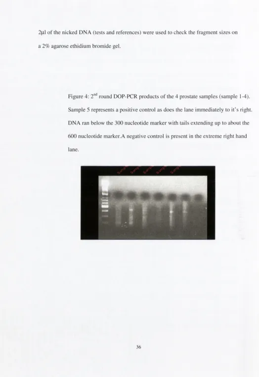
Figure 4:
2"^
round DOP-PCR products o f the 4 prostate samples (sample 1-4).
Sample 5 represents a positive control as does the lane immediately to it’s right
DNA ran below the 300 nucleotide m arker with tails extending up to about the
600 nucleotide marker.A negative control is present in the extreme right hand
2.3 CGH DNA Microarray
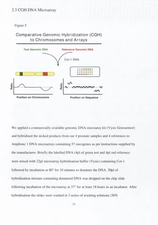
Figure 5
Com parative Genomic Hybridization (CGH)
to Chromosomes and Arrays
Test Genomic DNA
Reference Genomic DNA
C ot-1 DNA
o o o • o o o o
■
(0
cr
Position on Chromosome
- w V *I
Position on Sequence
We applied a com mercially available genomic DNA microaray kit (Vysis Genosensor)
and hybridised the nicked products from our 4 prostate samples and 4 references to
Amplionc 1 DNA microarrays containing 57 oncogenes as per instructions supplied by
the manufacturer. Briefly the labelled DNA (4|il o f green test and 4|il red reference
were mixed with 22|ll microarray hybridisation buffer (Vysis) containing Cot-1
followed by incubation at 80° for 10 minutes to denature the DNA. 30|J.l of
hybridisation mixture containing denatured DNA was dropped on the chip slide
[image:44.543.14.536.54.794.2]form am ide/2X SSC ) with incubations at 40° (10 m inutes each), and 4 series o f w ashing
solutions (IX SSC) at room tem perature and counterstained w ith 18|il 4 ’6-
diam idinophenylindole (D A PI) IV solution. (Vysis).
T he hybridised m icroarray slides w ere analysed w ith m icroarray reader and analysis
softw are (G enosensor A rray 300). G reen (test) to red (reference) (G /R) ratios were
autom atically determ ined for each sam ple and the norm alised G /R ratio w as taken to
represent the relative average num ber o f copies o f the sequence for those spots that
w ere selected as controls. Spots with G/R ratios m ore than the m ean plus three standard
deviations (-1 .3 ) were considered as am plifications or gains o f the indicated copy
num ber.
A control experim ent w as run in tandem w ith the experim ent using cell line DNA
(C osh) m ixture com prising 3 cell lines) provided by the m anufacturer with known
docum ented am plifications o f a variety o f oncogenes. O ur results w ere exactly
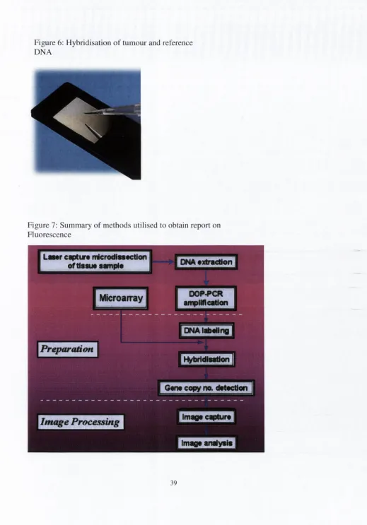
Figure 6: Hybridisation o f tum our and reference
DNA
Figure 7: Summary o f methods utilised to obtain report on
Fluorescence
LaMr captur* microdtn«ction
of tiSMM sanpi*
DNA •xtracUon
DOP^CR
amplM cation
Microarray
DNAI#>«llng
Preparation
Hybridlaatlon
Imaqa cipturt
[image:46.543.13.532.45.788.2]2.4 Evaluation of expression o f Topoisomerase gene (amplified on CGH
microarrays) by immunohistochemistry
Immunohistochemistry was used to evaluate the expression of one of the genes
(Topoisomerase II alpha gene) in which amplifications were detected on CGH microarrays.
Expression of this gene was evaluated in a larger cohort o f prostate cancer and benign prostatic
hyperplasia cases.
2.4.1 Patients and tumours used in immunohistochemical study
The immunohistochemical study included 100 patients with adenocarcinom a of the
prostate and 42 patients with benign prostatic hyperplasia (BPH). The adenocarcinoma
cases included 59 transurethral resection o f prostate (TURP) specim ens and 41 radical
prostatectom y specimens. The 42 BPH cases were transurethral resection of prostate
(TURP) specimens. These cases were diagnosed during the period 1999-2003 and were
identified by performing a SNOM ED code search of the Adelaide & M eath Hospital
Incorporating the National Children’s Hospital Cellular Pathology database. Formalin
fixed blocks from these cases were retrieved from the files. Tissue microarrays were
constructed as described previously.[l 11] 138 cases were present in triplicate and
included representative areas o f tum our or glandular tissue in cases o f BPH. Sufficient
reports which included clinical details were reviewed for all adenocarcinom a cases and
cases were classified into 4 categories benign prostatic hyperplasia (n=41), prostate
carcinom a Gleason Score 6 (n=28), prostate carcinoma Gleason Score 7-8 (n=35), and
prostate carcinoma Gleason Score 9-10 (n=37). We also compared prostate carcinomas
Gleason Score 8-10 under the categories o f hormone resistant (n=14) and hormone
responsive (n=14). The 14 cases categorised as hormone resistant were recurrent
prostate adenocarcinomas present in transurethral resection of prostate (TURP)
specim ens when clinical details indicated that the patient had received anti-androgen
treatment. These cases were Gleason Score 8-10. 14 cases of m atched Gleason Score
prostate carcinomas who had not received anti-androgen treatment were compared with
these cases using the U Mann Whitney test.
2.4.2 Immunohistochemical staining
Immunohistochemistry for Topoisom erase II-Alpha was performed using a monoclonal
anti- Topoisom erase II-Alpha antibody with a dilution o f 1:300. Sections o f 5|im
thickness were studied. Antigen retrieval was performed with dew axed sections using a
pressure cooker and the Trilogy system for 10 minutes. A standard avidin-biotin-
peroxidase complex technique (DAKO) was used for visualisation with
diam inobenzidine as a chromogen. Sections were counterstained with haematoxylin
and mounted. Tonsil samples from our routine files were used as positive controls for
2.4.3 Evaluation o f Immunohistochemistry
Im m unohistochem ical staining w as evaluated light m icroscopically using an X 40
objective and a M iller ocular. The M iller ocular eyepiece gave a square field, in the
com er o f w hich w as a sm aller ruled square, one-ninth the area o f the total square.
Nuclei w ith strong hom ogenous T opoisom erase II-A lpha staining w ere counted in the
large square and the total num ber o f nuclei, both positive and negative staining counted
in the sm all square. The num ber o f cells in the sm all square was m ultiplied by 9 to
obtain the total num ber o f cells in the large square. A m ean o f 240.5 epithelial cells
w ere counted for the prostate cancer cases and a m ean o f 88 epithelial cells counted for
the cases o f benign prostatic hyperplasia. 5 non-overlapping square fields w ere counted
for each tissue core, 3 in the y-axis and 2 in the x-axis. The percentage o f positively
staining nuclei w ithin the 5 large squares w as calculated for each core. In cases present
in triplicate an average result was obtained from 3 cores and in cases present in
duplicate an average was obtained from 2 cores. This result was the T opoisom erase II-
A lpha index for the case. This m ethod has been used in previous studies in the
evaluation o f im m unohistochem ical results in tissue m icroarrays.[ 1 12][113]
In addition each T opoisom erase II-A lpha index was obtained by im age analysis using
the Ariol system fo r tissue m icroarrays. An acceptable level o f concordance w as taken
as w ithin
10% o f TI index obtained using the M iller ocular.
O f 140 cases in total (138 present in triplicate and 2 present in duplicate) 14 cases
A second observer reviewed these discordant cases. This discordant group was
composed o f one Gleason score 7, two Gleason score 8, nine Gleason score 9, and two
Gleason score 10 cases. These 14 cases tended to have higher Topoisomerase indices
(mean TI o f 13%) using the Miller ocular than the average Topoisomerase index for all
99 carcinoma cases analysed (mean TI o f 3.69%).
Review of these discordant cases by a second observer using light microscopy and a
Miller ocular correlated within 10% of the original Topoisomerase indices obtained by
the first observer. The discrepancy between the Ariol image analysis results and the
light microscopy results was due to a difficulty by the image analyser in distinguishing
Topoisomerase negative tumour cells from stroma when there was infiltration o f the
stroma by less differentiated tumour. It therefore under estimated the number o f total
tumour cells within a field of analysis hence leading to an over-estimation o f % cells
positive for the Topoisomerase II alpha antibody.
Examples o f immunohistochemical staining results obtained are presented in Figures 8
A
C
E
•
V j f v .
vV.»
y - m r h
• V v V V .
-I
• /
^
• t*
.•*-
hB
'
•
•
•
♦
»
•
•
%
•
«\
D
1
i
*
%
I
f , ^ m
•
-•
1
*
1 *
*
‘ 1
Figure 8 A-F:
Im m unohistochem ical Staining for TOPO II-A lpha in
(A) Low pow er view o f TM A showing TO PO II-Alpha focal positivity in benign prostatic
hyperplasia (BPH).
(B) High pow er view o f a benign prostatic gland show ing focal TOPO II-Alpha nuclear positivity.
(C) Low pow er view o f TM A o f a prostatic adenocarcinom a (Gleason score 3-1-3) show ing focal
cell positivity for T O PO II-Alpha.
(D) High pow er view o f prostatic adenocarcinom a (Gleason score 3+3) showing focal discrete
nuclear TO PO II-A lpha positivity in prostatic acini.
(E)
Low pow er view o f TM A o f prostatic adenocarcinom a (Gleason score 9) showing m ultiple
cells positive for T O PO II-Alpha.
(F)
High pow er view o f prostatic adenocarcinom a (G leason score 9) showing discrete nuclear
[image:51.544.8.526.31.782.2]J* ,
•
>f
> s. •
'-^ i?
•
-
» • • ,
* ••
“
[image:52.542.7.533.42.778.2], ^.»» %
7 y
.
2.5 Statistics:
Data were entered on to a com puterised database and analysed by the SPSS statistical
CHAPTER THREE
Benign prostatic hyperplasia
Figure 10: M icroarrays
Localised prostate cancer
CGH MICROARRAYS
Androgen independent
prostate cancer
[image:55.544.11.537.13.781.2]3.1 ARRAY CGH ANALYSIS
Figure 11: CGH Microarray AmpliOnc
GenoSensor software
A report is generated for each array. This report tabulates among other variables the
assigned target number, target clone name, cytogenetic location and the normalised test
to reference intensity and a pooled correlation co-efficient. Normalised test to reference
intensities more than the mean plus three standard deviations (-1 .3 ) were considered as
am plications or gains of the indicated copy number.
One o f these cases (Gleason Score 9) had been treated with anti-androgens and was
[image:56.543.14.532.51.804.2]TURP surgical specimen with involvement o f prostate chippings by Gleason Score 7
prostate adenocarcinoma. The fourth sample was a TU RP surgical specimen with
benign prostatic hyperplasia (BPH) and no evidence o f prostatic carcinoma.
Figure 12: Example o f report of fluorescence ratios
generated by AmpliOnc GenoSensor software
A m p l i O n c ™ I
V i '
1 1 1 1 ■ I ■ I I ■ 1 1 1 1 1 1 1 I 1 1 1 ■ 1 1 1 1 1 1
[image:57.544.11.531.0.795.2]T able 5:
A m plifications o f the follow ing genes w ere identified for the case o f androgen
independent advanced prostate cancer:
G ene
N orm alised test to reference
intensity (fluorescence
ratio)
Pooled correlation co
efficient
M ET
(tyrosine kinase, am plified
in the transition betw een
prim ary tum ours and
m etastases)
4.27
0.91
HRAS
(Plays role in norm al
grow th and differentiation)
4.11
0.85
C B FA 2
(core binding factor runt
dom ain, involved in A M L)
3.96
0.93
TOP2A
(Involved in cell
replication)
3.63
0.85
FES
(feline sarcom a virus)
[image:58.542.20.533.49.776.2]Table 6:
A m plifications o f the follow ing genes were identified for the case o f m etastatic prostate
cancer:
G ene
N orm alised test to reference
intensity (fluorescence
ratio)
Pooled correlation co
efficient
FES
(feline sarcom a virus,)
3.42
0.86
M YB
(A lterations in M YB gene
in m ore than 1/3 o f hum an
solid tum ours)
2.19
0.87
B C L2 3 ’
(B locks the apoptotic death
o f cells ie. lym phoctes)
1.85
0.91
TOP2A
(Involved in cell
replication)
1.74
0.91
A R 5 ’
(Can facilitate tum our cell
grow th in low androgen
[image:59.545.5.528.21.782.2]
[image:60.543.27.537.52.677.2]
(61)