Original Article
Effects of L-carnitine on liver injury in rats and
its impact on blood lipids
Zhengguo Xia1,2*, Xixi Tian1*, Weichang Kong1, Xingzhao Li1, Junhui Song1, Chen Cai1, Qinglian Xu1
1Department of Burns, The First Affiliated Hospital of Anhui Medical University, Hefei City, Anhui Province, P. R.
China; 2Department of Burns and Plastic Surgery, The Fourth Affiliated Hospital of Anhui Medical University, Hefei
City, Anhui Province, P. R. China. *Equal contributors and co-first authors.
Received April 16, 2018; Accepted May 29, 2018; Epub September 15, 2018; Published September 30, 2018
Abstract: Objective: To explore mechanisms of anti-oxidant activity of L-carnitine on liver injury and the impact of L-carnitine on serum lipid levels in rats. Methods: Ninty normal male Wister rats were selected and randomly clas-sified into the blank control group (n=30), the placebo group (n=30) and the L-carnitine group (n=30). The rats in the L-carnitine group and the placebo group were intraperitoneally injected with endotoxin lipopolysaccharide at 0.1 mg/kg. Apart from that, those in the L-carnitine group also received concomitant L-carnitine at 1 g/kg by an intragastric route, while those in the placebo group were given equal amounts of normal saline by an intragastric route. Those in the blank control group were administered equal amounts of normal saline. The rats in the three groups were compared in the liver injury indicators, antioxidant-related indicators, inflammatory cytokines, and serum lipid content. Results: Compared with the blank control group, the serum aspartate aminotransferase (AST), alanine transaminase (ALT), malonialdehyde (MDA), low-density lipoprotein cholesterol (LDL-C), tetracycline (TC) and triglyceride (TG) levels in rats of the L-carnitine group and the placebo group were significantly elevated. Serum high-density lipoprotein cholesterol (HDL-C), superoxide dismutase (SOD), and and glutathione peroxidase (GSH-Px) lev-els were reduced. Nitric oxide (NO) content, nitric oxide synthase (NOS) activity, interleukine-6 (IL-6), interleukine-1β (IL-1β), and tumor necrosis factor-α (TNF-α) levels in hepatic tissue were markedly enhanced in rats of the L-carnitine group and the placebo group (all P<0.001). Compared to the rats in the placebo group, those in the L-carnitine group showed significantly lower serum AST, ALT, MDA, LDL-C, TC, and TG levels, considrably elevated serum HDL-C, SOD, and GSH-px levels, and reduced NO contents, NOS activity, IL-6, IL-1β, and TNF-α levels in hepatic tissue (all P<0.001). Conclusion: L-carnitine exerts a protective effect on endotoxin lipopolysaccharide-induced liver injury, and its protective mechanisms are associated with its anti-oxidant activity and reduced levels of inflammatory cytokines.
Keywords: L-carnitine, liver injury, antioxidation, blood lipid, inflammatory cytokine
Introduction
Liver injury is a complex pathophysiological pro-cess mediated by multiple factors. Increasing evidence shows that the pathogenesis of most liver disorders is related to involvement of reac-tive oxygen species (ROS) [1, 2]. Endotoxin acts on the body to produce a broad range of active molecules (such as ROS) which induce mem-brane damage and mitochondrial structure destruction of hepatocytes, and decreased liver metabolism, eventually leading to apopto-sis or necroapopto-sis of hepatocytes in rats [3, 4]. Multiple studies indicate that botulinum toxin can specifically bind to liver macrophages, transmit cell signals and irritate cells to
activa-tion of inflammatory pathways, ultimately lead -ing to liver injury.
L-carnitine is a water-soluble amino acid de- rivative that is widely found in the tissues of the body. It primarily functions to promote β-oxidation of long-chain fatty acids, and scav -enge free radicals as an anti-oxidant activity [7, 8]. If reactive oxygen species (ROS) production exceeds the scavenging capacity of the anti-oxidant system in the body, it may induce lipid peroxidation, protein denaturation, and gene mutation, eventually leading to oxidative dam-age to the cells. Previous evidence indicates the major mechanisms for carnitine to reduce liver injury are reducing the production and transport of tumor necrosis factor (TNF), pre -venting cell membrane damage, and improving cell permeability while blocking the activation of platelet activity factors, inhibiting the synthe-sis and release of vasoconstrictor substances and reducing portal hypertension [9-12]. However, mechanisms for carnitine antioxidant protection in endotoxin-induced liver injury are rarely reported. In this study, rat models of endotoxin-induced liver injury were construct- ed and assigned to receive L-carnitine and pla-cebo interventions, respectively; meanwhile, additional healthy rats were taken as blank controls to study the effect of L-carnitine on blood lipids and the mechanisms of anti-oxida-tion in rats with liver injury, with an aim to pro-vide experimental epro-vidence for the use of L-carnitine in treating liver injury.
Materials and methods
Experimental methods
A total of 90 normal male Wister rats with 12 weeks of age and a weight of 180-220 gra-ms were enrolled and randomly divided into the blank control group, the placebo group, and the L-carnitine group. The rats in the L-carni- tine group and the placebo group were intra-peritoneally injected with endotoxin lipopoly-saccharide at 0.1 mg/kg. Additionally, the rats in the L-carnitine group also received cocomi-tant L-carnitine at 1 g/kg by intragastric route, whereas those in the placebo group were intra-gastrically given equal amounts of normal saline. Those in the blank control group received equal amounts of normal saline. All the rats were administered once daily for 3 weeks.
Outcome measures
Determination of serological indicators: Three weeks after intervention, 2 ml of blood was drawn from the tail vein of each rat in all the groups after 12-hour fasting, placed in an anticoagulant tube, and centrifuged at 3000 r/min for 10 min. Subsequently, the serum was separated, and stored at -20°C for testing. The alanine transaminase (ALT), aspartate aminotransferase (AST), and blood lipid levels were detected with an automatic biochemi- cal analyzer (Olympus, Japan), while supero- xide dismutase (SOD), malondialdehyde (MDA), and glutathione peroxidase (GSH-Px) levels were determined by the biochemical enzyme assays. Assay kits were purchased from Sig-ma, USA, and were used strictly following the instructions.
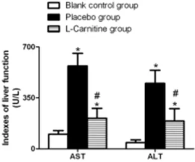
[image:2.612.90.286.73.236.2]Detection of nitric oxide (NO) content, nitric oxide synthase (NOS) activity, and inflammato-ry cytokines in liver tissue: Three weeks aft-er intaft-ervention, rats waft-ere sacrificed undaft-er anesthesia. Hepatic tissue samples were har-vested after laparotomy, weighed, homoge-nized at low temperature, and centrifuged at 4000 r/min for 10 minutes. Supernatant was removed and stored at -20°C for testing. The nitrate reductase-based colorimetric assay was employed for detection of the NO cont- ents. The NO kits were purchased from R&D Science, USA, and the testing procedures were strictly followed the instructions on the kits. Figure 1. Effect of L-carnitine on the serum ALT and
The NOS activity was detected with the use of a colorimetric assay, and the NO syntha- se kits were purchased from Sigma, USA. The procedures were performed according to the instructions on the kits. The absorban-ce was measured at a wavelength of 530 nm, and NOS activity was calculated on the base of the absorbance values. The levels of inflammatory cytokines were detected by the enzyme-linkedimmunosorbent assay (EL-ISA). Kits for interleukin-6 (IL-6), interleu kin-1β (IL-1β), and tumor necrosis factor-α (TNF-α) detection were purchased from R&D Science, USA.
and the placebo group were markedly higher than those of the rats of the blank control group (all P<0.001), and the serum ALT and AST levels in the L-carnitine group was remarakbly lower than those in the placebo group (P<0.001), as illustrated in Figure 1.
NO contents and NOS activity in hepatic tissue of rats
[image:3.612.97.520.72.243.2]Greater improvements in the NO contents and NOS activity in hepatic tissue of rats were noted in the L-carnitine group and the placebo group than the blank control group (all P< 0.001). Compared with the placebo group, the
Figure 2. Comparison of the NO contents and NOS activity in the hepatic tissue of rats of all groups. *P<0.001, compared to the blank control group; #P<0.001, compared to the placebo group.
Statistical analysis
All the data were analyzed using the SPSS software, ver-sion 18.0. Quantitative data with normal distribution are presented as mean ± sd, and the differences across the three groups were analyzed by oneway analysis of vari-ance (ANOVA) with post-hoc Bonferroni test. P<0.05 was deemed as significant.
Results
Effect of L-carnitine on serum ALT and AST levels in rats with liver injury
[image:3.612.90.379.305.363.2]Serum AST and ALT levels in rats of the L-carnitine group
Table 1. Serum SOD, MDA and GSH-px levels in rats of all groups
Groups SOD (U/mL) GSH-Px (U/mL) MDA (U/mL)
Blank control group 281.21±13.82 405.36±15.82 3.73±0.72 Placebo group 184.25±11.36* 318.3±13.19* 7.51±1.03* L-carnitine group 228.53±12.27*,# 376.9±12.45*,# 4.64±0.85*,#
Note: SOD denotes superoxide dismutase; GSH-Px, glutathione peroxidase; MDA malonialdehyde.*P<0.001, compared with the Blank control group; #P<0.001, compared with the placebo group.
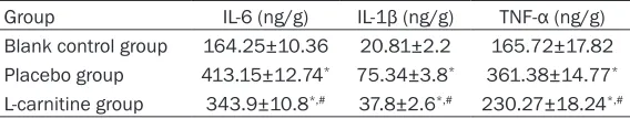
Table 2. Inflammatory cytokines levels in the hepatic tissue of rats of all groups
Group IL-6 (ng/g) IL-1β (ng/g) TNF-α (ng/g)
Blank control group 164.25±10.36 20.81±2.2 165.72±17.82 Placebo group 413.15±12.74* 75.34±3.8* 361.38±14.77* L-carnitine group 343.9±10.8*,# 37.8±2.6*,# 230.27±18.24*,#
[image:3.612.91.375.440.494.2]L-carnitine groups could significantly inhibit the increases in the NO contents and NOS activity (P<0.001; Figure 2).
Serum SOD, MDA and GSH-px levels of rats
Lower SOD and GSH-px levels, but higher MDA levels were observed in the placebo group than those in the blank control group and the L-carnitine group (all P<0.001). The rats in the L-carnitine group showed significant higher MDA levels than those in the blank control group, as well as lower SOD and GSH-px levels (all P<0.001; Table 1).
Levels of inflammatory cytokines in the hepatic tissue of rats
The levels of IL-6, IL-1β and TNF-α in hepatic tissue of rats in the placebo group were consid-erably higher than those in the blank control group (all P<0.001). The levels of IL-6, IL-1β and TNF-α were profoundly lower in the L-carnitine group than in the placebo group (all P<0.001;
Table 2).
Serum lipid levels in rats of all groups
Higher serum low-density lipoprotein choles-terol (LDL-C), tetracycline (TC), and triglyceride (TG) levels, and lower high-density lipoprotein cholesterol (HDL-C) levels were seen in the pla-cebo group than in the blank control group (all P<0.001). The L-carnitine group had significant -ly reduced serum LDL-C, TC and TG levels and elevated serum HDL-C levels compared to the placebo group (all P<0.001; Table 3).
Discussion
When various factors induce hepatocyte ne- crosis, the morphology and physiological fu- nctions of the liver membrane are destroyed, and various enzymes in the cells appear in the serum [13, 14]. It has been reported that
[15]. The results of the present study indicate that ASL and ALT levels are significantly elevat -ed in the placebo group, indicating that rat models of endotoxin-induced liver injury were successfully constructed. Abnormal blood li- pid metabolism is associated with liver injury [16]. Oxidative stress mediated by free fatty acids at high concentrations has shown to be closely linked to lipoapoptosis of liver cells [17]. In our current study, the TG, TC and LDL-C levels were significantly elevated and the HDL-C level was remarkably decreased in the placebo group, implying that impairment of liver func-tion affects the lipid metabolism in the body. Three weeks after L-carnitine medication by intragastric route, elevation of AST, ALT, TG, TC and LDL-C levels were significantly inhibited and the HDL-C levels were remarkably elevated in the rats of the L-carnitine group, implying that L-carnitine can protect the liver from dam-age, improve the lipid levels, and reduce lipotoxicity.
[image:4.612.90.414.85.139.2]Recent studies indicate that antioxidant me- chanisms play a crucial role in the protection of L-carnitine against tissue damage [18]. NO, a highly reactive free radical, acts as both a neurotransmitter and a second messenger to mediate endotoxin, TNF and other cytokines. NOS, a synthase that catalyzes NO produc- tion by L-arginine and O2, is extensively pre- sent in tissues including the liver. Evidence shows that endotoxin is the major cause for stimulating NO synthesis in liver cells [19]. In the current study, NO and NOS levels in rats with liver injury in the placebo group were remarkably higher than those in the blank control group, suggesting that NO and NOS were implicated in liver injury. Nevertheless, the NO and NOS levels of rats in the L-carni- tine group were substantially lower than those in the placebo group, implying a protective effect of L-carnitine on rats with liver injury might be related to the NO and NOS levels,
Table 3. Serum lipid levels in rats of all groups
Groups LDL-C (mmol/L) HDL-C (mmol/L) TC (mmol/L) TG (mmol/L) Blank control group 1.38±0.15 0.95±0.04 1.93±0.13 0.71±0.08 Placebo group 3.85±0.22* 0.25±0.03* 7.62±0.32* 1.27±0.11* L-carnitine group 2.41±0.18*,# 0.59±0.05*,# 4.23±0.25*,# 0.92±0.09*,#
Note: LDL-C denotes low-density lipoprotein cholesterol; HDL-C, high-density lipoprotein cholesterol; TC, tetracycline; TG, triglyceride. *P<0.001, compared to the Blank control group; #P<0.001, compared to the placebo group.
which is in line with the results reported by Babicova et al. [20].
Under normal conditions, the scavenging and production of free radicals maintain homeosta-sis in the body. Changes in the SOD activity, MDA contents, and GSH-Px activity can reflect the capacities of anti-oxidation and free radical scavenging [21, 22]. SOD, one of the most important antioxidant enzymes in the defense system against oxidative damage, can reflect the body’s ability to scavenge oxygen free radi-cals. MDA, an essential end product of lipid peroxidation, reflects the severity of damage to the tissues and cells by free radicals. GSH-Px, a dominant enzyme that catalyzes the decom-position of hydrogen peroxide, can specifically catalyze the reduction of hydrogen peroxide by reduced glutathione, thereby playing a role in protecting the structure and functions of the cell membrane. In this study, rats in the L-carnitine group had significantly lower MDA levels than those in the blank control group, as well as markedly higher SOD and GSH-px levels than those in the placebo group, suggesting that L-carnitine confers anti-oxidant protection in endotoxin-induced liver injury, which is simi-lar to the result reported in previous literature [23].
The liver is the most vulnerable organ in endo-toxemia patients. Endotoxins going into the liver along with the blood circulation can di- rectly induce the liver Kupffer cells to release a variety of inflammatory mediators and cyto -kines which include IL-6, IL-1β, TNF-α and free radicals. Inflammatory mediators and cyto -kines play decisive roles in the pathogenesis of the liver, and act together with endotoxin to induce hepatic stellate cells and vascular endo-thelial cells to further release more inflamma -tory mediators, leading to cascade amplifica -tion effect [24]. The results of the current study reveal that the use of L-carnitine considerably can reduce elevation of IL-6, IL-1β and TNF-α levels in endotoxin-induced liver injury, down-regulate over-expression of inflammatory cyto -kines, and reduce the inflammatory response
in vivo.
In summary, L-carnitine intervention for endo-toxin lipopolysaccharide-induced liver injury in rats, significantly improved the liver antioxidant contents, enhanced SOD content and GSH-PX activity, and reduced MDA, NO and NOS
expres-sion. Moreover, it also reduced expression of inflammation cytokines, providing more experi -mental evidence for the clinical treatment of liver injury.
Acknowledgements
This work was supported by the Key Techno-logy R&D Program of Anhui province (NO. 1604a0802083).
Disclosure of conflict of interest
None.
Address correspondence to: Qinglian Xu, Depart- ment of Burns, The First Affiliated Hospital of An-hui Medical University, No. 218 Jixi Road, Shushan District, Hefei 230022, Anhui Province, P. R. China. Tel: +86-0551-62923503; Fax: +86-0551-6292-3503; E-mail: qinglianxu05@163.com
References
[1] Amirtharaj GJ, Natarajan SK, Pulimood A, Bala-subramanian KA, Venkatraman A and Ram -achandran A. Role of oxygen free radicals, ni-tric oxide and mitochondria in mediating cardiac alterations during liver cirrhosis in-duced by thioacetamide. Cardiovasc Toxicol 2017; 17: 175-184.
[2] Gusdon AM, Song KX and Qu S. Nonalcoholic fatty liver disease: pathogenesis and thera-peutics from a mitochondria-centric perspec-tive. Oxid Med Cell Longev 2014; 2014: 637027.
[3] Zhou RJ, Ye H, Wang F, Wang JL and Xie ML. Apigenin inhibits d-galactosamine/LPS-in-duced liver injury through upregulation of he-patic Nrf-2 and PPARgamma expressions in mice. Biochem Biophys Res Commun 2017; 493: 625-630.
[4] Ouelaa W, Jegatheesan P, M’Bouyou-Boungou J, Vicente C, Nakib S, Nubret E and De Bandt JP. Citrulline decreases hepatic endotoxin-in-duced injury in fructose-inendotoxin-in-duced non-alcoholic liver disease: an ex vivo study in the isolated perfused rat liver. Br J Nutr 2017; 117: 1487-1494.
[5] Wang T, Wang Z, Yang P, Xia L, Zhou M, Wang S, Du J and Zhang J. PER1 prevents excessive innate immune response during endotoxin-in-duced liver injury through regulation of macro-phage recruitment in mice. Cell Death Dis 2016; 7: e2176.
mi-tochondrial oxidative phosphorylation. Bio-chem Biophys Res Commun 2016; 469: 1083-1089.
[7] Tamai I. Pharmacological and pathophysiolo- gical roles of carnitine/organic cation trans-porters (OCTNs: SLC22A4, SLC22A5 and Sl-c22a21). Biopharm Drug Dispos 2013; 34: 29-44.
[8] Tein I. Carnitine transport: pathophysiology and metabolism of known molecular defects. J Inherit Metab Dis 2003; 26: 147-169.
[9] Bodaghi-Namileh V, Sepand MR, Omidi A, Aghsami M, Seyednejad SA, Kasirzadeh S and Sabzevari O. Acetyl-l-carnitine attenuates arse-nic-induced liver injury by abrogation of mito-chondrial dysfunction, inflammation, and apoptosis in rats. Environ Toxicol Pharmacol 2018; 58: 11-20.
[10] Liu T, He W, Yan C, Qi Y and Zhang Y. Roles of reactive oxygen species and mitochondria in cadmium-induced injury of liver cells. Toxicol Ind Health 2011; 27: 249-256.
[11] Triggiani M, Oriente A, Golino P, Gentile M, Battaglia C, Brevetti G and Marone G. Inhibi-tion of platelet-activating factor synthesis in human neutrophils and platelets by propionyl-L-carnitine. Biochem Pharmacol 1999; 58: 1341-1348.
[12] Pugliese D, Sabba C, Ettorre G, Berardi E, An-tonica G, Godi L, Palasciano G, Lee SS and Al-bano O. Acute systemic and splanchnic hae-modynamic effects of L-carnitine in patients with cirrhosis. Drugs Exp Clin Res 1992; 18: 147-153.
[13] Bjorndal B, Alteras EK, Lindquist C, Svardal A, Skorve J and Berge RK. Associations between fatty acid oxidation, hepatic mitochondrial function, and plasma acylcarnitine levels in mice. Nutr Metab (Lond) 2018; 15: 10. [14] Shiraki M, Shimizu M, Moriwaki H, Okita K and
Koike K. Carnitine dynamics and their effects on hyperammonemia in cirrhotic Japanese pa-tients. Hepatol Res 2017; 47: 321-327. [15] Jia YN, Lu HP, Peng YL, Zhang BS, Gong XB, Su
J, Zhou Y, Pan MH and Xu L. Oxyresveratrol pre-vents lipopolysaccharide/d-galactosamine-in- duced acute liver injury in mice. Int Immuno-pharmacol 2018; 56: 105-112.
[16] Saied NM and Hamza AA. Selenium amelio-rates isotretinoin-induced liver injury and dys-lipidemia via antioxidant effect in rats. Toxicol Mech Methods 2014; 24: 433-437.
[17] Yamaguchi K, Yang L, McCall S, Huang J, Yu XX, Pandey SK, Bhanot S, Monia BP, Li YX and Diehl AM. Inhibiting triglyceride synthesis im-proves hepatic steatosis but exacerbates liver damage and fibrosis in obese mice with nonal -coholic steatohepatitis. Hepatology 2007; 45: 1366-1374.
[18] Sun R, Zhang J, Wei H, Meng X, Ding Q, Sun F, Cao M, Yin L and Pu Y. Acetyl-l-carnitine par-tially prevents benzene-induced hematotoxici-ty and oxidative stress in C3H/He mice. Envi-ron Toxicol Pharmacol 2017; 51: 108-113. [19] Wen Z, Liu Y, Li F and Wen T. Low dose of car
-bon monoxide intraperitoneal injection pro-vides potent protection against GalN/LPS-in-duced acute liver injury in mice. J Appl Toxicol 2013; 33: 1424-1432.
[20] Babicova A, Havlinova Z, Hroch M, Rezacova M, Pejchal J, Vavrova J and Chladek J. In vivo study of radioprotective effect of NO-synthase inhibitors and acetyl-L-carnitine. Physiol Res 2013; 62: 701-710.
[21] Yang GL, Jia LQ, Wu J, Ma YX, Cao HM, Song N and Zhang N. Effect of tanshinone IIA on oxida-tive stress and apoptosis in a rat model of fatty liver. Exp Ther Med 2017; 14: 4639-4646. [22] Lu Y, Luo Q, Cui H, Deng H, Kuang P, Liu H,
Fang J, Zuo Z, Deng J, Li Y, Wang X and Zhao L. Sodium fluoride causes oxidative stress and apoptosis in the mouse liver. Aging (Albany NY) 2017; 9: 1623-1639.
[23] Sener G, Paskaloglu K, Satiroglu H, Alican I, Kacmaz A and Sakarcan A. L-carnitine amelio -rates oxidative damage due to chronic renal failure in rats. J Cardiovasc Pharmacol 2004; 43: 698-705.