Original Article
Case analysis of triangular bracket from autogenous
nasal septal and auricular cartilage for the
correction of short nose
Li Wang1, Yueyuan Gan1, Hui Sun2, Xu Huang1
1Department of Cosmetic Surgery, Suzhou Belief Huamei Cosmetic Hospital, Suzhou 215000, Jiangsu Province,
China; 2Department of Dermatology, Nanjing Youyi Plastic Surgery Hospital, Nanjing Medical University, Nanjing,
Jiangsu Province, China
Received December 11, 2017; Accepted December 29, 2017; Epub February 15, 2018; Published February 28, 2018
Abstract: Objective: To evaluate the clinical efficacy of short nose correction which was performed by transplanting the autogenous nasal septal cartilage combination with auricular cartilage. Methods: One hundred women (from 18 to 40 years old) with short nose were enrolled in our hospital. All these participants showed short nasal tip, low and flat nasion, obtuse nostril, and short nasal dorsum. Flexible transplantation of autogenous nasal septal cartilage combination with auricular cartilage and implantation of nasal prosthesis were used to reconstruct the support structure, and provide enough forward and downward support forces for nasal tip. Also, this approach could increase the length of nose, and correct the topspin of nasal tip. Meanwhile, auricular cartilage with shield and cap-form grafts were employed to promote the shape of nasal tip. The clinical efficacy and complication were also assessed. Results: The elongated nasal dorsum and normal nasolabial angle were achieved in all participants. After 3 to 12 months following-up, participants showed stable nasal profile, except 2 cases without enough lengthen of nasal tip. There was no complication, including infection, prosthetic deflection, graft cartilage and prosthesis ex -posure, perforation of nasal septal, dorsal nasal sag, secondary deformities, and skin damage. Conclusion: Strong forward and downward support forces were needed for nasal tip, which could extend the short nose effectively. Reconstruction the bracing structure of the lower part of the nose was also necessary. The shield and cap-form of cartilage grafts could further promote the efficacy of correction.
Keywords: Short nose, nasal septal cartilage, auricular cartilage, transplantation, low nose
Introduction
Many Asian noses show characteristics inclu- ding low and flat nasion and nasal dorsum, bulbous nasal tip, clear nostril exposure, short nasal columella, soft nasal cartilage bracket, and sick skin soft tissue. The short nose ge- nerally means that, the length from nasion to nasal tip is below 1/3 of facial length [1]. In order to correct the malformation of short no- se, the nasal columella, alar, lateral and septal cartilage, bone, skin and mucous membrane
are involved in the operation. The difficulties
and key points of this technique are the con-struction of strong bracing structure from au- togenous cartilage, then achieving backspin and increasing bulges of nasal tip. At present,
mainly include septal extension, osteotherapy, and the cosmetic treatment [2-5]. As the plas- tic cartilage, the auricular cartilage is soft and has natural radian, which is used as graft in nasal tip with shield or cap-form. For the nasal septal cartilage, the harder and straightness features are suitable for the reconstruction of nasal tip with strong framework [6-8]. Thus, the nasal tip bracket from nasal septal cartilage and auricular cartilage for the correction of short nose was accepted by more and more clinicians.
However, as the main difficulty in the plastic
surgery for nasal, the correction of short nose
is hard to maintain the efficacy in long time
sal septal cartilage and auricular cartilage for the correction of short nose was employed. The height and projection of the nasal were increased, and the length of nasal dorsum was
extended significantly.
Materials and methods
Participant information
This study has got approval from local ethical committee. One hundred women (from 18 to 40 years old) with short nose were enrolled in our hospital from December 2015 to August
2017. The participants understood and sign- ed the informed consent. The diagnostic crite-ria included short nasal tip, low and flat na-sion and nasal dorsum, short nasal columel- la, obtuse nostril, overlarge nasolabial angle, and no obvious malformation on nasal bone. The inclusion criteria were consisted of (A) meeting the diagnostic criteria which was list- ed above, (B) health without underlying diseas-es, (C) tolerance for corrective surgery. The ex- clusion criteria were consisted of (A) non-com-pliance with the diagnostic criteria above, (B) surgical and anesthetic contraindications, (C) history of short nose correction.
Surgery program
The endotracheal general anesthesia or total intravenous anesthesia combined with block anesthesia for bilateral infraorbital foramen
and local infiltration anesthesia were
employ-ed in the surgery. The nasal columella and bi-
lateral nasal alar were dissected. Local
infil-tration anesthesia (epinephrine and 2% lido-caine buffer, with 1:200,000 dilution) was per-formed for the front boundary with marked lines of left ear and the nasal. The front boun- dary with marked lines was separated, and the cartilage (around 10 mm*20 mm) was ta- ken out (Figure 1). After electrocautery, the skin was stitched by 6/0 nylon lines disconti- nuously. Then 3/0 nylon lines were used as ear packing and compression bandage.
Then the bottom of nasal columella was divid-ed as W-form. Both ends were folddivid-ed up along the inner columella to 1/2 length of nasal alar. After throughout of the nasal column by scis-
sor and uncover the skin flap, inner, outer and
fornix parts of bilateral nasal alar cartilage we-
re exposed. The fiber linker between bilateral
[image:2.612.90.288.72.272.2] [image:2.612.90.290.314.499.2]fornix was separated. Throughout the inner of bilateral nasal alar cartilage, the trailing edge of nasal septal cartilage was revealed. The mucous membrane and perichondrium of bi- lateral trailing edge were separated. After se- parating the perpendicular plate of the eth- moid with clinging the perichondrium, the sep-tal cartilage was exposed completely. With the parallel cutting on 10 mm away from leading edge of nasal septal cartilage, the cartilage (10 mm*20 mm) was separated by rotary cutter (Figure 1). The integrity of nasal mucous me- mbrane needed to be protected in the surgery.
Figure 1. Cartilage graft example.
The separated cartilage was then fixed on the
top of remaining nasal septal cartilage by stit- ching with 5-0 PDS line. It would lengthen no- se, and provide a strong support force. After further processing, the separated auricular cartilages included two pieces of long strips (around 20 mm long), cap and shield-shaped cartilages. One piece of cartilage with long st-
rip was fixed on the bottom of nasal septal
cartilage, another piece was then embedded in the inner of nasal alar cartilage to heighten the nasal tip. The cap and shield-form cartilag-es could also transplant on the top of columel- la as needed, which could lengthen and hei- ghten the nasal tip furtherly. For the
partici-pants with low and flat bridge of the nose, the
augmentation was employed to assist
rhino-plasty. The skin flap of nasal tip and columella
was covered the area of nasal tip without ten-sion. The 6-0 nylon line was used to suture the incision discontinuously. The gelatin sponge was embedded between the two nostrils to in-
duce the septal and soft tissue fitting together. The nasal dorsum was fixed through tape
first-ly. Then nasal splint was prepared by the soft thermoplastic plate after warming. It would
pro-vide reliable fixation for the rhinoplasty (Figure 2).
Cold compress (4-5 times, 20 min for each ti- me) was employed after the surgery. Medicine was changed locally 24 h later. Then the nasal
fillings were taken out, and the incision was
cleaned 48 h later after the surgery. The ban-dage on ear was taken out 72 h later after the surgery. Routine antibiotic treatment was giv- en for 3-5 d. The stitches and nasal splint we- re taken out 7 d later after the surgery.
Outcome measures
The main outcome measures were the length extended of the participant’s nose, and the satisfaction of participant. The secondary out-come measures included the appearance ef-
ficacy of rhinoplasty, and occurrence rate of
Data analysis
The data analysis was performed by SPSS. 22.0. The measurement data were expressed as mean ± standard deviation and the com- parison adopted t test. P<0.05 considered a
significant difference.
Results
Evaluation of nasal elongation efficacy
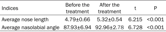
An elongation of 4-7 mm was observed in all 100 participants as the average nose leng- th was 4.79±0.66 before the treatment and
5.32±0.54 after the treatment with significant
difference (P<0.001). The exposure of the
nos-trils was significantly improved. The topspin
of nasal tips was corrected completely. The average nasolabial angle was 87.93±6.94 be- fore the surgery and 92.96±2.78 after the sur-gery, which was corrected to normal level (P< 0.001, Table 1). And the participants were sa-
tisfied with the nasal tip shape and height. After 6 to 12 months following-up, participants sh- owed stable nasal profile (especially for the nasal length), except 2 cases without enough lengthen of nasal tip.
Evaluation of complications
There was no complication, including infection, prosthetic deflection, graft cartilage and pros -thesis exposure, perforation of nasal septum, dorsal nasal sag, secondary deformities, and skin damage.
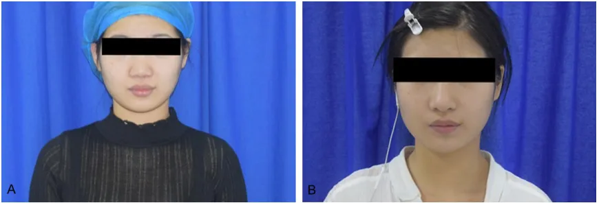
Typical case
As shown in Figure 3, the length and height of
nasal tip was significantly extended, which in-dicated the good efficacy of correction.
Discussion
[image:3.612.90.361.96.149.2]According to the requirement of nasal exten-sion, different correction methods were chosen Table 1. Comparison of the average nose length and
nasola-bial angle before and after the treatment
Indices Before the treatment treatmentAfter the t P Average nose length 4.79±0.66 5.32±0.54 6.215 <0.001 Average nasolabial angle 87.93±6.94 92.96±2.78 6.728 <0.001
for the short nose. The ideal ratio of nasal and middle face length is 0.67. Normally, compar- ed with ideal nasal length, 1.0-3.0 mm less of
that was defined as mild short nose. The clas-sical features include low, flat and obtuse na-sal tip, without low and flat nasion and nana-sal
dorsum. Moreover, 3.0-5.0 mm less was de-
fined as moderate short nose, which shows
drooping nasal tip, and the ratio of height and length of nose less than 1/2. The backspin of releasing lower lateral cartilage or other carti-lages (such as nasion, nasal tip, or dorsum)
can be employed as fillings for the nasal tip
structure adjustment to improve the nasal len- gth. For the moderate short nose (3.0-5.0 mm less of ideal nasal length obtuse nasal tip wi- th topspin, exposure of nostrils, low and flat of nasion and nasal dorsum severely) and severe short nose (5 mm or above), the exclusive use of previous approach cannot extend the leng- th of nasal dorsum, and promote nasolabial angle. The cartilage transplantation and nasal framework reconstruction are necessary for the correction. To achieve the successful rhino-plasty, the lower framework of nasal needs to be reconstructed to improve the support force on up and front of nasal tip. The inner, middle, and outer linkers of nasal alar cartilage, and the connections between alar cartilage and lat-eral nasal cartilage were loosen completely in the surgery to provide enough space for the nasal extension [9-12].
Using cartilage transplantation for the recon-struction of the nasal support structure, the nasal tip could be moved forward, then the short nose could be extended. As the most commonly used method, the nasal septal car-
tilage transplantation was employed in short
nose extension [13-15]. After fixing the graft
cartilage on the septum, the extended cartila- ge would suffer tension from soft tissue of nasal tip. The remaining L-form structure is the thinnest part when taking out the middle and top side of the nasal septal cartilage. If there is not enough support force on nasal cartilage framework, the graft would be distorted easily [16, 17]. One of the morphological features of Asian nasal cartilage framework is the small, thin, and short septal cartilage. The extension graft would induce the unstable of nasal car- tilage when the support force is not enough
(thin, soft or deflected framework). Meanwhile,
[image:4.612.89.524.71.219.2]the drooping nasal tip, deviation of nasal alar, and distortion of nasal columella would appear if the framework was unstable [18, 19]. The L and X-form cartilage grafts were designed in the previous studies, which resulted in good performance for nasal tip elevation and exten-sion [16, 20]. With the best structural streng- th in theory, the stability of triangle bracket is believed as the best. Distortion or deviation is not easily appeared on this support arm for nasal tip reconstruction, then the most stable structure was formed to avoid the deviation of nasal tip framework after surgery. In order to keep the support function of septal cartilage in forward part of nose, the limited cartilage could be taken out. It also limited the cons- truction of framework if only using septal car- tilage. Thus, combined the auricular cartilage with septal cartilage were employed together for the construction of nasal tip framework, which resulted in good performance in this study.
In addition, malleability of mucous and carti-lage membranes in nasal cavity were evaluat- ed seriously before the surgery. The nasal so- ft tissue was separated and loosed widely fr- om the cartilage membrane. The fully loosed nasal skin with mucous and cartilage mem-brane would prolong the nasal tip effectively and avoid the damage from overlarge pressure
for skin of nasal tip. Moreover, the fiber
con-nections between nasal alar cartilage and lat-eral cartilage also needed to loosen adequa- tely. The fully loosing between soft tissue and cartilage was an important foundation for na- sal tip extension.
In conclusion, the triangular bracket from au- togenous nasal septal cartilage and auricular
cartilage showed good efficacy for the correc -tion of short nose. This approach not only reconstructed the framework of nasal tip with
good efficacy, but also avoid the excessive cut -ting the nasal septal cartilage, which reduced the complication and increased the safety of short nose extension surgery. As the limited participants and surgeries, the conclusion still need to be investigated in the future studies. Disclosure of conflict of interest
None.
Address correspondence to: Xu Huang, Depart- ment of Cosmetic Surgery, Suzhou Belief Huamei Cosmetic Hospital, No.658 Donghuan Road, Su- zhou 215000, Jiangsu Province, China. Tel: +86-13041489669; E-mail: [email protected]
References
[1] Jung DH. Modern rhinoplasty in Korean. Liaon-ing Science and Technology PublishLiaon-ing House 2005; 26-27.
[2] Xu JM. The application of auricular cartilage with EPTFE in rhinoplasty. Chinese Medical Cosmetology 2016; 6: 3-5.
[3] Liu WT, Wu XM, Zhang YF. EPTFE combined with aural cartilage graft to lengthen and aug-ment nasal tip. Chinese Journal of Aesthetic Medicine 2017; 26: 58-61.
[4] Lobar PC, Adams WP, Mitchell CA. Lengthening the short nose. Clin Plast Surg 2010; 37: 327-33.
[5] Wang T, Chen XP, Lin JD, Zheng XY, Shi CL. Aes-thetic lengthening procedure for foreshorted nose. Chinese Journal of Aesthetic and Plastic Surgery 2011; 22: 456-459.
[6] Xiao XY, Li D, Mo HY, Liang J, Liang HJ. Flexible strategy for lengthening short nose with nasal septal cartilage and auricular cartilage. Chi-nese Journal of Aesthetic Medicine 2016; 25: 22-26.
[7] Xiong JW, Xu WJ. Application and efficacy of
combined nasal septal cartilage with aural car-tilage for Asian nasal tip. Chinese Medical Cos-metology 2016; 6: 20-22.
[8] Liang G, Wang SS, Zhang ZH, Yi H, Hou C. The effect study of nasal septal cartilage applica-tion in rhinoplasty and repair augmentaapplica-tion rhinoplasty surgery. Medical Innovation of Chi-na 2017; 6: 29-32.
[9] Jang YJ, Yu MS. Rhinoplasty for the Asian nose. Facial Plastic Surgery 2010; 26: 93-101. [10] Chen WL, Fan JC. Deformity and treatment of
short nose. Chinese Journal of Aesthetic and Plastic Surgery 2015; 26: 626-628.
[11] Zou YH, Gao S, Tian FX, Zeng G. Autogenous auricular cartilage combined with expanded
polytetrafluoroethylene short nasal deformity
correction. Chinese Journal of Aesthetic Medi-cine 2013; 22: 2345-2347.
[12] Wang XC. Surgery strategy of deformities for short nose. The 17th National Academic Semi-nar of Society of Aesthetic Plastic Surgery in Chinese Medical Association 2014.
[13] Daniel RK. Middle Eastern rhinoplasty: anato-my, aesthetics, and surgical planning. Facial Plas Surg 2010; 26: 110-118.
[14] Chen XP, Wang X, Lin JD, Shi CL, Zheng XY. Lengthening the short nose with bilateral sep-tal spreader graft and columellar strut. Chi-nese Journal of Medical Aesthetics and Cos-metology 2013; 19: 8-11.
[15] Wang L, Zhu Y, Chen MJ, Dong B, Liu LB. Re-shaping the nasal tip with autologous nasal septum cartilage combined with ePTFE grafts and autologous auricular cartilage grafts. Zhonghua Er Bi Yan Hou Tou Jing Wai Ke Za Zhi 2012; 47: 594-6.
[16] Xiao XY, Li D, Zhou X, Mo HY, Li J, Liang HJ. Ap-plication of L-shaped silicone implant com-bined with auricular cartilage in the rhinoplas-ty. Chinese Journal of Aesthetic Medicine 2016; 25: 1-4.
[17] Zhao YF, Guo DC, Tao HY, Dong HH, Shi YY, Wang RR. Use of L-shaped cartilage bracket building with auricular concha in the nose comprehensive plastic surgery. Chinese Jour-nal of Aesthetic Medicine 2015; 12: 24-26. [18] Wang SY, Jiang XQ, Zhang ZY, Zhang XL.
[19] Li ZJ, Zhang B, Liu Q, Jin S, Ren YY, Wang C, Liang CK, Yao W, Guo HL. Repair of unilateral cleft lip combined with nasal deformity with nasal cartilages restructuring and septal carti-lage grafting on 25 cases. Chinese Journal of Aesthetic and Plastic Surgery 2011; 22: 734-736.
[20] Wang X, Chen XP, Lin JD, Shi CL, Zheng XY,