warwick.ac.uk/lib-publications
Original citation:
García-Rodríguez, Luis J., De Piccoli, Giacomo, Marchesi, Vanessa, Jones, Richard C.,
Edmondson, Ricky D. and Labib, Karim. (2015) A conserved Polϵ binding module in Ctf18-RFC
is required for S-phase checkpoint activation downstream of Mec1. Nucleic Acids Research,
43 (18). pp. 8830-8838.
Permanent WRAP URL:
http://wrap.warwick.ac.uk/85208
Copyright and reuse:
The Warwick Research Archive Portal (WRAP) makes this work of researchers of the
University of Warwick available open access under the following conditions.
This article is made available under the Creative Commons Attribution 4.0 International
license (CC BY 4.0) and may be reused according to the conditions of the license. For more
details see: http://creativecommons.org/licenses/by/4.0/
A note on versions:
The version presented in WRAP is the published version, or, version of record, and may be
cited as it appears here.
A conserved Pol
⑀
binding module in Ctf18-RFC is
required for S-phase checkpoint activation
downstream of Mec1
Luis J. Garc´ıa-Rodr´ıguez
1, Giacomo De Piccoli
2, Vanessa Marchesi
1, Richard C. Jones
3,
Ricky D. Edmondson
4and Karim Labib
5,*1Cancer Research UK Manchester Institute, University of Manchester, Wilmslow Road, Manchester M20 4BX, UK, 2Division of Biomedical Cell Biology, Warwick Medical School, University of Warwick, Coventry CV4 7AL, UK,3MS
Bioworks, 3950 Varsity Drive, Ann Arbor, MI 48108, USA,4Myeloma Institute for Research and Therapy, University of
Arkansas for Medical Sciences, 4301 W Markham #776, Little Rock, AR 72205, USA and5MRC Protein
Phosphorylation and Ubiquitylation Unit, Sir James Black Centre, College of Life Sciences, University of Dundee, Dow Street, Dundee DD1 5EH, UK
Received May 29, 2015; Revised July 24, 2015; Accepted July 27, 2015
ABSTRACT
Defects during chromosome replication in eukary-otes activate a signaling pathway called the S-phase checkpoint, which produces a multifaceted response that preserves genome integrity at stalled DNA repli-cation forks. Work with budding yeast showed that the ‘alternative clamp loader’ known as Ctf18-RFC acts by an unknown mechanism to activate the checkpoint kinase Rad53, which then mediates much of the checkpoint response. Here we show that bud-ding yeast Ctf18-RFC associates with DNA poly-merase epsilon, via an evolutionarily conserved ‘Pol
⑀ binding module’ in Ctf18-RFC that is produced by interaction of the carboxyl terminus of Ctf18 with the Ctf8 and Dcc1 subunits. Mutations at the end of Ctf18 disrupt the integrity of the Pol⑀binding module and block the S-phase checkpoint pathway, downstream of the Mec1 kinase that is the budding yeast ortho-logue of mammalian ATR. Similar defects in check-point activation are produced by mutations that dis-place Pol⑀from the replisome. These findings indi-cate that the association of Ctf18-RFC with Pol ⑀ at defective replication forks is a key step in activation of the S-phase checkpoint.
INTRODUCTION
Chromosome replication poses a major threat to genome integrity. This can be due to mutations that result from er-rors during DNA synthesis, or genome rearrangements that originate from defective DNA replication forks, driven by
the exposure of single-strand DNA or by the unprotected ends of nascent DNA molecules. For these reasons, defects in chromosome replication play an important role in the early development of human cancer (1), and eukaryotic cells have evolved many adaptive mechanisms that help to pre-serve genome integrity during the process of DNA replica-tion.
One of the best characterized of these pathways is the S-phase checkpoint response (2–5), which is activated by an increased exposure of single-strand DNA at replication forks, resulting from the combination of defects in DNA synthesis and the ongoing action of the DNA helicase that is responsible for fork progression. The ATR (ATR=ATM related) checkpoint kinase (6) is recruited to areas of in-creased single-strand DNA (7), by interaction of its reg-ulatory subunit with the single-strand DNA binding pro-tein known as RPA (RPA=Replication Protein A). The recruitment of other checkpoint proteins to the same sites then leads to local activation of ATR and the phospho-rylation of a range of downstream targets (5). One of the most important consequences of ATR activation at defec-tive replication forks is the recruitment and activation of a downstream checkpoint kinase called Chk1 in higher eu-karyotes or Rad53 (Rad=radiation sensitive) in budding yeast. The downstream kinase then diffuses away from the defective replication forks and induces a wide variety of cel-lular responses that help to maintain fork stability and thus preserve genome integrity (2–5). Amongst others, these re-sponses include the inhibition of late-firing origins of DNA replication (8,9), the transcription of replication and repair factors (10,11), and the stimulation of dNTP production by regulation of ribonucleotide reductase (12–16).
*To whom correspondence should be addressed. Tel: +44 1382 384108; Email: [email protected]
Present address: Luis J. Garc´ıa-Rodr´ıguez, College of Life Sciences, University of Dundee, Dow Street, Dundee DD1 5EH, UK.
C
The Author(s) 2015. Published by Oxford University Press on behalf of Nucleic Acids Research.
Nucleic Acids Research, 2015, Vol. 43, No. 18 8831
Activation of the downstream checkpoint kinase at de-fective replication forks is driven by ATR but also requires an ‘adaptor’ known as Claspin or Mrc1 (Mrc=Mediator of the Replication Checkpoint), which associates with the replisome at replication forks via factors around the DNA helicase and by interaction with the leading strand DNA polymerase⑀(17–22). Claspin/Mrc1 is phosphorylated by ATR and serves as a scaffold that recruits the downstream checkpoint kinase, promoting activation of the latter by auto-phosphorylation (23–25).
More enigmatically, work with budding yeast has shown that another conserved replication factor called
Ctf18-RFC (Ctf=Chromosome Transmission Fidelity; RFC=
Replication Factor C) is also needed for activation of the Rad53 downstream checkpoint kinase at defective replica-tion forks (26–29). Ctf18-RFC is one of four ‘clamp loader complexes’, which each contain five related ATPases that serve to load ring-shaped ‘clamps’ around the junctions of primers with template DNA at replication forks (30). All forms of RFC share a common core comprising Rfc2–4, but each also contains a unique largest subunit that con-fers specificity of action. Rfc1-RFC is essential to load the trimeric Pol30/PCNA clamp around the junction of primer and template DNA at replication forks, where it serves as a processivity factor for DNA polymerases (30). In contrast, Elg1-RFC (Elg=Enhanced Level of Genome Instability) is thought to unload PCNA from nascent DNA after the passage of replication forks (31,32), whereas Rad24-RFC helps to activate the DNA damage checkpoint by loading a trimeric ‘checkpoint clamp’ at sites of damaged DNA (33,34). The action of Ctf18-RFC as a clamp loader is still understood poorly; it has been shown like Rfc1-RFC to be important in vivofor efficient association of PCNA with replicating chromatin (27,35), butin vitroCtf18-RFC was found to serve principally as an unloader for PCNA (36). At present, the molecular mechanism by which Ctf18-RFC mediates activation of the DNA replication checkpoint is not understood.
In addition to being required for activation of the S-phase checkpoint, Ctf18-RFC is important during chro-mosome replication for the establishment of cohesion be-tween sister chromatids (37,38). This is likely to involve the PCNA-dependent recruitment to replication forks of the Eco1 acetyltransferase (Eco=Establishment of cohe-sion), which acetylates the Smc3 subunit of the cohesion complex and thus counteracts the ‘anti-establishment’ ac-tivity of Rad61/Wpl1, which otherwise would destabilise the cohesin ring that encircles pairs of sister chromatids (39). Ctf18-RFC is also required by an unknown mecha-nism for correct positioning of telomeric chromatin at the nuclear periphery (40).
Uniquely amongst the four clamp loader complexes, all the known roles of Ctf18-RFC also require two additional subunits of the complex called Ctf8 and Dcc1 (Dcc= De-fective in sister chromatid cohesion). These are unrelated to Rfc1–5/Elg1/Rad24/Ctf18 and form a heterodimer that associates with Ctf18 (37). Until now, the only clue to the molecular role of Ctf8-Dcc1 in any species came from a study of the human Ctf18-RFC complex, which was found to interact with DNA Pol ⑀ that synthesizes the leading strand at DNA replication forks (41). Association of human
Ctf18-RFC with Pol⑀requires not only Ctf8-Dcc1 but also Ctf18, which together form a Pol ⑀-binding module (41). However, the significance of this Pol⑀-binding module for Ctf18-RFC function in human cells still remains to be ex-plored. Moreover, it was unclear until now whether the as-sociation of human Ctf18-RFC with Pol⑀represented an evolutionarily conserved feature in other species. Here we report that budding yeast Ctf18-RFC associates with Pol
⑀ in a very similar manner to the interaction of the hu-man proteins. Importantly, we show that activation of the S-phase checkpoint in budding yeast, downstream of the Mec1 checkpoint kinase, is not only dependent upon the Pol⑀-binding module of Ctf18-RFC, but also requires the incorporation of Pol⑀ into the replisome. These findings indicate that Pol⑀serves as a hub for S-phase checkpoint signaling at defective DNA replication forks.
MATERIALS AND METHODS
Yeast strains and growth
Supplementary Table S1 lists theSaccharomyces cerevisiae
strains that were used in this study. Cultures were grown in rich media (YPD) that contained yeast extract (1%), pep-tone (2%) and glucose (2%). When required, cells were syn-chronized in G1 by addition of 7.5g/ml alpha-factor mat-ing pheromone and released into S phase by washmat-ing twice with fresh YPD media. To inhibit ribonucleotide reductase and slow progression through S-phase, hydroxyurea (HU; Sigma-Aldrich H8627) was added to a final concentration of 150mM in solid medium or 200mM in liquid cultures. Cells were arrested in G2-phase by addition of 5 g/ml nocodazole (Sigma-Aldrich M1404) to the culture medium for one generation time.
For transformation and selection, synthetic complete dropout medium (SC-media) was used with the required supplements. For selection of ura- cells, 5-Fluoroorotic acid (5-FOA; F5001, Melford Laboratories) was added to a fi-nal concentration of 1% in SC medium supplemented with uracil.
To make thectf18–2Aallele with theW736A W740A mu-tations, theURA3cassette was introduced into one copy of theCTF18locus in a diploid strain. We then transformed the resultant diploid with a polymerase chain reaction (PCR) product corresponding to the ctf18–2A allele fol-lowed by theKluveromyces lactis TRP1marker (K.l.TRP1), with homology at both ends of the PCR product to genomic
CTF18sequences either side of the inserted URA3 gene. Transformants expressing theK.l.TRP1marker gene were selected and checked for loss of theURA3gene using SC plates supplemented with 5-FOA. 5-FOA resistant clones were sequenced to confirm the formation of the newctf18– 2Alocus. From the resulting heterozygous diploid strains we generated by tetrad analysis the final haploidctf18–2A strain in which theCTF18locus was identical to control cells, except for presence of the W736Aand W740A mu-tations, and theK.l.TRP1marker inserted at the 3end of
ctf18–2A. In parallel, we made an equivalent control strain with theK.l.TRP1marker inserted at the 3end of wild type
Plasmids
Appendix Supplementary Table S2 lists plasmids that were used in this study. Two-hybrid plasmids were made by re-combination in budding yeast, by co-transforming digested versions of pGADT7 (Gal4-activation domain-HA tag; Clontech) or pGBKT7 (Gal4-DNA binding domain-MYC tag; Clontech) into yeast cells, together with PCR products that contained the test sequence flanked by 50 bp homol-ogy to the digested vector. Subsequently the correctly re-combined plasmids were recovered from yeast and retrans-formed in order to confirm the resulting phenotypes.
Yeast two-hybrid assays
Two-Hybrid analysis based on the Gal4 transcription fac-tor was performed by co-transformation of derivatives of pGADT7 (Gal4 activation domain;LEU2marker) and
pG-BKT7 (Gal4 DNA binding domain; TRP1 marker) into
the yeast strains PJ69–4A (wild type two-hybrid strain), YLG60 (ctf8Δversion of PJ69–4A) or YLG63 (dcc1Δ ver-sion of PJ69–4A). For each assay, five independent trans-formed colonies were mixed together in PBS medium and used to make serial dilutions, before spotting 10-fold di-lutions from 50 000 to 50 cells onto SC medium lacking tryptophan and leucine (selective for pGADT7 and pG-BKT7, but non-selective for the two-hybrid interaction) or SC medium lacking tryptophan, leucine and histidine (se-lective for the two-hybrid interaction).
Immunoprecipitation and immunoblotting of proteins from yeast cell extracts
Cells extracts were obtained from 250 ml culture samples (about 2.5×10⁹ cells) in the presence of 100 mM
potas-sium acetate (or 50 mM potaspotas-sium acetate for the experi-ment in Figure3D), as previously described (42,43), using a SPEX SamplePrep 6850 Freezer/Mill. For the digestion of chromosomal DNA, extracts were incubated for 30 min at 4◦C with 800 units of benzonase (71206–3, Merck Bio-sciences). We isolated tagged proteins by immunoprecipita-tion using magnetic Dyna- beads M-270 Epoxy (Invitrogen) coupled to rabbit IgG (Sigma S-1265) or M2 anti-FLAG monoclonal antibody (Sigma F3165). We analysed samples by SDS-PAGE and typically loaded 4l of cell extracts and 12l of IP samples.
The TAP tag was detected using Peroxidase:Anti-Peroxidase complex (Sigma P-2026). Other proteins were detected by immunoblotting using polyclonal antibodies previously described (18), polyclonal anti-FLAG antibody (Sigma F-7425), 9E10 anti-MYC antibody (Cancer Re-search UK), polyclonal anti-Rad53 antibody (Santa Cruz sc-6749) and polyclonal antibody specific for a histone H2A peptide containing phosphorylated Serine 129 (Ab-cam ab15083). Rad53 and␥-H2A immunoblotting was per-formed with protein samples obtained by trichloroacetic acid precipitation (44). For the experiment in Figure 5A, the signals for hyperphosphorylated and hypophosphory-lated Rad53 were quantified using ‘ImageJ’ software.
Purification of protein complexes and analysis by mass spec-trometry
TAP-tagged proteins were purified from 4 l cultures, as de-scribed previously (18). For mass spectrometry analysis of protein content, each sample was run in a gel lane that was then cut into 60 bands (Supplementary Figure S1A), or else run for about 2 cm in a gel lane that was cut into 10 bands (Supplementary Figure S1B), before in-gel digestion of pro-teins with trypsin. The digested peptides were then analysed by nano-liquid chromatography tandem mass spectrometry (MS Bioworks) with an Orbitrap Velos (Thermo Fisher Sci-entific). Product ion data were searched against the Saccha-romyces Genome Database (SGD;www.yeastgenome.org), using the Mascot search engine v2.0.04 (Matrix Science, London, UK) via Mascot Daemon v.2.0.0 (Supplementary Figure S1A).
Cohesion assays
Cohesion analyses were performed using strains expressing the repressor fused to GFP and an array of the Tet-operator sites at theura3locus. Cells were arrested in the G2-M phase by the addition of nocodazole and fixed with 8% formaldehyde. We used a Zeiss Axiovert 200M micro-scope and a Cool Snap HQ camera (Photometrics), con-trolled via Metamorph acquisition software (Molecular De-vices).
RESULTS
The association of Ctf18-RFC with DNA polymerase epsilon is conserved from humans to yeast
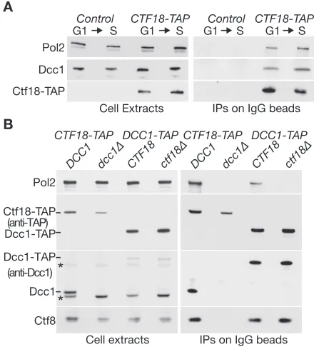
In a systematic analysis of yeast protein complexes by mass spectrometry (45), Pol2 and Dpb2 (the largest two subunits of Pol⑀; Dpb=DNA polymerase B subunit 2) were found to co-purify with TAP-tagged Ctf18, but Ctf18-RFC was not observed in purifications of Dpb2-TAP or Dpb3-TAP. We repeated these purifications, and not only detected pep-tides of Pol2 in samples of purified Ctf18-TAP (Supple-mentary Figure S1A), but also found that peptides from Ctf18-Ctf8-Dcc1 were specifically enriched in samples of purified Dpb2-TAP (Supplementary Figure S1B). We con-firmed these interactions by immunoblotting after isola-tion of Ctf18-TAP, and found that Ctf18-RFC can associate with Pol⑀not only in extracts of S-phase cells, but also dur-ing G1-phase (Figure1A).
Ctf18 did not co-purify with Pol2 in the absence of the Dcc1 subunit of Ctf18-RFC (Figure1B,CTF18-TAP dcc1Δ). Similarly, the Dcc1-Ctf8 heterodimer only co-purified with Pol2 in the presence of Ctf18 (Figure1B, com-pareDCC1-TAPandDCC1-TAP ctf18Δ). It thus appears that Ctf18 and Ctf8-Dcc1 are jointly required for Ctf18-RFC to associate with Pol⑀in budding yeast, mirroring the interaction of the human proteins.
in-Nucleic Acids Research, 2015, Vol. 43, No. 18 8833
B
Dcc1 Ctf18-TAP
Cell Extracts IPs on IgG beads
A
Pol2
G1 S G1 S
G1 S G1 S
CTF18-TAP
Control Control CTF18-TAP
Cell extracts IPs on IgG beads Pol2
Dcc1-TAP Dcc1 Ctf8 (anti-Dcc1) (anti-TAP)
*
*
CTF18-TAP DCC1-TAP
dcc1∆
DCC1 CTF18 ctf18 ∆
CTF18-TAP DCC1-TAP
dcc1∆
DCC1 CTF18 ctf18∆
[image:5.612.67.287.69.312.2]Dcc1-TAP Ctf18-TAP
Figure 1. Ctf18 and Ctf8-Dcc1 are required for budding yeast Ctf18-RFC to associate with DNA polymerase epsilon. (A) Control cells (YLG98) and CTF18-TAP(YLG100) were synchronized at 24◦C in the G1-phase of the cell cycle by addition of mating pheromone, before release into S phase for 30 min. Cell extracts were incubated with magnetic beads coupled to rab-bit IgG, and the immunoprecipitated proteins were then monitored by im-munoblotting. (B) Asynchronous cultures ofCTF18-TAP(YGDP1970), CTF18-TAP dcc1Δ(YGDP1971),DCC1-TAP(YGDP1972) and DCC1-TAP ctf18Δ(YGDP1973) were grown at 24◦C, and then processed as in (A). Asterisks in the immunoblots denote non-specific bands.
teract with the Dpb2–3–4 subunits of Pol⑀(Supplementary Figure S2; data not shown for Dpb2). Ctf18 also interacted in the same assay with the carboxy terminal half of Pol2 (Figure2A, Pol2CT=1128–2222), potentially indicating a second binding site for Ctf18 within Pol⑀. However, Pol2CT interacts with itself in the two-hybrid system (Supplemen-tary Figure S3), and we thus cannot exclude that endoge-nous Pol2 serves as a bridge between Pol2CT and Ctf18 in this assay (with Ctf18 binding the amino terminal domain of endogenous Pol2, and Pol2CT binding to the carboxy-terminal half of endogenous Pol2).
The interaction of Ctf18 with Pol2NT required both Ctf8 and Dcc1 (Figure2B), indicating that the assay reflected the specific association of Ctf18-RFC with Pol ⑀as described above. Analysis of truncated versions of Pol2NT indicated that the interaction with yeast Ctf18-RFC was mediated by a small region at the amino terminus of Pol2, overlapping partially with the exonuclease but distinct from the DNA polymerase domain (Figure2C).
Mutations in the extreme carboxyl terminus of Ctf18 prevent association with Ctf8-Dcc1 and DNA polymerase epsilon
Truncations of yeast Ctf18 implicated the unique carboxy terminal half of the protein, beyond the ‘RFC box’ that is shared with other large subunits of RFC complexes, as be-ing required for interaction with Pol⑀(Figure3A). Align-ment of this region of Ctf18 in orthologues from closely
re-B
3 days at 30°C
Non selective medium Selective medium
AD DBD
Ctf18 Pol2-NT Ctf18 Pol2-NT Ctf18 Pol2-NT Hof1 Inn1 Hof1 Inn1 Hof1 Inn1
(dcc1∆) (ctf8∆) (control) (control)
(dcc1∆) (ctf8∆)
C Pol2
1 2223
1 227
1 396
1 933
1265 1
214
+++
1265
392
-1265 -1265 627
930 1265 -++
+++ +++
DBD-Pol2 (1-227) DBD-Pol2 (1-396)
AD-Ctf18 Non-selective Selective (-His)
DBD-Pol2 (1-1265)
DBD
3 days at 30°C DBD-Pol2 (214-1265)
DBD-Pol2 (392-1265) DBD-Pol2 (627-1265) DBD-Pol2 (930-1265) DBD-Pol2 (1-933)
Interaction with Ctf18? Exo
-nuclease
Polymerase Zinc finger A
Non selective medium Selective medium
AD DBD
(positive control) Ctf18
Ctf18 Pol2-NT
-3 days at -30°C Hof1 Inn1
Pol2-NT
-(1-1265) Pol2-CT
[image:5.612.324.561.72.618.2]
-(1128-2222) Ctf18 Pol2-CT
A
AD-Ctf18 (1-116) AD-Ctf18 (1-374)
DBD-Pol2-NT Non-selective Selective (-His)
AD-Ctf18 (1-494) AD-Ctf18 (1-595) AD-Ctf18 (1-657) AD-Ctf18 (1-741) AD-Ctf18 (369-741) AD-Ctf18 (484-741) AD
3 days at 30°C
369 741 +++
484 741 +++
1 116
-4 7 3 1 -494 1 -5 9 5 1 -657 1 -741 1 +++ Ctf18 1 4 7 1 ‘RFC box’ B (i) ctf18-2A (W736A W740A) S.mikatae Ctf18
S.cerevisiae Ctf18 S.bayanus Ctf18 S.kluyveri Ctf18 S. castellii Ctf18
739 717 714 701 715 763 741 738 725 739
*
*
*
*
*
*
*
*
**
(ii) Interaction with Pol2-NT CDBD-Pol2-NT Non-selective Selective (-His) AD-Ctf18 (484-741)
AD-Ctf18-2A (484-741) AD
3 days at 30°C (W736A W740A)
Cell extracts IPs of Dcc1-TAP Pol2
Dcc1-TAP
Ctf8 Ctf18
Control ctf18∆ ctf18-2A Control ctf18∆ ctf18-2A
D
X.l. Ctf18 H.s. Ctf18
A.t. Ctf18 D.m. Ctf18 S.c. Ctf18 S.p. Ctf18
*
*
*
*
*
*
*
*
*
1000 1002 954 993 741 960 975 977 978 930 970 717 935 952 [image:6.612.69.261.68.568.2]O.s. Ctf18
Figure 3. Mutations in a conserved motif at the carboxyl terminus of Ctf18 disrupt the interaction of budding yeast Ctf18-RFC with DNA polymerase epsilon. (A) The indicated truncations of Ctf18 were tested for their abil-ity to interact with Pol2-NT in the two-hybrid assay. (B) (i) Alignment of the carboxyl terminus of Ctf18 from each of the indicated budding yeast species, generated with ClustalW and BOXSHADE software. As-terisks denote 10 conserved hydrophobic residues within this region. (ii) An analogous alignment of the end of the Ctf18 protein from diverse eu-karyotic species, showing conservation of 9/10 hydrophobic residues (X.l. =Xenopus laevis; H.s=Homo sapiens; O.s.=Oryza sativa; A.t.= Ara-bidopsis thaliana; D.m.=Drosophila melanogaster; S.c.=Saccharomyces cerevisiae; S.p.=Schizosaccharomyces pombe). (C) TheW736A W740A mutations prevent interaction of yeast Ctf18 484–741 with Pol2NT in the yeast two-hybrid assay. (D) Asynchronous cultures ofDCC1-TAP( Con-trol, YLG301),DCC1-TAP ctf18Δ(ctf18Δ, YVM850) andDCC1-TAP ctf18–2A(ctf18–2A, YLG303) were grown at 24◦C, and then processed as in Figure1A.
lated budding yeast species showed that the final 25 amino acids of Ctf18 are particularly well conserved, and contain 10 invariant hydrophobic residues (Supplementary Figure S4A), which might contribute to protein–protein interac-tions. This region of Ctf18 is also well conserved in or-thologues of Ctf18 from more distantly related eukaryotic species such as humans, including nine of the 10 hydropho-bic residues from the C-terminal tail of yeast Ctf18 (Supple-mentary Figure S4B). Notably, the C-terminal 23 residues of human Ctf18 were found to be sufficientin vitroto pro-duce a ternary complex with Ctf8-Dcc1 and Pol⑀(41).
To try and identify an allele of yeastCTF18that would specifically disrupt the putative Pol-epsilon interaction module, allowing us to investigate its functional significance for the first time in any species, we mutated each of the last 25 amino acids of Ctf18 (Figure 3B) to alanine, ex-cept for A729 that we mutated to glycine or threonine. Us-ing the two-hybrid assay, we found that none of the sUs-ingle mutations abolished the interaction of Ctf18 with Pol2NT or Dcc1, but mild defects in both interactions were pro-duced by mutation of either W740 or E741 at the extreme carboxyl terminus of Ctf18 (Supplementary Figure S5). As aromatic residues can be particularly important in mediat-ing protein–protein interactions, we combined mutation of W740 with mutation of the neighbouring aromatic residue W736 (Figure 3B), and found that Ctf18 484–741 with the W736A W740A mutations was unable to interact with Pol2NT in the two-hybrid assay (Figure3C). These data in-dicate that the carboxyl terminus of Ctf18 contains a Pol
⑀interaction motif that has been conserved from yeast to humans.
The W736A W740A mutations were then introduced into the endogenousCTF18locus of budding yeast cells to create thectf18–2Aallele (see Materials and Methods). The Dcc1-Ctf8 heterodimer was unable to interact with Ctf18–2A or with Pol⑀inctf18–2Acells (Figure3D, IPs of Dcc1-TAP), confirming that the W736A W740A mutations in Ctf18 dis-rupted the Pol-epsilon binding module of Ctf18-RFC, de-spite the Ctf18–2A protein being expressed to a similar level as wild type Ctf18 (Figure3D, Cell extracts).
Integrity of the Pol⑀ binding module of Ctf18-RFC is re-quired downstream of the Mec1 protein kinase for activation of the S-phase checkpoint
Cells lacking Ctf18 are highly sensitive to DNA replica-tion stress induced by treatment the ribonucleotide reduc-tase inhibitor hydroxyurea, and have a strong defect in the establishment of cohesion between sister chromatids during chromosome replication (28,37).ctf18–2Acells are also sen-sitive to growth in the presence of hydroxyurea (Figure4A and Supplementary Figure S6) and are defective in sister chromatid cohesion (Figure4B), but both defects are less se-vere than those observed in the complete absence of Ctf18. This might indicate that Ctf18–2A is partially defective in the various functions of wild type Ctf18, or that the mu-tated Ctf18–2A protein is specifically defective in a subset of functions. We thus tested whetherctf18–2Ashared other reported phenotypes ofctf18Δcells, by crossingctf18–2A
Nucleic Acids Research, 2015, Vol. 43, No. 18 8835
A
ctf18-2A Control
rad53∆
ctf18∆
Control
YPD YPD +150mM HU
2 days at 30°C
B
Phase TetR-GFP Phase TetR-GFP
C
2 days at 30°C
*
ctf18-2Amrc1∆ YPDctf18-2A / CTF18 mrc1∆ / MRC1
*
*
*
*
*
*
*
*
*
*
*
*
* *
*
*
*
*
D
ctf18-2A / CTF18 ctf4∆ / CTF4
2 days at 30°C
*
ctf18-2Actf4∆*
*
* *
*
*
*
YPD
*
*
*
*
*
10 20 30 40 50
ctf18∆ ctf18-2A Control
[image:7.612.91.260.67.551.2]% cells with cohesion defects Cells arrested in G2-M phase
Figure 4. Phenotypes of thectf18–2Aallele. (A) Serial dilutions of control cells (W303–1a),rad53Δ(YAC53),ctf18Δ(YVM164),CTF18::K.l.TRP1 (wild typeCTF18but with theK.l.TRP1marker inserted into the ge-nomic locus, after the STOP codon ofCTF18; YLG316) andctf18–2A (also with theK.l.TRP1marker inserted after the STOP codon ofCTF18; YLG320) were plated on YPD medium, or YPD supplemented with 150mM hydroxyurea (HU), then grown for 2 days at 30◦C. (B) Control cells (CTF18-K.l.TRP1;YLG445),ctf18–2A (ctf18–2A K.l.TRP1; YLG447) andctf18Δ::K.l.TRP1(YLG449), all expressing the Tet-repressor fused to GFP and with an array of the Tet-operator sites at theura3locus, were grown at 30◦C and then arrested in G2-M phase by addition of noco-dazole. Defects in sister chromatid cohesion were scored microscopically, by examining 100 cells and determining the percentage with two dots of TetR-GFP instead of one (examples of each class are shown in the upper panels). (C) Diploid cells with the genotypectf18–2A/CTF18 mrc1Δ / MRC1(YLG292) were sporulated and then subjected to tetrad analysis on YPD medium. The image was taken after two days growth at 30◦C. (D) Tetrad analysis of the meiotic progeny ofctf18–2A/CTF18 ctf4Δ /CTF4 (YLG263).
B
Mcm4 Mcm2 Cdc45
IPs of Mcm4 Extracts of S-phase
cells (+HU)
CTF18 ctf18-2Actf18∆ mrc1∆ CTF18 ctf18-2Actf18∆ mrc1
∆
Psf1 Psf1-P A
Rad53
Control ctf18∆ ctf18-2A
0 Release into S-phase +0.2M HU (minutes)20 40 60 0 20 40 60 0 20 40 60
0 20 40 60 80 100
60 40 20 0
[image:7.612.341.546.73.318.2]% Rad53 phosn. 0 20 40 60 0 20 40 60
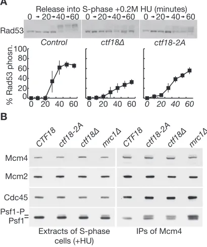
Figure 5. Disruption of the Pol⑀binding module of Ctf18-RFC blocks activation of the S-phase checkpoint, downstream of Mec1. (A) Control cells (W303–1a),ctf18–2A(YLG249) andctf18Δcells (YVM164) were arrested in G1-phase at 24◦C and then released into fresh medium con-taining 0.2M hydroxyurea for the indicated times. Rad53 hyperphospho-rylation was monitored by immunoblotting (upper panels), and the data from three such experiments were then quantified (lower panels; ‘% Rad53 phosphorylation’ was calculated from the ratio of hyperphosphorylated to hypophosphorylated Rad53). (B) Control cells (YLG426),ctf18–2A (YLG423),ctf18Δ(YLG421) andmrc1Δ(YGDP993) were arrested in G1-phase at 24◦C, before release for 90 min into S-phase in the presence of 0.2M hydroxyurea. Mcm4–5FLAG was immunoprecipitated from cell ex-tracts and the indicated proteins monitored by immunoblotting.
cause synthetic lethality (29,46). Strikingly, bothctf18–2A mrc1Δ(Figure4C) andctf18–2A ctf4Δ(Figure4D) were found to be inviable. This raised the possibility that at least one function of Ctf18-RFC might be lost when the integrity of the Pol-epsilon binding module is disrupted.
Finally, we tested whether the S-phase checkpoint path-way is defective inctf18–2Acells. Whenctf18Δormrc1Δ
cells enter phase in the presence of hydroxyurea, the S-phase checkpoint pathway cannot be activated at the defec-tive replication forks that are established from early repli-cation origins, leading to increased DNA damage and the firing of late replication origins (17,26–27). Activation of the Rad53 checkpoint kinase is defective but is not abol-ished inctf18Δormrc1Δcells under such conditions, since the unprotected replication forks activate a DNA-damage branch of the checkpoint pathway, which is independent of Ctf18 and Mrc1 (26,27). We found that Rad53 activation was defective whenctf18Δorctf18–2Acells entered S-phase in the presence of hydroxyurea (Figure5A), and this was associated with increased phosphorylation of Serine 129 of histone H2A, which provides a marker for DNA damage (Supplementary Figure S7). These data suggested that acti-vation of the S-phase checkpoint is defective when the Pol
To confirm these findings and explore which stage of the S-phase checkpoint pathway is defective in ctf18–2A
cells, we examined the Mec1-dependent phosphorylation of the CMG helicase (42). When rad53Δ, mrc1Δ or ctf18Δ
cells are treated with hydroxyurea, we found previously that Mec1-signalling at defective replication forks is increased, as reflected by increased Mec1-dependent phosphorylation of the Psf1 subunit of the Cdc45-MCM-GINS (CMG) DNA helicase (42). Importantly, the same effect is observed in hydroxyurea-treatedsld3-A dbf4–4Acells (42), in which the checkpoint kinases are activated normally but the key Rad53 phosphorylation sites in the initiation factors Sld3 and Dbf4 have been mutated (47), leading to the firing of late origins and an increase in DNA replication stress. This means that accumulation of the CMG helicase with hyper-phosphorylated Psf1 is a sensitive marker for the aberrant firing of late-origins in cells treated with hydroxyurea (42), reflecting a specific failure to activate the S-phase check-point response at some check-point downstream of the Mec1 ki-nase.
We synchronized wild type cells, ctf18Δ, mrc1Δ and
ctf18–2A in G1-phase at 24◦C by treatment with mating pheromone, before release into S-phase for 90 min in the presence of 0.2M hydroxyurea. Critically, the Psf1 subunit of the CMG helicase accumulated in a hyperphosphory-lated form inctf18–2Acells as well as inctf18Δandmrc1Δ
(Figure5B). This was particularly evident when the isolated CMG helicase was monitored in the immune-precipitates of
Mcm4–5FLAG (Figure5B, Psf1 in IPs of Mcm4–5FLAG),
but could also be detected in the cell extracts (Figure 5B, Psf1 in extracts of S-phase cells +HU), reflecting the greater proportion of total GINS that is present at replication forks when early and late origins fire under conditions of replica-tion stress. These data indicate that the integrity of the Pol-epsilon binding module of Ctf18-RFC is required for acti-vation of the S-phase checkpoint in budding yeast, down-stream of Mec1 activation.
S-phase checkpoint activation requires incorporation of Pol⑀ into the replisome
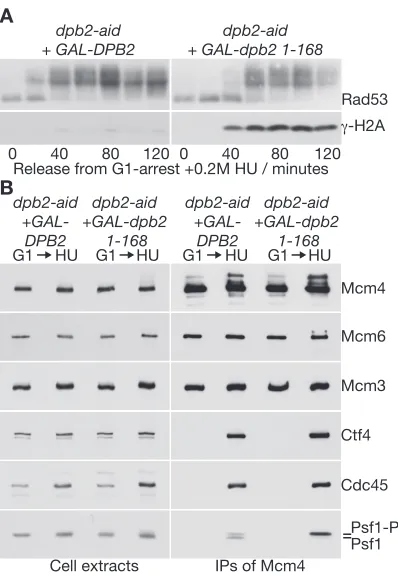
Previous work indicated that DNA polymerase epsilon is important for activation of the S-phase checkpoint (48–50). However, the mechanism was unclear and it was not known whether incorporation of Pol-epsilon into the replisome was important for checkpoint activation. We and others previ-ously showed that Dpb2 links Pol ⑀to the CMG helicase within the replisome, by interaction of the amino-terminal domain of Dpb2 with GINS, and the remainder of Dpb2 with Pol2 (51). Although depletion of Dpb2 blocks CMG assembly during the initiation of chromosome replication, we found that this could be rescued by over-expression of Dpb2NT, producing a replisome that lacks DNA poly-merase epsilon, despite the presence of wild type Pol2 (51). Therefore, we used this experimental system to test directly whether S-phase checkpoint activation requires the incor-poration of DNA polymerase epsilon into the replisome.
Using cells in which the endogenous DPB2 gene was
fused to the auxin-inducible degron (52), we expressed ei-ther wild type Dpb2 or the N-terminal fragment comprising Dpb2 1–168 in G1-phase cells and then depleted Dpb2-aid.
dpb2-aid
+GAL-DPB2
dpb2-aid +GAL-dpb2
1-168
G1 HU G1 HU G1 HU G1 HU
dpb2-aid
+GAL-DPB2
dpb2-aid +GAL-dpb2
1-168
Psf1 Psf1-P Cdc45 Mcm6 Mcm4
Cell extracts IPs of Mcm4
Mcm3 Ctf4 A
Release from G1-arrest +0.2M HU / minutes 0 40 80 120 0 40 80 120
dpb2-aid + GAL-DPB2
dpb2-aid + GAL-dpb2 1-168
Rad53
[image:8.612.333.532.66.354.2]γ-H2A B
Figure 6.Incorporation of DNA polymerase epsilon into the replisome is required for activation of the S-phase checkpoint, downstream of Mec1. (A) Controldpb2-aid GAL-DPB2(YCS394) anddpb2-aid GAL-dpb2 1– 168(YCS396) were arrested in G1-phase at 24◦C in YPRaff medium. Cells were then switched for 35 min to YPGal medium containing mating pheromone, before addition of 0.5 mM auxin (indole-3-acetic acid) and in-cubation for 60 min. Samples were then washed into fresh YPGal medium lacking mating pheromone but containing auxin. Samples were taken at the indicated times, and cell extracts were used to monitor the phospho-rylation of Rad53 and Histone H2A by immunoblotting. (B) Cells were grown as in (A) and released from into S-phase in the presence of hydrox-yurea for 90 min. Mcm4–5FLAG was immunoprecipitated from cell ex-tracts and the indicated proteins monitored by immunoblotting.
Upon release into S-phase in the presence of hydroxyurea, checkpoint activation was normal in cells expressing wild type Dpb2, as reflected by the rapid hyper-phosphorylation of Rad53 (Figure6A,dpb2-aid GAL-DPB2). In cells ex-pressing Dpb2 1–168, however, the activation of Rad53 was delayed and occurred concomitantly with the induction of
Nucleic Acids Research, 2015, Vol. 43, No. 18 8837
DISCUSSION
The Ctf8-Dcc1 heterodimer and the extreme C-terminus of Ctf18 together form a Pol⑀-binding module in Ctf18-RFC (41), which our data show to have been conserved from yeast to humans. In budding yeast, the Pol⑀-binding mod-ule of Ctf18-RFC is required for activation of the S-phase checkpoint in response to defects in DNA replication (Fig-ure5). Previous studies indicate that Ctf18-RFC is required for activation of the Rad53 checkpoint kinase at stalled DNA replication forks (26–29), downstream of Mec1 (42). These features of Ctf18-RFC are analogous to the role of Mrc1 in checkpoint activation, and it will be important in future studies to explore how Ctf18-RFC contributes to the recruitment and auto-activation of the Rad53 checkpoint kinase under such conditions.
Our data highlight two important new aspects of S-phase checkpoint activation: incorporation of DNA polymerase epsilon into the replisome and recruitment of Ctf18-RFC to the amino terminus of Pol2, adjacent to the exonuclease domain. These features should position Ctf18-RFC close to the ssDNA that forms an important signal for the check-point upon replication stress, and would thus bring Ctf18-RFC into the proximity of the activated Mec1 checkpoint kinase. It is striking that Mrc1 also associates with Pol ⑀ (21), although it has not yet been possible to test directly whether the association of Mrc1 with Pol ⑀ is required for activation of the S-phase checkpoint. One interesting model for future investigation would be that Pol ⑀serves as a scaffold upon which both Mrc1 and Ctf18-RFC re-cruit Rad53, facilitating auto-phosphorylation of the latter
in trans. Other possibilities could also be envisaged at this stage, and ultimately it will be important to try and estab-lish biochemical systems with which to reconstitute Rad53 activation by Mrc1 and Ctf18-RFC.
Since Ctf8 and Dcc1 appear to be required for all the known roles of Ctf18-RFC at DNA replication forks (37,40,53), in addition to activation of the S-phase check-point (26,29), it will be interesting in future studies to ex-plore how other functions of Ctf18-RFC might be modu-lated by association with Pol⑀. For example, our data indi-cate that the integrity of the Pol⑀-binding module of Ctf18-RFC is also important for sister chromatid cohesion (Fig-ure 4B), although the cohesion defect of ctf18–2Ais less severe than that observed in ctf18Δ cells. Recruitment of Ctf18-RFC to the leading strand DNA polymerase might facilitate (without being essential for) the loading of PCNA around the nascent leading strand DNA, which otherwise might receive less PCNA than the nascent lagging strand that is synthesized by a process involving repeated prim-ing and PCNA loadprim-ing events. Such a mechanism might aid the recruitment of the Eco1 acetyltransferase to both of the nascent sister chromatids, as well as potentially help-ing to stimulate other PCNA-linked processes such as chro-matin assembly on the newly synthesised leading strand DNA (54). It thus seems clear that future studies of Ctf18-RFC will still have much to contribute to our understanding of the some of the most enigmatic features of chromosome replication in eukaryotic cells.
SUPPLEMENTARY DATA
Supplementary Dataare available at NAR Online.
FUNDING
Cancer Research UK, the Medical Research Council and the Wellcome Trust [097945/B/11/Z for flow cytome-try; 102943/Z/13/Z for Senior Investigator Award to K.L.]. Cancer Research UK Career Development Fellow-ship [C44595/A16326 to G.D.P.]. Funding for open access charge: Wellcome Trust.
Conflict of interest statement.None declared.
REFERENCES
1. Gorgoulis,V.G., Vassiliou,L.V., Karakaidos,P., Zacharatos,P., Kotsinas,A., Liloglou,T., Venere,M., Ditullio,R.A. Jr, Kastrinakis,N.G., Levy,B.et al.(2005) Activation of the DNA damage checkpoint and genomic instability in human precancerous lesions.Nature,434, 907–913.
2. Errico,A. and Costanzo,V. (2012) Mechanisms of replication fork protection: a safeguard for genome stability.Crit. Rev. Biochem. Mol. Biol.,47, 222–235.
3. Labib,K. and De Piccoli,G. (2011) Surviving Chromosome
Replication: the many roles of the S-phase checkpoint pathway.Phil. Trans. R. Soc. B,366, 3554–3561.
4. Yekezare,M., Gomez-Gonzalez,B. and Diffley,J.F. (2013) Controlling DNA replication origins in response to DNA damage - inhibit globally, activate locally.J. Cell Sci.,126, 1297–1306.
5. Zou,L. (2013) Four pillars of the S-phase checkpoint.Genes Dev.,27, 227–233.
6. Bentley,N.J., Holtzman,D.A., Flaggs,G., Keegan,K.S.,
DeMaggio,A., Ford,J.C., Hoekstra,M. and Carr,A.M. (1996) The Schizosaccharomyces pombe rad3 checkpoint gene.EMBO J.,15, 6641–6651.
7. Zou,L. and Elledge,S.J. (2003) Sensing DNA damage through ATRIP recognition of RPA-ssDNA complexes.Science,300, 1542–1548.
8. Santocanale,C. and Diffley,J.F.X. (1998) A Mec1- and
Rad53-dependent checkpoint controls late-firing origins of DNA replication.Nature,395, 615–618.
9. Shirahige,K., Hori,Y., Shiraishi,K., Yamashita,M., Takahashi,K., Obuse,C., Tsurimoto,T. and Yoshikawa,H. (1998) Regulation of DNA-replication origins during cell-cycle progression.Nature,395, 618–621.
10. Bruin,D. (2009) All eukaryotes: Before turning off G1-S transcription, please check your DNA.Cell Cycle,8, 214–217. 11. Zhou,Z. and Elledge,S.J. (1993) DUN1 encodes a protein kinase that
controls the DNA damage response in yeast.Cell,75, 1119–1127. 12. Huang,M., Zhou,Z. and Elledge,S.J. (1998) The DNA replication
and damage checkpoint pathways induce transcription by inhibition of the Crt1 repressor.Cell,94, 595–605.
13. Lee,Y.D., Wang,J., Stubbe,J. and Elledge,S.J. (2008) Dif1 is a DNA-damage-regulated facilitator of nuclear import for ribonucleotide reductase.Mol. Cell,32, 70–80.
14. Nestoras,K., Mohammed,A.H., Schreurs,A.S., Fleck,O., Watson,A.T., Poitelea,M., O’Shea,C., Chahwan,C., Holmberg,C., Kragelund,B.B.et al.(2010) Regulation of ribonucleotide reductase by Spd1 involves multiple mechanisms.Genes Dev.,24, 1145–1159. 15. Wu,X. and Huang,M. (2008) Dif1 controls subcellular localization of ribonucleotide reductase by mediating nuclear import of the R2 subunit.Mol. Cell. Biol.,28, 7156–7167.
16. Zhao,X., Muller,E.G. and Rothstein,R. (1998) A suppressor of two essential checkpoint genes identifies a novel protein that negatively affects dNTP pools.Mol. Cell,2, 329–340.
17. Alcasabas,A.A., Osborn,A.J., Bachant,J., Hu,F., Werler,P.J., Bousset,K., Furuya,K., Diffley,J.F., Carr,A.M. and Elledge,S.J. (2001) Mrc1 transduces signals of DNA replication stress to activate Rad53.Nat. Cell Biol.,3, 958–965.
association of Cdc45 with MCM in replisome progression complexes at eukaryotic DNA replication forks.Nat. Cell Biol.,8, 358–366. 19. Katou,Y., Kanoh,Y., Bando,M., Noguchi,H., Tanaka,H.,
Ashikari,T., Sugimoto,K. and Shirahige,K. (2003) S-phase
checkpoint proteins Tof1 and Mrc1 form a stable replication-pausing complex.Nature,424, 1078–1083.
20. Kumagai,A. and Dunphy,W.G. (2000) Claspin, a novel protein required for the activation of Chk1 during a DNA replication checkpoint response in Xenopus egg extracts.Mol. Cell,6, 839–849. 21. Lou,H., Komata,M., Katou,Y., Guan,Z., Reis,C.C., Budd,M.,
Shirahige,K. and Campbell,J.L. (2008) Mrc1 and DNA polymerase epsilon function together in linking DNA replication and the S phase checkpoint.Mol. Cell,32, 106–117.
22. Nedelcheva,M.N., Roguev,A., Dolapchiev,L.B., Shevchenko,A., Taskov,H.B., Stewart,A.F. and Stoynov,S.S. (2005) Uncoupling of unwinding from DNA synthesis implies regulation of MCM helicase by Tof1/Mrc1/Csm3 checkpoint complex.J. Mol. Biol.,347, 509–521.
23. Kumagai,A. and Dunphy,W.G. (2003) Repeated phosphopeptide motifs in Claspin mediate the regulated binding of Chk1.Nat. Cell Biol.,5, 161–165.
24. Osborn,A.J. and Elledge,S.J. (2003) Mrc1 is a replication fork component whose phosphorylation in response to DNA replication stress activates Rad53.Genes Dev.,17, 1755–1767.
25. Zhao,H., Tanaka,K., Nogochi,E., Nogochi,C. and Russell,P. (2003) Replication checkpoint protein Mrc1 is regulated by Rad3 and Tel1 in fission yeast.Mol. Cell. Biol.,23, 8395–8403.
26. Crabbe,L., Thomas,A., Pantesco,V., De Vos,J., Pasero,P. and Lengronne,A. (2010) Analysis of replication profiles reveals key role of RFC-Ctf18 in yeast replication stress response.Nat. Struct. Mol. Biol.,17, 1391–1397.
27. Kubota,T., Hiraga,S., Yamada,K., Lamond,A.I. and Donaldson,A.D. (2011) Quantitative proteomic analysis of
chromatin reveals that Ctf18 acts in the DNA replication checkpoint. Mol. Cell. Proteomics,10, doi:10.1074/mcp.M110.005561.
28. Naiki,T., Kondo,T., Nakada,D., Matsumoto,K. and Sugimoto,K. (2001) Chl12 (Ctf18) forms a novel replication factor C-related complex and functions redundantly with Rad24 in the DNA replication checkpoint pathway.Mol. Cell. Biol.,21, 5838–5845. 29. Pan,X., Ye,P., Yuan,D.S., Wang,X., Bader,J.S. and Boeke,J.D. (2006)
A DNA integrity network in the yeast Saccharomyces cerevisiae. Cell,124, 1069–1081.
30. Majka,J. and Burgers,P.M. (2004) The PCNA-RFC families of DNA clamps and clamp loaders.Prog. Nucleic Acid Res. Mol. Biol.,78, 227–260.
31. Kubota,T., Nishimura,K., Kanemaki,M.T. and Donaldson,A.D. (2013) The Elg1 replication factor C-like complex functions in PCNA unloading during DNA replication.Mol. Cell,50, 273–280. 32. Shiomi,Y. and Nishitani,H. (2013) Alternative replication factor C
protein, Elg1, maintains chromosome stability by regulating PCNA levels on chromatin.Genes Cells,18, 946–959.
33. Green,C.M., Erdjument-Bromage,H., Tempst,P. and Lowndes,N.F. (2000) A novel Rad24 checkpoint protein complex closely related to replication factor C.Curr. Biol.,10, 39–42.
34. Majka,J. and Burgers,P.M. (2003) Yeast Rad17/Mec3/Ddc1: a sliding clamp for the DNA damage checkpoint.Proc. Natl. Acad. Sci. U.S.A.,100, 2249–2254.
35. Lengronne,A., McIntyre,J., Katou,Y., Kanoh,Y., Hopfner,K.P., Shirahige,K. and Uhlmann,F. (2006) Establishment of sister chromatid cohesion at the S. cerevisiae replication fork.Mol. Cell, 23, 787–799.
36. Bylund,G.O. and Burgers,P.M. (2005) Replication protein A-directed unloading of PCNA by the Ctf18 cohesion establishment complex. Mol. Cell. Biol.,25, 5445–5455.
37. Mayer,M.L., Gygi,S.P., Aebersold,R. and Hieter,P. (2001) Identification of RFC(Ctf18p, Ctf8p, Dcc1p): an alternative RFC complex required for sister chromatid cohesion in S. cerevisiae.Mol. Cell,7, 959–970.
38. Spencer,F., Gerring,S.L., Connelly,C. and Hieter,P. (1990) Mitotic chromosome transmission fidelity mutants in Saccharomyces cerevisiae.Genetics,124, 237–249.
39. Borges,V., Smith,D.J., Whitehouse,I. and Uhlmann,F. (2013) An Eco1-independent sister chromatid cohesion establishment pathway in S. cerevisiae.Chromosoma,122, 121–134.
40. Hiraga,S., Robertson,E.D. and Donaldson,A.D. (2006) The Ctf18 RFC-like complex positions yeast telomeres but does not specify their replication time.EMBO J.,25, 1505–1514.
41. Murakami,T., Takano,R., Takeo,S., Taniguchi,R., Ogawa,K., Ohashi,E. and Tsurimoto,T. (2010) Stable interaction between the human proliferating cell nuclear antigen loader complex
Ctf18-replication factor C (RFC) and DNA polymerase{epsilon}is mediated by the cohesion-specific subunits, Ctf18, Dcc1, and Ctf8.J. Biol. Chem.,285, 34608–34615.
42. De Piccoli,G., Katou,Y., Itoh,T., Nakato,R., Shirahige,K. and Labib,K. (2012) Replisome stability at defective DNA replication forks is independent of S phase checkpoint kinases.Mol. Cell,45, 696–704.
43. Maric,M., Maculins,T., De Piccoli,G. and Labib,K. (2014) Cdc48 and a ubiquitin ligase drive disassembly of the CMG helicase at the end of DNA replication.Science,346, doi:10.1126/science.1253596. 44. Foiani,M., Marini,F., Gamba,D., Lucchini,G. and Plevani,P. (1994) The B subunit of the DNA polymerase alpha-primase complex in Saccharomyces cerevisiaeexecutes an essential function at the initial stage of DNA replication.Mol. Cell. Biol.,14, 923–933.
45. Gavin,A.C., Bosche,M., Krause,R., Grandi,P., Marzioch,M., Bauer,A., Schultz,J., Rick,J.M., Michon,A.M., Cruciat,C.M.et al. (2002) Functional organization of the yeast proteome by systematic analysis of protein complexes.Nature,415, 141–147.
46. Tong,A.H., Lesage,G., Bader,G.D., Ding,H., Xu,H., Xin,X., Young,J., Berriz,G.F., Brost,R.L., Chang,M.et al.(2004) Global mapping of the yeast genetic interaction network.Science,303, 808–813.
47. Zegerman,P. and Diffley,J.F. (2010) Checkpoint-dependent inhibition of DNA replication initiation by Sld3 and Dbf4 phosphorylation.Nature,467, 474–478.
48. Navas,T.A., Zhou,Z. and Elledge,S.J. (1995) DNA polymerase epsilon links the DNA replication machinery to the S phase checkpoint.Cell,80, 29–39.
49. Navas,T.A., Sanchez,Y. and Elledge,S.J. (1996) RAD9 and DNA polymerase epsilon form parallel sensory branches for transducing the DNA damage checkpoint signal in Saccharomyces cerevisiae. Genes Dev.,10, 2632–2643.
50. Puddu,F., Piergiovanni,G., Plevani,P. and Muzi-Falconi,M. (2011) Sensing of replication stress and Mec1 activation act through two independent pathways involving the 9–1–1 complex and DNA polymerase epsilon.PLoS Genet.,7, e1002022.
51. Sengupta,S., van Deursen,F., de Piccoli,G. and Labib,K. (2013) Dpb2 integrates the leading-strand DNA polymerase into the eukaryotic replisome.Curr. Biol.,23, 543–552.
52. Nishimura,K., Fukagawa,T., Takisawa,H., Kakimoto,T. and Kanemaki,M. (2009) An auxin-based degron system for the rapid depletion of proteins in nonplant cells.Nat. Methods,6, 917–922. 53. Gellon,L., Razidlo,D.F., Gleeson,O., Verra,L., Schulz,D.,
Lahue,R.S. and Freudenreich,C.H. (2011) New functions of Ctf18-RFC in preserving genome stability outside its role in sister chromatid cohesion.PLoS Genet.,7, e1001298.