University of Warwick institutional repository: http://go.warwick.ac.uk/wrap
A Thesis Submitted for the Degree of PhD at the University of Warwick
http://go.warwick.ac.uk/wrap/62781
This thesis is made available online and is protected by original copyright. Please scroll down to view the document itself.
Effect of Aging on the Planning and Execution of
Sit-to-Stand Movement
by
Kanokwan Srisupornkornkool
A thesis submitted in partial fulfillment of the requirements for the
degree of
Doctor of Philosophy in Psychology
University of Warwick, Department of Psychology
Table of Contents
Table of Contents ii
List of Tables vii
List of Illustrations viii
Acknowledgements xiii
Summary xiv
Abbreviations xv
Chapters
1 Introduction 11.1. Sit-to-stand movement 2
1.1.1. Definition of sit-to-stand movement 2
1.1.2. Biomechanics of sit-to-stand movement 3
1.1.3. Determinants of sit-to stand movement 5
1.1.4. Improvement on sit-to-stand movement 14
1.2. Motor imagery 19
1.2.1. Definition of motor imagery 19
1.2.2. Factor influencing motor imagery 20
1.2.3. Brain area involved in motor imagery 24
1.2.4. Implication of motor imagery 27
1.3. Attention to movement 42
1.3.1. Definition of Attention 42
1.3.2. Focus of attention and motor performance 43
1.3.3. Internal versus external attentional focus during 44
performance and learning 1.3.4. Attentional focus and motor imagery 52
1.5. Overview of the following chapters 60
1.5.1. Chapter 2 60
1.5.2. Chapter 3 60
1.5.3. Chapter 4 60
1.5.4. Chapter 5 61
1.5.5. Chapter 6 61
1.5.6. Chapter 7 62
2 Common Methodology 63
2.1. Introduction 64
2.2. Method 64
2.2.1. Apparatus 64
2.2.2. Experimental procedures 64
2.3. Measures, Design and Data Analysis 66
2.4. Further experiments 70
2.4.1. Experiment 2 70
2.4.2. Experiment 3 70
2.4.3. Experiment 4 71
3 Experiment 1: Attentional focus during physical and imagined 72
standing up affects young and older adults in divergent ways 3.1. Introduction 73
3.2. Method 80
3.2.1. Participants 80
3.2.2. Experimental procedures 81
3.3. Results and Discussion 84
3.3.1. Duration of postural transition and stability 84 of transition
3.3.2. Self-reported movement times and vividness 87
of imagery 3.3.3. Muscular activation during imagined STS 91
movements 3.4. Conclusion 95
4 Experiment 2: Seat height affects STS movements performed 97
under internal and external attentional focus 4.1. Introduction 98
4.2. Method 100
4.2.1. Participants 100
4.2.2. Experimental procedures 101
4.2.3. Measures, Design and Data Analysis 101
4.3. Results and Discussion 105
4.3.1. Duration of postural transition and stability 105
of transition 4.3.2. Self-reported movement times 113
4.3.3. Muscular activation during imagined STS 118
movements 4.4. Conclusion 122
5 Experiment 3: A secondary manual task differentially 124
affects STS movements performed under different foci of attention 5.1. Introduction 125
5.2. Method 127
5.2.1. Participants 127
5.2.2. Experimental procedures 128
5.3. Results and Discussion 133
5.3.1. Duration of postural transition and stability 133
of transition 5.3.2. Self-reported movement times 138
5.3.3. Muscular activation during imagined STS 140
movements 5.4. Conclusion 141
6 Experiment 4: Effects of attentional focus on learning through 143
motor imagery practice 6.1. Introduction 144
6.2. Method 145
6.2.1. Participants 145
6.2.2. Experimental Procedures 147
6.2.3. Measures, Design and Data Analysis 150
6.3. Results and Discussion 152
6.3.1. Duration of postural transition and stability 152
of transition 6.3.2. Lateral symmetry of ground reaction force 156
6.4. Conclusion 162
7 General Discussion 163
7.1 Goals of the project 164
7.2 Overview of experiment results 165
7.3. Analysis of the effects of attentional focus on 172
physical performance 7.4. Analysis of the effects of attentional focus on 174
imagery performance 7.5. Clinical implications 178
References 184
Appendix 232
Appendix 1. Overview of studies using motor imagery 233 and attentional focus
Appendix 2. Questionnaires 267
Appendix 3. Instructions 277
List of Tables
Table 1-1 Demonstrates similarity and dissimilarity aspects between 55
attentional focus and motor imagery
List of Illustrations
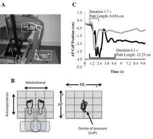
Figure 2-1 Experimental setup and measurement conventions. 69
A. Participants’ sitting position at the start of each STS
movement. B. Schematic representation of the conventions
used in recording CoP data from the force platform.
C. Sample data of a young (light grey) and an older
(dark grey) adult, showing the postural transition durations
identified by the analysis algorithm
Figure 2-2 Sample data of the AP component of ground reaction 70
force from a young (light grey) and an older (dark grey)
adults during imagined movement
Figure 3-1 The summary protocol of Experiment 1 83
Figure 3-2 Effect of age on the duration of postural transition of 84
young and older adults
Figure 3-3 Effect of attentional focus and age on the duration of 85
postural transition of young and older adults
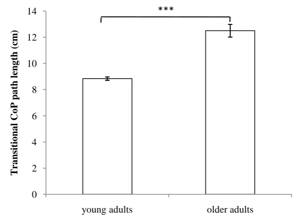
Figure 3-4 Effect of age on the stability of transition of young 86
and older adults
Figure 3-5 Effect of attentional focus and age on the stability of 86
transition of young and older adults
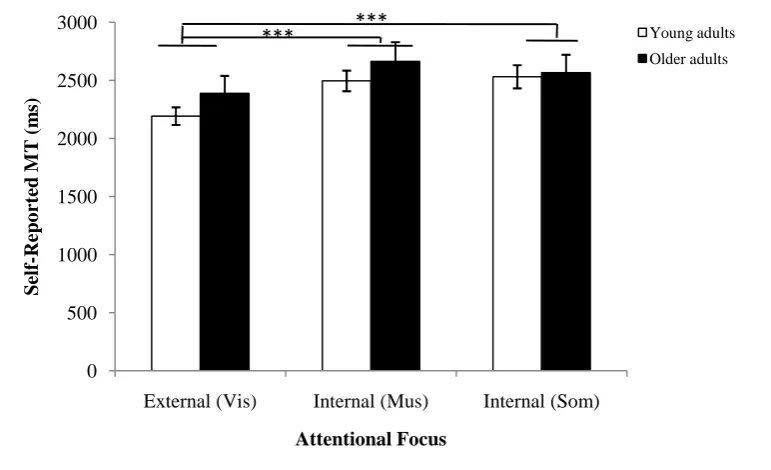
Figure 3-6 Effect of attentional focus on self-reported movement 88
times of young and older adults
Figure 3-8 Effect of attentional focus and time judgement 89
on vividness
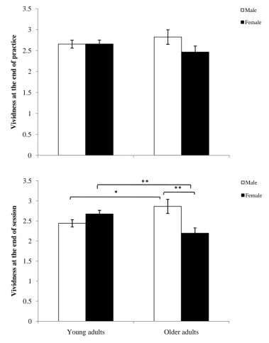
Figure 3-9 Effect of age and gender on vividness at the end 90
of practice (top panel) and at the end of seesion (bottom panel)
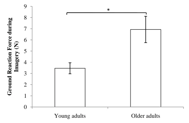
Figure 3-10 Effect of age on the ground reaction force of 92
young and older adults
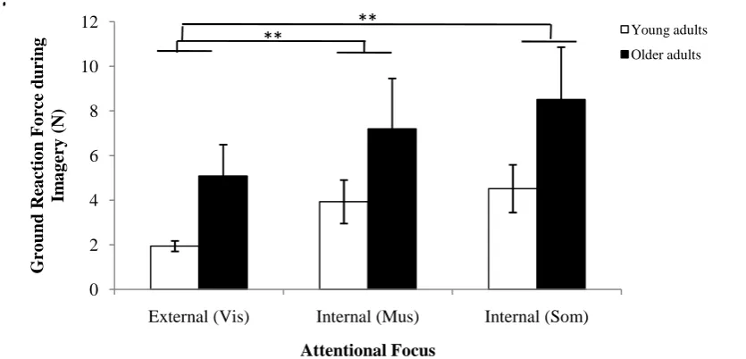
Figure 3-11 Effect of attentional focus on the ground reaction 93
force of young and older adults
Figure 4-1 The summary protocol of Experiment 2 103
Figure 4-2 Effect of age on the duration of postural transition 106
of young and older adults
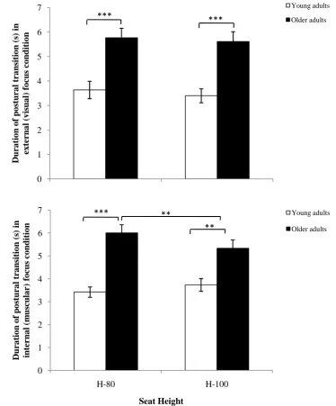
Figure 4-3 Effect of seat height and age on the duration of postural 107
transition in external (top panel) and internal (bottom panel) condition
Figure 4-4 Effect of seat height and age on the duration of postural 108
transition of young and older adults
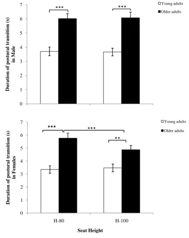
Figure 4-5 Effect of seat height and age on the duration of postural 109
transition of male (top panel) and female (bottom panel) participants
Figure 4-6 Effect of age on the stability of transition of young 111
and older adults
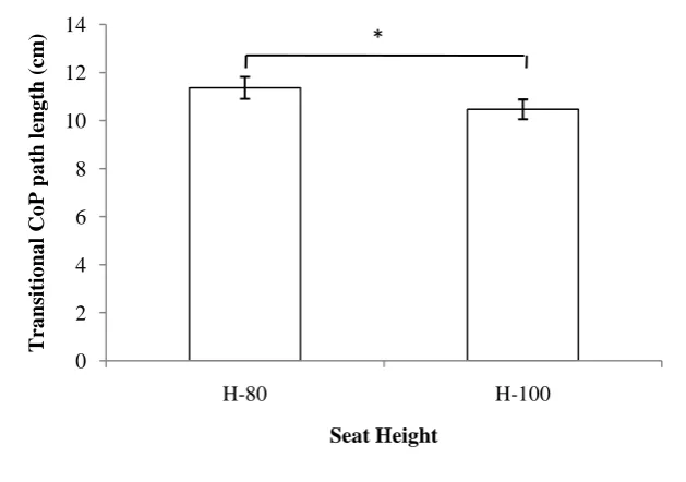
Figure 4-7 Effect of seat height on the stability of transition of 111
young and older adults
Figure 4-8 Effect of seat height and age on the stability of 112
Figure 4-9 Effect of attentional focus and seat height on the 112
stability of transition of young and older adults
Figure 4-10 Effect of seat height on self-reported movement times 115
of young and older adults
Figure 4-11 Effect of movement conditions on self-reported 115
movement times of young and older adults
Figure 4-12 Effect of attentional focus on self-reported movement 116
times of young and older adults
Figure 4-13 Effect of attentional focus and age on self-reported movement 117
times ofmale (top panel) and female (bottom panel) participants
Figure 4-14 Effect of age on the ground reaction force of young 120
and older adults
Figure 4-15 Effect of seat height on the ground reaction force of 120
young and older adults
Figure 4-16 Effect of seat height and age on the ground reaction force 121
in external (top panel) and internal (bottom panel) condition
Figure 5-1 The summary protocol of Experiment 3 131
Figure 5-2 The 500 ml juice bottle (A). The bottle was filled to 400 ml 133
(B), and a tissue was placed on the inside of the cap (C)
Figure 5-3 Effect of age on the duration of postural transition of 134
young and older adults
Figure 5-4 Effect of attentional focus on the duration of postural 135
Figure 5-5 Effect of manual task on the duration of postural 135
transition of young and older adults
Figure 5-6 Effect of manual task and age on the duration of 135
postural transition of young and older adults
Figure 5-7 Effect of age on the stability of transition of young 137
and older adults
Figure 5-8 Effect of movement conditions on self-reported 138
movement times of young and older adults
Figure 5-9 Effect of attentional focus on self-reported movement 139
times of young and older adults
Figure 5-10 Effect of manual task on self-reported 139
movement times of young and older adults
Figure 5-11 Effect of gender on the ground reaction force of 140
young and older adults
Figure 5-12 Effect of attentional focus on the ground reaction force 141
of young and older adults
Figure 6-1 The summary protocol of Experiment 4 151
Figure 6-2 Effect of age on the duration of postural transition of 153
young and older adults in anteroposterior and
mediolateral components
Figure 6-3 Effect of time of test on the duration of postural transition 153
of young and older adults in anteroposterior and
Figure 6-4 Effect of time of test and age on the duration of 154
postural transition of young and older adults in
anteroposterior component
Figure 6-5 Effect of time of test and type of training on the 154
duration of postural transition of young and older
adults in anteroposterior component
Figure 6-6 Effect of age on the stability of transition of young 155
and older adults in anteroposterior and mediolateral
components
Figure 6-7 Effect of age on the ground reaction force of young 157
and older adults
Figure 6-8 Effect of time of test on the ground reaction force of 157
young and older adults
Figure 6-9 Effect of time of test and type of training on the 158
ground reaction force of young and older adults
Figure 6-10 Effect of type of training on the ground reaction force 158
of young and older adults
Figure 6-11 Effect of type of training and age on the ground 159
reaction force of young and older adults
Figure 6-12 Effect of time of test and age on the ground reaction force 159
Acknowledgements
I gratefully acknowledge the advice and support of my supervisor, Dr. Suvobrata
(Joy) Mitra and Professor James Tresilian. My special thanks to Joy for the great opportunity
to work on a project so closely relative to my physiotherapy interests. He is a good principal
supervisor and source of energy to the thesis I am deeply indebted, because without his
guidance, as a physiotherapist without psychology background, I cannot cross border of such
a complex and mysterious field. My deepest thanks to Joy again for his worthy advice,
proofreading and editing of this thesis. My very good thanks to Professor James Tresilian for
helpful comments.
My appreciative thanks to Professor Elizabeth Maylor for arranging a
community-based volunteer panel and Nicola Doherty for her help with recruiting older participants. My
thanks to Hayley Boulton, PhD students and staff of Psychology Department, University of
Warwick for their support and warmest encouragement. I extend my thanks to Dr. Adrian
von Muhlenen (University of Warwick) and Dr. David Punt (University of
Birmingham) for acting as examiners to the thesis, and for helpful comments and
suggestions to the work.
My special thanks also extend to all my participants for their excellent
cooperation.The work presented in this thesis would not have been possible without a
kindness of participants who took part to the experiments. My sincrest thanks to Royal Thai
government and Science and Technology Ministry for financial support of my PhD study.
My very good thanks to my cousin, Ms Natda Chanakul, and staff in Thai Dusit (Coventry)
and Sabai Sabai (Leamington Spa) Restaurant for their support throughout the study in
University of Warwick. I extend my very sincrest thanks to my family for their love, special
care, understanding and constant source of my inspiration being.
Summary
Whole-body coordination such as in sit-to-stand (STS) movements is an important activity of independent daily living that is affected by decreased muscular strength and postural control due to ageing and also as a result of neurological diseases such as stroke. Recent research has taken an interest in using motor imagery for rehabilitation and training because it has many features in common with movement execution without some of the practical difficulties of repeated physical practice. Imagery tends to be more effective when it takes a first person perspective and focuses on kinesthetic aspects of movement. On the contrary, research in exercise science shows that movement execution is more fluent when attention is focused on body-external perceptual consequences of movement. How ageing affects this difference in the impact of attentional focus is not well understood. This thesis examines the effects of body-external (visual) and body-internal (muscular or somatosensory) attentional focus on STS movement execution and imagery in healthy young and older adults.
The thesis reports four experiments comparing execution and imagery performance in young and older adults. Experiment 1 was designed to clarify the impact of attentional focus on motor performance and imagery in young and older adults. Experiment 2 examined the impact of changing the level of effort (by manipulating the starting seat height) on the effects of attentional focus. Experiment 3 measured the impact of unimanually balancing a load in the hand on the role of attentional focus in physical and imagined STS movements. Experiment 4 studied the role of attentional focus in a training protocol employing motor imagery practice. Postural transition duration and transition stability during physical trials, self-reported movement times during physical and imagined trials, and ground reaction force and vividness of imagery during imagined trials were recorded.
Abbreviations
ABC Activities-specific Balance Confidence
ADL Activities of daily living
AP Anteroposterior component of ground reaction force
BBS Berg Balance Sclae
CNV Contingent negative variation
CoM Centre of Mass
CoP Centre of pressure
EMG Electromyography
fMRI functional Magnetic Resonance Imaging
KVIQ Kinesthetic and Visual Imagery Questionnaire
MI Motor imagery
MIQ Movement Imagery Questionnaire
MIQ-R Movement Imagery Questionnaire-Revised
ML Mediolateral component of ground reaction force
MP Mental practice
MT Movement time
PD Parkinson disease
PET Positron Emission Tomography
RMS Root mean square
ROM Range of motion
RT Reaction time
SCI Spinal cord injury
STS Sit-to-stand
TBI Traumatic brain injury
TMS Transcranial Magnetic Stimulation
1.1. Sit-to-stand movement
1.1.1. Definition of sit-to-stand movement
Sit-to-stand (STS) movements can be considered a fundamental motor skill, a
common motor activity with a specific pattern that involves different body parts,
namely the head, the trunk, the arms and the legs, for transitioning from a sitting to a
standing position. The nature of STS movements is dynamic and destabilizing
because of a rapid change of the body from a stable seat position to a small base of
support and a higher centre of mass (CoM) position (Nevitt, Cummings, & Hudes,
1991). Precisely defining standard STS movements seems to be difficult because
people have unique and distinctive movement styles. Thus, the definition of STS can
depend on the aim of the study in question. For example, Roebroeck, Doorenbosch,
Harlaar, Jacobs, and Lankhorst (1994) defined the STS movement as moving the
body’s CoM upward from a sitting position to a standing position without losing
balance. Vander Linden, Brunt, and McCulloch (1994) stated that the STS
movement was a transitional movement to the upright posture requiring movement
of the CoM from a stable position to a less stable position over extended lower
extremities.
Several daily activities are performed from a standing position because this
posture is not only vital for proper functioning of many organs, but also helps to
maintain proper bone loading and prevent excessive bone demineralization (Leo,
1985; Krebs, Ragnarsson, & Tuckman, 1983; Lukert, 1982). The ability to perform
STS movements is a fundamental ability to achieve normal activities of daily living
and also a prerequisite of locomotion (walking) activity. Moreover, the unassisted
(Ragnarsson et al., 1981; Igaroski & Black, 1985; Paulus, Straube, & Brandt, 1984;
Kerr, White, Barr, & Mollan, 1994; Lee, Wong, Tang, Cheng, & Ling, 1997;
Mathiyakom, McNitt-Gray, Requejo, & Costa, 2005; Janssen, Bussmann, & Stam,
2002; Etnyre & Thomas, 2007). Therefore, an understanding of the standing up in
healthy individuals provides fundamental knowledge about how the task is organized
and enables identification of abnormal STS movements when it occurs.
1.1.2. Biomechanics of sit-to-stand movement
The STS movement is a discrete task beginning from a seated position and
then transitioning to a standing position. Giving a standardized description of human
biomechanics is difficult because people do not only have individual and distinctive
movement styles, but also tend not to repeat movements in exactly the same way
(Etnyre & Thomas, 2007). Moreover, numerous studies have used varying methods
to explain biomechanics of STS movements. The majority of studies have utilized
kinematic (e.g., using videotape recordings, electrogoniometers and accelerometers)
and kinetic (mainly using force platform) measurement analysis to identify the
structure of STS movements. The effective analysis of STS events is generally
described in two ways: either flexion and extension phases, or momentum, torque
and velocity event changes. These events have often served as the major variables of
most studies on STS movements (see Etnyre & Thomas, 2007 for a review).
To clarify STS movements, the events were subdivided into four phases,
including a flexion-momentum, a momentum-transfer, an extension and a
stabilization phases. The flexion-momentum phase starts with initiation of
movement and ends just before lifting the buttocks from the seat. This phase is called
momentum-transfer phase (seat-off) starts with lifting the buttocks and ends with
achieving maximal ankle dorsiflexion. The extension phase begins after achieving
maximal ankle dorsiflexion and ends when one stops hip, leg and trunk extension.
The stabilization phase begins after achieving the hip extension and ends when all
motion is completed (Schenkman, Berger, Riley, Mann, & Hodge, 1990; Shepherd
& Gentilel, 1994).
This description emphasized the movement of body segments during the
performance of flexion and extension movements more than momentum changes.
However, events of STS movements should be unequivocal for any performance.
Riley, Schenkman, Mann, and Hodge (1991) divided STS events into three phases in
terms of momentum, including an initial, a transitional and an extension phases. The
initial phase is to generate upper body momentum. The transitional phase starts when
momentum from upper body movement is transferred to the total body, as the
momentum of body’s CoM changes from forward to vertical. The extension phase
takes place during the vertical ascent of the body. In addition, STS events are often
divided into seat off, beginning of movement and end of movement phases. The seat
off event is a transitional point, changing from a stable to unstable base of support.
There are different definitions of transitional point, depending on the objectives of
studies, such as the time at peak horizontal force (Doorenbosch, Harlaar, Roebroeck,
& Lankhorst, 1994; Gross, Stevenson, Charette, Apyka, & Marcus, 1998), peak
vertical force (Kaya, Kerbs, & Riley, 1998), initial vertical force (Riley et al., 1991;
Schenkman et al., 1990) and a point of the thigh’s separation from the seat (Kotake,
Dohi, Kajiwara, Sumi, Koyama, & Miura, 1993; Moxley Scarborough, Kerbs, &
Harris, 1999; Tully, Fitoohabadi, & Galea, 2005; Vander et al., 1994). The beginning
(Kralj, Jaeger, & Minih, 1990) or initiation of trunk flexion displacement or
momentum (see Etnyre & Thomas, 2007 for a review). Although the end of
movement is difficult to describe because there is postural sway during quiet
standing, this event is simply defined as a point of fully standing up. Moreover, the
end of movement is described, by monitoring displacement, velocity or momentum
in horizontal direction, as related to minimal movement of the head, the spine, the
shoulders or the hips (see Etnyre & Thomas, 2007 at follow review).
Although there is a wide range of studies involving analysis of STS
movements, it seems that there is no consensus on a standardized method for
analyzing STS events because of the diversity of movements between and within
individuals, relating to influential factors on the movement such as unique pattern of
movement, constrained conditions of studies’ purpose, and varied strategies during
performing STS movements. Studying STS movements also requires knowledge of
the potential factors influencing how the movement is performed. Thus, it is
necessary to conduct further research on STS movements.
1.1.3. Determinants of sit-to stand movement
STS movements are common physical tasks that are frequently used (ranging
between 45-65 times a day in independently living people) to change to locomotor or
other functional activities (Bohannon et al., 2008; Dall & Kerr, 2010). A clearly
understanding of the features of STS movements requires a basic knowledge of
determinants that influence how the movement is performed. Generally, the
determinants are often divided into two domains–constraint-related and
1.1.3.1 Constraint-related
In studies of STS movements, positional constraints are commonly
divided to chair-related and strategy-related.
Chair-related:
The literature indicates that a chair has an influence on the ability to
perform STS movements (Janssen et al., 2002). Chair-related determinants, such as
the height of a chair, type of a chair and use of a backrest, have been observed,
however, most studies have focused on the height of a chair (e.g., Schenkman, Riley,
& Pieper, 1996; Arborelius, Wretenberg, & Lindberg, 1992; Hughes & Schenkman,
1996; Hughes, Weiner, Schenkman, Long, & Studenski, 1994; Hughes, Myers, &
Schenkman, 1996; Itakazu, Uemura, Aoki, & Takatsu, 1998; Kawagoe, Tajima, &
Chosa, 2000; Munro, Steele, Bashford, Ryan, & Britten, 1998; Munton, Ellis, &
Wright, 1984; Rodosky, Andriacchi, & Andersson, 1989; Su, Lai, & Hong, 1998;
Weiner, Long, Hughes, Chandler, & Studenski, 1993), whereas a few studies have
tried to clarify the influence of a type of chair that is designed for making STS
movements easy (e.g. Munro et al., 1998; Burdett, Habasevich, Pisciotta, & Simon,
1985; Wheeler, Woodward, Ucovich, Perry, & Walker, 1985; Bashford, Steele,
Munro, Westcott, & Jones, 1994; Ellis, Seedhom, Amis, Dowson, & Wright, 1979).
For example, Ellis et al. (1979) claimed that the knee moment and joint loading force
decreased during STS movements when using a motorised chair that assists the
postural transition. Similarly, use of an ejector chair (a type of motorized chair) has
been reported to assist the STS transfer (Bashford et al., 1994; Munro et al., 1998).
Surprisingly, there are no experimental studies concerned with the influence of a
backrest in order to standardize the STS movement starting position (e.g., Munro et
al., 1998; Hughes et al., 1994; Weiner et al., 1993).
Evidence suggests that changing the height of a seat affects the
maximal moment needed at the knee and the hip (e.g., Hughes & Schenkman, 1996;
Rodosky et al., 1989; Su et al., 1998; Arborelius et al., 1992; Schenkman et al.,
1996; Riley et al., 1991). These findings suggest that using a higher chair leads to a
decrease in moments and joint loading forces acting at the knee level (up to 60%)
and at the hip level (up to 50%), whereas using a lower chair increases the need for
momentum generation and more repositioning of the feet. Hughes et al. (1994)
described the repositioning of the feet as a movement strategy to lower moments
used for STS movements. They called this a “stabilization strategy”. Although
comparison of results among studies is difficult because of differences in study
design and reference points of seat height, the findings could be summarized by
noting that an unsuitable chair height changes biomechanical demands or alters
strategies of STS movements. This is due to the imposed biomechanical demand due
to different foot, trunk, or arm positioning. Despite reports in the literature that chairs
should be of adequate height, and have sufficient space under the seat (Kawagoe et
al., 2000), people usually need to engage in this task from different chair heights that
typically vary from 30.5-45.7 cm (Weiner et al., 1993). It is a challenge to
understand how individuals accommodate to these changing conditions.
Strategy-related:
Despite the absence of a standardized method for studying STS
movements, previous studies have often analysed STS movements while restricting
control variability among individuals. There are several strategy-related determinants
that have been studied in order to gain insight into the influence of the determinants
on STS movements, such as foot position, speed of movement, use of the arms, and
attention. Firstly, the position of the foot is one of the common parameters that is
frequently controlled before starting the STS movement in experimental studies.
Abundant evidence supports the fact that different foot placements can influence the
strategy of STS movements. For example, some studies have reported a shorter
movement time of the pre-extension phase, and lower maximum extension moment
at the hip, with the feet placed posterior than in neutral or anterior position (e.g.,
Shepherd & Koh, 1996; Kawagoe et al., 2000; Khemlani, Carr, & Crosbie, 1999;
Raina, Stevermer, & Gillette, 2005; Gillett & Stevermer, 2012). Previous studies
have also reported less the head movement and lower ground reaction forces when
the lower leg is in the preferred position while performing STS movements (Stevens,
Bojsen-Moller, & Soames, 1989). These findings provide information on how to
choose suitable foot placement to help decrease force requirements for people with
weakness or disability.
Secondly, speed of movement is generally known as having an
influence on the strategy of movement. Many studies did not allow participants to
stand up at their self-selected speed; speed of movement was sometimes controlled
by synchronization of movement with a metronome (e.g., Roebroeck et al., 1994;
Pai, Naughton, Chang, & Roger, 1994; Pai & Rogers, 1990). For instance, some
experiments constrained movement by regulating the speed of movement during
rising, demonstrating that rising speed increases moments of the hip flexion, the
knee extension and the ankle dorsiflexion (Pai & Roger, 1991) and decreases the
However, the constrained speed condition is not the usual way to perform STS
movements.
Thirdly, based on the literature, use of the arm while performing STS
movements appears to influence performance. Carr (1992) reported that arm position
has an influence on the position of the body’s CoM. The CoM moves forward at the
end of the STS movement with the arm point the arm pointed forward, whereas
restricting the arm leads to a different pattern of angular displacement. More than
half of previous research did not allow participants to use the arm during STS
movements; participants were instructed to perform the movements with their arms
by the side, on the lap, crossed on the chest or placed on armrests (e.g., Alexander,
Schultz, & Warwick, 1991; Etnyre & Thomas, 2007; Gillette, Stevermer, & Hall,
2012). Some studies reported that use of the arm during STS movements is very
common among older adults and even among young people (Durward, 1994;
Wheeler et al., 1985). Moreover, several previous studies showed that the hip and the
knee extension moment and joint loading force were decreased when using a chair
arm-rest support (Gillette et al., 2012; Burdett et al., 1985; Arborelius et al., 1992;
Seedhom & Terayama, 1976; Bahrami, Riener, Jabedar-Maralani, & Schmidt, 2000;
Schultz, Alexander, & Ashton-Miller, 1992). Accordingly, Etnyre and Thomas
(2007), for example, reported that using an armrest while performing STS
movements produced less average force and had longer time to vertical peak force
than three conditions (with the arm free, the hand on the knee and the arm crossed),
but there was no significant difference between the arm-use conditions in STS times.
However, to our knowledge, there are no experimental studies to clarify the impact
of functional arm motion during STS movements, particularly standing up while
Finally, it is generally known that attention to movement is able to
modulate motor responses. For instance, directing attention away from cued
movements is able to shorten reaction time (e.g., Wulf, McNevin, & Shea, 2001) and
increase frequency of movement adjustments (e.g., McNevin, Shea, & Wulf, 2003).
Previous research did not directly emphasise the effect of focus of attention on STS
performance. Usually, they only provided verbal instructions to keep the movement
correct, and inhibit habitual postural adjustments. For example, Gillette et al. (2012)
used different verbal commands to correct the movement while using different
movement strategies, including momentum, stabilization and vertical strategies. The
results showed that the knee extension moment increased with the vertical strategy.
Stevens et al. (1989) compared the effects of guidance to inhibit head and neck
postural adjustment on STS movements. Their finding showed that there was
significant decrease in the head movement in the guided movement. It also found
that ground reaction force and electromyography (EMG) activity were decreased
with the guided movement.
Surprisingly, little experimental research has addressed the influence
of attending to the movement on the performance of STS movements. The strategy
for STS movements can differ considerably with the participant’s attention to the
movement under specific instructions. For example, Yamada and Demura (2005)
found that the peak value of the ground reaction force was higher and movement
time (MT) was faster under the assigned-speed condition (i.e., stand up as fast as
possible) than in the self-paced condition (stand up without any speed instructions).
In another study, Sato, Mizuma, Kawate, Kasai, and Wada (2012) asked participants
to perform STS movements while paying attention to their bilateral symmetry. They
participants remaining aware of the need for bilateral symmetry during STS
movements. It can be seen from these examples that instructions were not precisely
detailed and oriented towards directing attention to the STS movement. Therefore, it
is worth knowing how beneficial focus instructions are to the performance of STS
movements before attempting to utilize them in enhancing the STS skill.
Indeed people have to perform STS movements under widely
different circumstances, particularly under different seat height conditions and
different body positions (e.g., depending on how the hands are used). There are no
reports of the effects of changing seat height or simultaneously using the hands
under different attentional instructions during STS performance. Thus, to understand
the STS movement skill, it is necessary to explore the ability to perform STS
movements while coping with different constraint conditions, and use that
information to interpret the performance. In the last section of the introduction, the
implications of attention to movement are discussed again.
1.1.3.2 Participant-related
The ability to perform STS movements is essential for activities of
daily living, especially in older people, because this transitional movement is among
the most challenging co-ordinations of daily life. When rising from a chair, the lower
extremities, the lower body joints and the leg muscles have to be used to transfer the
body up from a seated position (Eriksrud & Bohannon, 2003; O’Meaea & Smith,
2006). Thus, the transitional movements require significant muscular strength and
postural control. STS movements register high moments of as much as 4.7 times
body weight across joints in the lower limbs (Khemlani et al., 1999), which can pose
movements also pose a significant balancing challenge because of the rapid upward
shift of the body’s CoM to a position of reduced stability (Roebroeck et al., 1994;
Vander Linden et al., 1994).
STS movements among different populations have been described in
several previous studies. Older people and those with disabilities have particular
difficulty in performing transitional movements. Age-related differences appear to be
important during transitional movements because it is widely accepted that aging and
a decline in numerous physical performance measures are linked. Some investigators
have claimed that strategies of the STS task were slightly different between healthy
young and older adults (e.g., Ikeda, Schenkman, & Riley, 1991; Pai et al., 1994).
According to Feland, Hager, and Merrill’s study (2005), rising power decreases with
increasing age, whereas weight transfer time and centre of gravity sway remain
similar regardless of age. However, much STS research have been carried out on
deconditioned older people, because older people often have functional limitations,
leading to difficulty in achieving extension of the hips, the legs and the trunk, and
they move more slowly (due to decreased trunk flexion angular velocity). For
example, Papa and Cappozzo (2000) reported that their elder group showed a trend
to flex the trunk more before the seat-off phase, resulting in bringing the CoM closer
to the base of support and gaining a higher momentum. Older people also rotated the
body forward after the seat-off phase, bringing the CoM over the base of support and
then standing up. However, maximal speed during STS movements was lower in
older people than young adults. In addition, a study by Mourey, Pozzo,
Rouhier-Marcer, and Didier (1998), suggested that older participants spent more time
completing the sitting down movement and adjustment of velocity appeared in the
Evidence suggests that the difference in physical movement outcomes
between young and either older adults or disabled people may the result of changes
in any of three areas of movement control, including execution, control and planning
systems (Blevins, Hecker, Bigler, Boland, & Hayes, 1994; Johnson et al., 1994;
Lewis & Shaw, 1997; Ketcham & Stelmach, 2001; Butler, Lord, Rogers, &
Fitzpatrick, 2008). For example, the mechanical properties of muscles and tendons,
the effectiveness of sensory systems (e.g., visual, proprioceptive and vestibular) and
the selection of joint trajectories to perform a task (planning system) deteriorate with
age. Because older adults have often experienced a change in the mechanical
properties of muscles and tendons (execution system), such as a decrease in muscle
strength, a decrease in the rate of force production and an increase in tendon
stiffness, there are two important strategies adopted during STS movements in older
adults, including a momentum-transfer strategy and a stabilization strategy (Riley et
al., 1991; Hughes et al., 1994; Schultz et al., 1992). The momentum-transfer strategy
is characterized by a movement of the upper part of the body to generate momentum.
The stabilization strategy is characterized by the placement of the CoM closer to
base of support before the seat-off. Hughes et al. (1994) reported that older people
often use the stability strategy during STS movements.
Because of deterioration in the ability to perform the basic transitional
movements in older adults, impaired functioning and mobility in activities of daily
living normally occurs (Guralnik et al., 1994; Guralnik, Ferrucci, Simonsick, Salive,
& Wallace, 1995). As a result of functional limitations, older people’s STS
performance should differ from young people if determinants change (Ikeda et al.,
1991; Hughes et al., 1994; Pai et al., 1994; Schenkman et al., 1996). For example,
angular velocities were changed in older adults at the lower chair height. The authors
also suggested that older adults have to change their performance when they face
more demanding tasks. Likewise, Mazza, Benvenuti, Bimbi, and Stanhope (2004)
claimed that functional abilities and difficulty of the task have an influence on the
effectiveness of the ability to stand up from a chair. The results showed that
participants in the middle functional ability group had to swing their arm during STS
movements at the lower seat height, whereas participants in the least functional
group were not able to stand up at all at the lowest height of seat. In addition to
changing the arm-use determinant, Leung and Chang (2009) investigated three
posture-transfer strategies (no support, chair-arm, and cane) during STS movements
in older people. They found that the no-support strategy had the smallest value of
hip-compressed angle during STS movements, whereas there was no significant
difference between chair-arm and cane-use strategies. Subsequently, the trunk
flexion when using no support was greater than when using other strategies,
suggesting that these two strategies may be seen as adaptive mechanisms to decrease
the risk of anterior disequilibrium in older adults. People need to accomplish STS
movements under widely varying circumstances in daily life, including different
surfaces, different seat heights, different chair configurations and different body
positions (e.g., where the feet are during the task, whether the arms are used). Each
of these conditions should be evaluated especially regarding age-related differences
before interactions of all constraints on STS movements are fully appreciated.
1.1.4. Improvement on sit-to-stand movement
During STS movements, significant leg muscle strength and a wide range of
joint motion are involved, which presents a considerable challenge to dynamic
strength is associated with a diminished ability to perform functional activities (Brill,
Maccera, Davis, Blair, & Gordon, 2000; Lauretani et al., 2003; Purser, Pieper, Poole,
& Morey, 2003; Salem, Wang, Young, Marion, & Grendale, 2000; Adams,
Gandevia, & Skuse, 1990; Canning, Ada, & O’Dwyer, 1987) and a higher risk of
falling during movements (Blevins et al., 1994; Johnson et al., 1994; Lewis & Shaw,
1997; Guralnik et al., 1994; Guralnik et al., 1995; Cheng et al., 1998). Thus, this
motor act is a good indicator of mobility and frailty of older people
(Shumway-Cook, Brauer, & Woollacott, 2000; Studenski et al., 2003). It is also often included
in assessment and rehabilitation programs for people who present the inability to
perform this basic skill, due to impaired functioning and mobility in activities of
daily living (ADL).
The question of how to help these populations change position from sitting to
standing more easily and safely should be considered. Fortunately, the ability to
achieve STS movements is possible to develop with sufficient training. Physical
practice and MI training are current issues for improvement in functioning and
mobility in activities of daily living (ADL).
1.1.4.1. Physical practice
Muscular strength and postural control are essential to perform STS
movements independently. If the lower limb strength is reduced by weakness or
sedentary life style, balance and postural control will decline, with the consequence
that STS movements become difficult to carry out (Alexander et al., 1995; Doherty,
2003; Frontera et al., 2000; Alexander et al., 1991; Gross et al., 1998). Research
suggests that the ability to achieve STS movements can be improved with adequately
Knutzen, Poole, & Mrotek, 2003). Considerable effort has been focused on how to
improve muscular strength and postural control in order to achieve STS movements
more effectively. Numerous studies have shown positive effects of resistance
training or exercise intervention to enhance motor performance, including muscle
strength, balance, muscle mass, flexibility and aerobic capacity (e.g., Keysor & Jette,
2001; Singh, 2002; Seynnes et al., 2004; Schlicht, Camaione, & Owen, 2001). As a
result of gains in strength and balance, improvement in independent performance of
STS movements can be achieved. Although resistance training is safe and effective
for improving performance, it requires close supervision, especially during provision
of high-intensity resistance training. One solution for reducing the level of
supervision would be to lower intensities of the exercise programme. Moreover,
evidence suggests that strength training alone does not appear to improve STS
performance (Schlicht et al., 2001).
Using a task-specific function to provide a low-intensity resistance
exercise has been little attended. Repeating the STS movement may be sufficient to
improve lower limb muscle strength and then improve STS performance because by
repeating the STS movement, one can gain the quadriceps strength required to
generate the knee extension to stand up (Hughes et al., 1996). Some evidence
indicates the effectiveness of task-specific training on STS performance. For
instance, Rosie and Taylor (2007) compared six weeks of repeated STS exercise
with a progressive-resistance knee extension exercise using ankle weights. They
found that functional abilities measured by the Berg Balance Scale (BBS) in the
repeated sit-to-stand exercise group showed more improvement than in the other
group. Canning et al. (2003) investigated the effect of intensive task-specific training
(TBI). The study showed that training of STS and step-up combined with usual
rehabilitation programme resulted in an improvement in STS performance, probably
because of increases in lower limb muscle strength and endurance, and increases in
inter-segmental co-ordination. Furthermore, Mak and Hui-Chan (2008) stated that
peak horizontal velocity during STS movements increased more after two weeks of
task-specific training than conventional exercise training in people who suffered
from Parkinson’s disease (PD), and after four weeks of task-specific training, peak
horizontal and vertical velocity increased and movement time decreased when
compared with conventional exercise training. It appears that task-specific training is
better than conventional or resistance exercise training in improving STS
performance in older adults and disable patients.
1.1.4.2. Motor imagery (MI) practice
The majority of studies of STS movements have focused on four
major applications, including chair design, analysis of normal and abnormal STS
movements, biomechanical modeling and intervention method (Aissaoui &
Dansereau, 1999). Surprisingly, although there have been a range of studies on
potential interventions for enhancing STS performance, to our knowledge, only a
few studies have investigated the effectiveness of MI practice on the STS task.
Imagined movement represents a covert access to motor representations, and then
overt and covert movements facilitate a common network of cortical areas (e.g.,
Bonnet, Decety, Requin, & Jeannerod, 1997; Clark, Tremblay, & Ste-Marie, 2004).
It has been demonstrated that MI alone is able to affect movement execution and MI
training can induce a reactivation of the neural networks involved in the
representation of action (e.g., Page, 2001; Decety et al., 1994; Jackson, Lafleur,
2006). Moreover, MI is practical to provide and low in cost and time, so this may be
used in rehabilitation as an alternative or additional technique combined with other
techniques, such as physical practice. The present study expects that MI may be able
to improve the STS skill. It is likely to be successful if researchers and clinicians
understand how healthy people accommodate to MI practice prior to applying this
knowledge to people who have had impairments and functional limitations.
To date, MI has been used either as a way of accessing higher-level control
of complex body movements, or as an alternative to physical practice in clinical
settings. It seems to be valuable to clarify the impact of factors that influence how
the movement is performed during motor imagery. Particularly, the effects of such
determinants of performance as seat height or concurrent use of an arm, while
performing STS movements under different types of attentional focus need to be
established. Although the use of motor imagery is a rapidly growing area of focus in
motor rehabilitation (Dijkerman, Ietswaart, & Johnston, 2010; Oh, J. S. Kim, S. Y.
Kim, Yoo, & Jeon, 2010; Skoura, Papaxanthis, Vinter, & Pozzo, 2005), to the best of
our knowledge, differences due to changes in performance determinants (e.g., such
as seat height or concurrent use of an arm), especially under different focus of
attention, and in different age groups, has not been considered. Studying these
effects will provide valuable practical information about the conditions under which
MI practice might be effective.
Therefore, in the next section, motor imagery and motor imagery practice
will be defined and the effects of various factors that influence MI will be examined.
Then, evidence of brain activation during MI and brain reorganization following MI
improving motor performance in healthy individuals and retraining motor tasks
involving upper and lower extremities in people with disability will be discussed.
1.2. Motor imagery
1.2.1. Definition of motor imagery
Imagery has been considered as a means of accessing motor networks and
restoring motor function without executing actions (Gabbard & Cacola, 2009).
Imagery is defined as a complex cognitive process that refers to the creation or
recreation of experience in the mind using perception and sensory representations,
including auditory, olfactory, gustatory, tactile, visual and kinesthetic sensations
(Dickstein & Deutsch 2007; Jackson et al., 2001). When imagery is of human
movements, it is called motor imagery.
Motor imagery (MI) is the ability to imagine performing movement (without
any overt movement) using a cognitive organization that requires memory and
spatial attention. It is self-generated using sensory and perception processes,
enabling the reactivation of specific motor actions within working memory (Guillot
& Collet, 2005; Solodkin, Hlustik, Chen, & Small, 2004; Annett, 1995; Kosslyn,
Ganis, & Thompson, 2001; Jeannerod, 1995; Decety & Grezes, 1999). Thus, three
major components of neural processing are required for MI– sensory-perceptual,
memory and motor mechanisms–such that persons engaged in imagery are
consciously aware of it and are able to report the contents of the imagined acts
1.2.2. Factor influencing motor imagery
In the context of rehabilitation in general, research on motor imagery has not
only considered the effects of motor impairment severity (Kwakkel, Kollen, Van Der
Grond, & Prevo, 2003), and time since impairment (Liu, Chan, Lee, & Hui-Chan,
2004; Mueller, Butefisch, Seitz, & Homberg, 2007), but also imagery ability
(Malouin, Richards, Durand, & Doyon, 2008), cognitive deficits (Malouin,
Belleville, Richards, Desrosiers, & Doyon, 2004), and imagery characteristics such
as first or third-person perspective (Jackson, Meltzoff, & Decety, 2006), and visual
or kinesthetic imagery modality (Dickstein & Deutsch, 2007). Therefore, studies
involving MI should be concerned with the factors that influence MI, such as
imagery perspective, imagery modality, and imagery ability.
1.2.2.1. Imagery perspective
Imaging of motor actions requires the ability to form internal representations
of specific motor activity. Movement representation can be categorized as having
external (third-person) perspective or internal (first-person) perspective (Mahoney &
Avener, 1977; Callow & Hardy 2004; Malouin, Richards, Jackson, & Doyon, 2010;
Dijkerman et al., 2010; Jackson et al., 2001; Bakker, de Lange, Stevens, Toni, &
Bloem, 2007; Solodkin et al., 2004). External perspective corresponds to imagining
another person’s movement and implies only visual representation of the motor task,
whereas internal perspective engages imagination of one’s own movement and can
involve both visual and kinesthetic representation of the imagined movement
(Malouin et al., 2010; Malouin & Richards, 2010).
One issue in MI practice is the selection of perspective. There is
imagery perspective (Dijkerman et al., 2010). Several studies have claimed that
performance on motor tasks were improved by first-person MI training (Page, 2000;
Page, Levine, Sisto, & Johnston, 2001 a; Page, Levine, & Leonard, 2007; Crosbie,
McDonough, Gilmore, & Wiggam, 2004; Dijkerman, Letswaart, Johnston, &
MacWalter, 2004; Muller et al., 2007). Moreover, evidence from behavioural,
neurophysiological and neuroimaging studies suggests that MI using first-person
perspective engages the motor system more than MI using third- person perspective
(Jackson et al., 2006; Jackson et al., 2001; Bakker et al., 2008; Guillot et al., 2008;
Fourkas, Avenanti, Urgesi, & Aglioti, 2006; de Lange, Helmich, & Toni, 2006;
Guillot et al., 2009; Stinear, Byblow, Steyvers, Levin, & Swinnen, 2006; Vargas et
al., 2004). These findings suggest that the internal, first-person perspective shares
more physiological characteristics with movement execution.
1.2.2.2. Imagery modality
There can be two modalities of mental representation in imagined
action–kinesthetic or visual (Deiber et al., 1998; Ruby & Decety, 2001). Kinesthetic
imagery corresponds to the intense sensation of kinesthetic representation of the
action from inside the person. A key difference between kinesthetic and visual
imagery is that kinesthetic imagery involves imaging one’s own movement, whereas
visual imagery is associated with spatial coordination of a movement in the
environment, which could be one’s own or another person’s movement (Stevens,
2005). Evidence from functional brain imaging studies suggest that visual and
kinesthetic MI promote different but overlapping neural networks (Guillot et al.,
2009). Visual MI activates occipital and the superior parietal area, whereas
MI practice always involves maintenance and manipulation of visual
and kinesthetic information in memory (Malouin et al., 2004), but evidence from
psychology and sport science suggests that the modality used in MI can depend on
the type of task and the stage of learning (Fery, 2003). Fery (2003) suggested that at
the early stages of learning, visual MI is more suited to the task as it focuses on the
form of the movement. However, movement timing and coordination are better
learned by kinesthetic MI. Moreover, Rodrigues et al. (2003) suggested that visual
MI is more effective than kinesthetic MI in improving stance stability and learning
open motor skills, but kinesthetic MI is more effective for learning closed motor
skills (Hall, Buckolz, & Fishburne, 1992). So, a challenge in using MI is to ascertain
that people are using the imagery modality that best facilitates activation of the
targeted neural networks. Consequently, a key consideration during MI is the
instructions that direct attention to different aspects of the task. Focus of attention
instruction will be discussed in the last section. We consider imagery ability next.
1.2.2.3. Imagery ability
Because MI requires the representation of an action within working
memory (Decety & Grezes, 1999), the effectiveness of MI can be modified by the
ability to form internal representation of motor acts (Dickstein & Deutsch, 2007;
Malouin & Richards, 2010). Before using MI, MI ability could be determined in
order to obtain optimum benefits from MI practice. MI ability is difficult to assess,
however, and there are three main approaches used in clinical settings: mental
rotation, mental chronometry and questionnaires. Mental rotation is a sort of inner
motor simulation; it is used to measure the accuracy of motor representations
(Johnson, 2000; Johnson, Sprehn, & Saykin, 2002). Mental chronometry is the
tasks; it is used to determine temporal organization of imagined actions (Decety &
Boisson, 1990; Malouin, Richards, Desrosiers, & Doyon, 2004; Sirigu et al., 1995;
Sirigu et al., 1996; Stinear, Fleming, Barber, & By-blow, 2007). Finally,
questionnaires are commonly used to clarify details of the images and the intensity
of the sensations (vividness) perceived during imagined movement.
MI ability is usually evaluated by individual responses to ordinal
rating scales. Although there are several assessment tools for determining the ability
to engage in MI, two are most commonly used: the Movement Imagery
Questionnaire (MIQ) (Hall, Pongrac, & Buckloz, 1985) and the Kinesthetic and
Visual Imagery Questionnaire (KVIQ) (Malouin et al., 2007). It has been shown that
MIQ is useful for examining the imagery ability of healthy people, while KVIQ was
developed for assessing imagery ability (vividness of MI from a first-person
perspective) in people with disabilities. KVIQ consists of ten visual and ten
kinesthetic imagery items addressing different body parts (e.g., the head, the
shoulders, the trunk, and the upper and lower extremities). It has five point scales to
rate the clarity of the image (visual subscale) and the intensity of the sensations
(kinesthetic subscale) (Malouin et al., 2007). However, KVIQ has not been of much
help when used with healthy people (Malouin, Richards, Durand, & Doyon, 2008).
MIQ was developed for use in motor learning and control research
(Hall et al., 1985; Goss, Hall, Buckolz, & Fishburne, 1986) and then has been
extensively used in sport research (Rodgers, Hall, & Buckolz, 1991; Gregg, Hall, &
Nederhof, 2005). It has been shown to have high reliability and validity. MIQ is
composed of nine visual and nine kinesthetic imagery items involving the arm, the
leg and the whole body movement. Each item requires four steps, including the
or difficulty of the imagining movement on a seven-point scale (7= very ease to
picture/feel, 1= very difficult to picture/feel). MIQ was revised by Hall and Martin
(1997) in order to be feasible to use for a wide range of people, as is the case for the
Movement Imagery Questionnaire-Revised (MIQ-R). MIQ-R consists of four visual
and four kinesthetic items (see Appendix 2), which are completed in the same
manner as MIQ. It has been found that MIQ-R is a useful substitution for MIQ when
used in non-athletes.
Because of the subjective nature of self-reported ratings, the validity
of MI questionnaires has been questioned. However, several studies have examined
the relationship between the imagery ability scale and brain activation patterns
(Alkadhi et al., 2005; Cramer, Orr, Cohen, & Lacourse, 2007; Hotz-Boendermaker et
al., 2008) or motor cortex excitability (Lotze, Scheler, Tan, Braun, & Birbaumer,
2003; Lotze, Flor, Grodd, Larbig, & Birbaumer, 2001; Fourkas, Bonavolonta,
Avenanti, & Aglioti, 2008). These studies have shown a strong relationship between
imagery vividness scores and either brain activation patterns or motor cortex
excitability during MI, suggesting that imagery questionnaire scores can be good
indicators of the ability to generate strong activation of motor areas in the brain. The
present study was interested in imagery performance and training in healthy people,
so it attempted to determine the ability to generate clear imagery of movements.
MIQ-R was used for examining imagery ability as baseline individual information.
1.2.3. Brain areas involved in motor imagery
It is generally known that an action has an overt stage and a covert stage.
Every overtly executed action implies the existence of a covert stage while a covert
that consists of the purpose of the action, the information needed to practice it, and
the possible outcomes. This stage includes not only self-intending action that will
become eventually executed action, but also imagined action and recognizing tools
(Jeannerod, 2001). A clear understanding of MI comes from behavioural studies as
well as neurophysiological and neuroimaging studies (e.g., measured by Positron
Emission Tomography (PET), functional Magnetic Resonance Imaging (fMRI),
Transcranial Magnetic Stimulation (TMS)) examining the similarities between overt
and covert motor activities.
The process of imagining body movements is so similar to the act of
performing them that imagined actions are thought to be simulations of their
physical counterparts (Jeannerod, 2006; Jackson et al., 2001). Evidence for this
comes from behavioural studies showing that imagined actions adhere to the same
temporal regularities that are observed in corresponding physical actions, such as
temporal scaling of movement duration to distance (e.g., Papaxanthis, Schiepatti,
Gdentili, & Pozzo, 2002; Sirigu et al., 1996), the speed-accuracy tradeoff expressed
in Fitts’ law (e.g., Decety & Jeannerod, 1996; Stevens, 2004), adherence to
biomechanical constraints (e.g., Frak, Paulignan, & Jeannerod, 2001; Johnson,
2000), and the same pattern of simulated effort (e.g., Cerritelli, Maruff, Wilson, &
Currie, 2000). Likewise, neurophysiological evidence supports a unitary mechanism
for action representation and execution (e.g., Bonnet et al., 1997; Clark et al., 2004),
and brain imaging also points to common loci of cortical activation between motor
imagery and execution (de Lange, Hagoort, & Toni, 2005; Grèzes & Deecety, 2001;
Orr, Lacourse, Cohen, & Cramer, 2008; Miyai et al., 2001; Malouin, Richards,
Jackson, Dumas, & Doyon, 2003; Bakker et al., 2008; Iseki, Hanakawa, Shinozaki,
1999; Wagner et al., 2008; Jahn et al., 2004; Szameitat, Shen, & Sterr, 2007) and
similar the excitability of the corticomotor pathway, in term of temporal and spatial
characteristics between motor imagery and actual movements (Hashimoto &
Rothwell, 2003). These studies suggest that the similarities of cortical network
activation between imagined and real movements apply to simple or complex body
movements (e.g., locomotor skills and transitional movement). In addition, the
literature indicates that corticospinal effects of more complex limb or body MI
movement could be predicted from corticospinal effects of a simple MI task
involving the same muscle (Bakker et al., 2008).
As a result of the above, it has been suggested that the benefits of MI training
should be linked to the activation of neural networks that are comparable to those
activated during physical execution. There is some evidence that MI training induces
the use of brain organization that represents functional movement (Page, 2001;
Decety et al., 1994; Jackson et al., 2001), and also activates the same neuromuscular
structures as physical practice (Ito, 1993). Moreover, it has been found that MI
training is able to induce changes in brain activation patterns (brain reorganization)
(e.g., Lotze & Cohen, 2006; Lotze & Halsband, 2006). Sacco et al. (2006), for
instance, reported that people who mentally rehearsed sequences of leg movements
showed a decrease in the visuospatial activation in the posterior cortex, suggesting
that MI training was able to decrease the role of visual imagery processes in favor of
motor-kinesthetic processes. Accordingly, Jackson, Lafleur, Malouin, Richards, and
Doyon (2003) illustrated that brain reorganization was found in the medial aspect of
the orbitofrontal cortex (increase) and cerebellum (decrease) after intense MI
training of a sequence of foot movements for 5 days, supporting that MI training was
(Pascual-Leone et al., 1995). In addition, recent brain imaging research has strongly suggested
that using MI training can enhance motor learning. In Lafleur et al. (2002), for
example, early and late phases of learning of a sequence of foot movements showed
a similar pattern of changes in neuronal activity during physical and MI training.
Also, MI may not generate overt movements, but it has been shown to
produce specific, patterned, and level-attenuated EMG activity in the involved
muscles (e.g., Guillot et al., 2007; Lebon, Rouffet, Collet, & Guillot, 2008). The
absence of movement execution during imagery despite the many similarities
between imagined and physical movements (and the common patterns of cortical
activation, including in the primary motor cortex) is thought to be the result of an
inhibition mechanism that acts downstream of the motor cortex, possibly at the brain
stem or spinal level (Jeannerod, 2006), and potentially arising in the posterior
cerebellum (Lotze et al., 1999), or descending from the premotor cortex in parallel
with corticospinal excitation (Prut & Fetz, 1999). EMG activity occurring during MI
might originate from an incomplete motor command inhibition (Jeannerod, 1994),
leading to tiny muscular contractions (Bonnet et al., 1997). Moreover, the content of
the MI has been found to be reflected in the magnitude of EMG activity. That is,
internal imagery showed higher muscular excitation than external imagery (Harris &
Robinson, 1986; Bakker, Boschker, & Chung, 1996).
1.2.4. Implication of motor imagery
Mental practice (MP) refers to the systematic application of imagery
techniques for improving output (Dickstein, Dunsky, & Marcovitz, 2004). Thus, MP
is a voluntary training or rehearsal while a person performs a task, whereas motor