RESEARCH ARTICLE
Improved cardiac filling facilitates the postprandial elevation of
stroke volume in
Python regius
Sanne Enok1,*, Gabriella S. P. C. Leite2, Cléo A. C. Leite2, Hans Gesser1, Michael S. Hedrick3and Tobias Wang1
ABSTRACT
To accommodate the pronounced metabolic response to digestion, pythons increase heart rate and elevate stroke volume, where the latter has been ascribed to a massive and fast cardiac hypertrophy. However, numerous recent studies show that heart mass rarely increases, even upon ingestion of large meals, and we therefore explored the possibility that a rise in mean circulatory filling pressure (MCFP) serves to elevate venous pressure and cardiac filling during digestion. To this end, we measured blood flows and pressures in anaesthetized Python regius. The anaesthetized snakes exhibited the archetypal tachycardia as well as a rise in both venous pressure and MCFP that fully account for the approximate doubling of stroke volume. There was no rise in blood volume and the elevated MCFP must therefore stem from increased vascular tone, possibly by means of increased sympathetic tone on the veins. Furthermore, although both venous pressure and MCFP increased during volume loading, there was no evidence that postprandial hearts were endowed with an additional capacity to elevate stroke volume. In vitro measurements of force development of paced ventricular strips also failed to reveal signs of increased contractility, but the postprandial hearts had higher activities of cytochrome oxidase and pyruvate kinase, which probably serves to sustain the rise in cardiac work during digestion.
KEY WORDS: Mean circulatory filling pressure, Cardiac growth, Blood volume, Venous tone, Digestive regulation, Blood flow
INTRODUCTION
The renowned rise in oxygen consumption (V̇O2) during digestion
in pythons (McCue et al., 2005; Overgaard et al., 1999; Secor et al., 2000; Wang et al., 2002) is attended by an equally impressive elevation of cardiac output (Q̇), governed by concomitant increases of both heart rate (fH) and stroke volume
(VS) (Secor and White, 2010; Secor et al., 2000). While the
tachycardia stems from a combination of NANC (non-adrenergic-non-cholinergic) factors and a withdrawal of cholinergic tone (Enok et al., 2012; Skovgaard et al., 2009), the rise inVSremains
more enigmatic. The postprandial increase ofVSin pythons was
originally ascribed to a rapid and extensive growth of the heart allowing for an elevated end-diastolic volume (Andersen et al., 2005; Secor and White, 2010), but a number of recent studies
clearly demonstrate that heart mass does not increase during digestion (Hansen et al., 2013; Henriksen et al., 2014; Jensen et al., 2011; Slay et al., 2014).
VSis determined by the difference between diastolic and
end-systolic volumes, and either increased filling or increased ejection fraction must therefore contribute to the postprandial augmentation ofVS. In fasting reptiles, however, ejection fraction is already very
high (Burggren et al., 2013), leaving little capacity for further reductions in end-diastolic volume. Enhanced cardiac filling during diastole is therefore a more likely contributor, a view that is supported by magnetic resonance imaging, demonstrating substantial increases of end-diastolic heart volume during digestion in pythons and anacondas (Hansen et al., 2013; Zerbe et al., 2011). The increased cardiac filling must occur despite the shortened filling time that results from the postprandial tachycardia, and must therefore stem from increased venous return. The overall haemodynamic state dictating venous return can be assessed from measurements of mean circulatory filling pressure (MCFP), the pressure within the cardiovascular system that prevails when the heart is stopped, which reflects a combination of blood volume and venous tone (Guyton et al., 1955, 1954).
In the present study, we investigate whether an increased MCFP contributes to the rise in VS during digestion in snakes. We
specifically address the possibility that increased postprandialVSis
due to an increase in cardiac filling, caused by a higher MCFP in digesting snakes, and whether this elevation is due to an increase of venous tone or a rise in blood volume. Furthermore, to evaluate the possible involvement of an augmented ejection fraction on the increased stroke volume during digestion, we address whether the contractile propertiesin vitro and enzymatic properties of the ventricles are influenced by digestion.
MATERIALS AND METHODS Experimental animals
Ball pythons [Python regius(Shaw 1802)] were purchased from a commercial supplier and kept in vivaria furnished with a heating system (25–32°C) and a shelter. The snakes had free access to water and were fed mice each week, but food was withheld for at least 3 weeks prior to experimentation. The animals grew during captivity and appeared healthy. All experiments were conducted in accordance with Danish national authorities.
Instrumentation for haemodynamic measurements in anaesthetized snakes
Six fasting and seven digesting snakes (ingestion of 20% of their body mass 48 h prior to measurements) were anaesthetized by an intraperitonial injection of 30 mg kg−1 pentobarbital
(Sygehusapotekerne, Denmark), placed on a heating pad to ensure a body temperature of approximately 30°C, and intubated for artificial ventilation at 5 breaths min−1 and a tidal volume of
50 ml kg−1using a Harvard Apparatus mechanical ventilator.
Received 12 May 2016; Accepted 18 July 2016
1
Zoophysiology, Department of Bioscience, Aarhus University, Aarhus 8000, Denmark.2Department of Physiological Sciences, Federal University of São Carlos, São Paulo 13565-905, Brazil.3Department of Biological Sciences, California State University, East Bay, Hayward, CA 94542, USA.
*Author for correspondence ([email protected]) S.E., 0000-0003-4157-3617
Journal
of
Experimental
After exposing the heart through a ventrolateral incision, the pericardium was opened to place an occluder around the left and right aortic arches (LAo and RAo, respectively) as well as the pulmonary artery. The vertebral artery and a major branch of the jugular vein were occlusively cannulated with P50 catheters containing heparinized saline (50 IU); the venous catheter was advanced into the sinus venosus to obtain central venous blood pressure (PCV). Both catheters were connected to a disposable
pressure transducer (PX600; Baxter Edwards, Irvine, CA, USA) calibrated daily against a static water column. The signals were amplified using an in-house-built preamplifier and recorded with a Biopac MP100 data acquisition system (Biopac Systems, Goleta, CA, USA) at 200 s−1.f
Hwas derived from the pulsatile pressure
signal. Two transit-time ultrasonic blood flow probes (Transonic Systems, New York, NY, USA) were placed around the LAo and RAo, and connected to a Transonic dual-channel blood flow meter (T206), and recorded at 200 s−1. With an occluded vertebral
artery, flows from the LAo and RAo (Q_LAoandQ_RAo, respectively)
represent total systemic blood flow (Q_sys) with the exception of carotid blood flow.
Experimental protocol to determine MCFP in response to volume loading
Haemodynamic values (Psys, PCV, Q_sys and fH) were allowed to
stabilize for 30 min after instrumentation. Then, MCFP was measured as the stable PCV that developed within 10–30 s upon
occlusion of flows in all arteries. The procedure was repeated three times under each condition, and haemodynamic variables normally returned to normal within a few minutes after releasing the occlusion. Following this, 15% of estimated blood volume (i.e. 5.5% of body mass) (Lillywhite and Smith, 1981; Smeller et al., 1978; Smits and Lillywhite, 1985; Thorson, 1968) was withdrawn into a heparinized syringe, and all haemodynamic variables including MCFP were measured after 10 min (denoted ‘−15%’). All haemodynamic measurements were repeated upon infusion of the blood to re-establish normal blood volume (‘0%’) and yielded similar (not significantly different as revealed by at-test) values as before volume depletion, thus data were pooled. The animals were then subjected to volume loading with isosmotic saline containing the synthetic colloid Voluven (308 mOsmol l−1,
Sygehusapotekerne, Denmark), in steps of 10% of the estimated blood volume. All variables were measured 5 min after each injection (10, 20, 30, 40 and 50%). All animals were killed by an
injection of pentobarbital upon completion of the volume expansion and the hearts were harvested for measurements of atrial and ventricular masses.
Determination of blood volume in fully recovered snakes Eleven snakes (337±20 g) were anaesthetized by inhalation of 5% isoflurane, placed on a heating pad to achieve a body temperature of 30°C, and intubated for artificial ventilation (10 breaths min−1
and a tidal volume 50 ml kg−1) with 2% isoflurane. A 5 cm
ventrolateral incision posterior to the kidneys gained access to occlusively cannulate the dorsal aorta with PE50 containing heparinized saline (50 IU ml−1). The catheter was exteriorized
and secured to the skin.
Snakes were left to recover for 24 h after surgery (Olesen et al., 2008) before measuring blood volume using Evans Blue (EB) (Dawson et al., 1920). Six snakes were measured 48 h after ingestion of a 20% meal, while fasted snakes were given an additional 48 h recovery, so both groups were measured 72 h after surgery, at a body temperature of 30°C. After recovery a 1 ml blood sample was taken in a heparinized syringe, transferred to an Eppendorf tube and centrifuged to separate red blood cells and plasma. The plasma was then saved for an individual standard curve. The red blood cells were dissolved in Voluven (Sygehusapotekerne, Denmark) and re-injected into the catheter. After 10 min, a 100μl blood sample was taken in a heparinized haematocrit tube to obtain a blank plasma sample and haematocrit. EB (0.5 mg ml−1dissolved
in saline) was then injected in the catheter at a concentration of 0.5 mg kg−1. Additional blood samples of approximately 100μl
were then collected in heparinized haematocrit tubes every 5–10 min over 2 h after EB injection (1.5 ml blood in total). Haematocrit was measured again in the last blood sample. All collected blood samples were centrifuged in a haemofuge at ∼20,000g (Sepatech Herareus, Hanau, Germany) for 1 min to ensure separation of the red blood cells and plasma. The plasma was then diluted four to six times in MilliQ water to reach a volume of at least 100μl and analyzed on a spectrophotometer equipped with eight-multicell transport (Diodearram HP 8453, 1 cm cuvette) at 627 and 720 nm.
Before data analysis, a standard curve for each individual snake provided the relationship between EB concentration and absorbance. EB was added to three to five blank plasma samples at a concentration between 0.0 and 0.125 mg ml−1EB (from the
0.5 mg ml−1saline stock solution) and diluted four times in MilliQ
water and measured at 627 and 720 nm. The measured absorbances at both wavelengths were corrected for dilution and background absorbance was eliminated (Wamberg et al., 2002), so plasma volume (Vp) could be determined as:
Vp¼
A
C0; ð
1Þ
whereAis the amount of EB injected andC0is the theoretical dye
concentration at time 0.C0 is obtained by plotting the measured
absorbance versus time and extrapolating the fitted exponential one-phase decay curve to Y0, obtaining the theoretical absorbance at
time=0. To identify which concentration of EB the absorbance atY0
corresponds with, a standard curve was made, in which the absorbance of five different EB concentrations were measured and a linear regression was fitted. Any absorbance could now be matched to a specific concentration of EB. Knowing the concentration of EB in the blood, the injected amount of EB, and Vp, we can thus
List of symbols and abbreviations
EB Evans Blue
fH heart rate LAo left aortic arch
MCFP mean circulatory filling pressure NANC non-adrenergic-non-cholinergic
PCV central venous pressure
Psys mean arterial pressure _
QLao left aortic flow _
QRAo right aortic flow _
Qsys cardiac output RAo right aortic arch
Rsys systemic resistance
Rven venous resistance
Vb blood volume
Vp plasma volume
VS stroke volume
V̇O2 rate of oxygen consumption
Journal
of
Experimental
calculate blood volume (Vb) as:
Vb¼
Vp
ð1HctÞ: ð2Þ
Twitch force and contractile performance
Hearts from five fasted and five fed snakes (48 h after ingesting 30% of their body mass) were harvested after deep anaesthesia with 5% Halothane and subsequent decapitation. The excised hearts were placed in a Ringer solution (115 mmol l−1NaCl, 2.5 mmol−1KCl,
1 mmol l−1MgSO
4, 25 mmol l−1NaHCO3, 1 mmol l−1NaH2PO4,
2 mmol l−1 CaCl
2 and 5 mmol l−1 glucose), and the mass of the
other visceral organs was determined. Longitudinal strips were cut from the ventricle and mounted vertically between a force transducer (Statham UC 2, Oxnard, CA, USA) and a platinum stimulation electrode, with the other platinum electrode just above the preparation. The preparation was submerged in a 50 ml organ bath at 30°C with a Ringer solution equilibrated with 2% CO2and
98% O2 ( pH 7.45). Contractions were electrically stimulated at
0.4 Hz (8 ms pulses; Grass SD 9 stimulator, Quincy, MA, USA) at a voltage yielding maximal contractile response. Preparations were then stretched with a micrometre screw to reach maximal force of contraction and left to stabilize for 45 min before stepwise increments in adrenaline concentrations (0.1, 0.5, 5 and 20 µmol l−1). After the highest concentration of adrenaline, the
combined effect of adrenaline and calcium (7 mmol l−1) was
assessed, whereupon the Ringer solution was replaced several times until twitch force returned to initial values. Then, calcium concentration was increased in steps from 2 to 5 and 7 mmol l−1,
followed by addition of adrenaline (20 µmol l−1). At the end of each
experiment, length and wet mass of the strip was measured to express twitch force relative to cross-sectional area, assuming a tissue density of 1.0 mg mm−3and a uniform thickness.
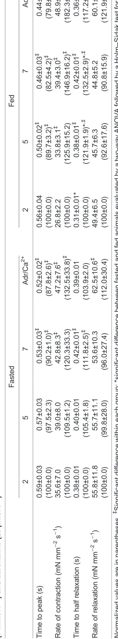
Time to peak was calculated as the time between onset of the contractile cycle and peak force. Time for 50% relaxation is the interval from peak force to 50% relaxation. Contraction and relaxation rates were calculated as the change in force per unit time.
Ventricular enzyme activities
Ventricular muscle tissue was homogenized at 5°C in a solution containing 50% glycerol, 20 mmol l−1 sodium phosphate buffer,
5 mmol l−1 β-mercaptoethanol, 0.5 mmol l−1 EDTA and 0.04%
BSA (Chi et al., 1983) and stored at−80°C. The same technique was used for tissue protein quantification, although BSA was emitted from the solution.
Enzymatic activities were measured spectrophotometrically at 25°C. Pyruvate kinase activity (µmol min−1 mg−1 protein) as an
estimate of glycolytic capacity was measured in a two-step reaction. The first step with pyruvate kinase is rate limiting, as the second step is catalysed by lactate dehydrogenase (LDH) in excess. Here,
–15 0 10 20 30 40 50 0
0.5 1.0 1.5
VS
(ml kg
–1
)
*
‡ ‡ ‡ ‡–15 0 10 20 30 40 50 0
20 40 60 80 100 120
Qsys
(ml min
–1
kg
–1
)
*
*
*
*
–15 0 10 20 30 40 50 0
1 2 3
PCV
(kPa)
*
*
*
*
*
*
‡ ‡
‡
‡ ‡
‡
‡ ‡
–15 0 10 20 30 40 50 0
20 40 60 80 100
fH
(min
–1
)
*
*
*
*
*
*
*
–15 0 10 20 30 40 50 0
2 4 6 8 10 12
Psys
(kPa)
‡
–15 0 10 20 30 40 50 0
1 2 3 4
Change in volume (%)
MCFP
(kPa)
*
*
*
*
*
*
‡ ‡
‡ ‡ ‡
‡ ‡ ‡
A
B
C
D
E
F
[image:3.612.56.559.358.683.2]•
Fig. 1. Effects of volume loading in steps of 10% body mass and a 15% blood volume depletion on various parameters inPython regius.(A) Stroke volume (VS), (B) heart rate (fH), (C) systemic flow (Q_sys), (D) central venous pressure (PCV), (E) mean circulatory filling pressure (MCFP) and (F) mean arterial pressure (Psys) in fasted pythons (white,N=6) and in pythons fed a rodent meal equivalent to 20% of body mass (black;N=7). Baseline values (0%) are indicated by red symbols.‡Significant difference from baseline; *significant difference from fasted values evaluated by a two-way ANOVA followed by a
Holm–Sidak multiple comparison test (P<0.05). Values are means±s.e.m.
Journal
of
Experimental
pyruvate is reduced to lactate with the concomitant oxidation of NADH being recorded at 340 nm in a solution of 50 mmol l−1 triethanolamine, 75 mmol l−1 KCl, 8 mmol l−1
MgSO4, 0.8 mmol l−1 phosphoenolpyruvate, 1 mmol l−1 ADP
and 0.3 mmol l−1NADH and LDH in excess ( pH was adjusted to
7.45 with NaOH when necessary). The activity of cytochrome oxidase as an estimate of aerobic capacity was measured at 550 nm on the basis of the relative decrease in the concentration of reduced cytochromec. The reaction was performed in a pH 7.45 solution containing 13 mmol l−1sodium phosphate and 0.4 mgl−1reduced
cytochrome c. After addition of homogenate, the cytochrome c concentration was recorded for 10 min; thereafter, potassium hexacyanoferrate (III) was added to complete the oxidation of cytochrome c. The activity was expressed by the first-order rate constant, i.e. the relative decrease in concentration of reduced cytochrome c per minute. Thus, tissue cytochrome activity was calculated by the rate constant divided by the tissue concentration expressed as ml g−1protein min−1.
Calculation of vascular parameters
Systemic flow was the sum of flow in the LAOand the RAO. Heart
rate was derived from the pressure of the vertebral artery, and stroke volume was calculated as Q_sys=fH (Wang et al., 2000).Systemic
vascular resistance (Rsys) was calculated as the difference in
systemic and venous pressure, divided by the cardiac output [Rsys¼ ðPsysPCVÞ=Q_sys], while venous resistance (Rven) was
calculated as the difference in MCFP and central venous pressure, divided by cardiac output [Rven¼ ðMCFPPCVÞ=Q_sys] (Skals
0 1 2 3
0 20 40 60 80 100
0 1 2 3
0 20 40 60 80
0 1 2 3
0 0.5 1.0
1.5
A
B
C
0 1 2 3
0 1 2 3 4
MCFP
(kPa)
0 1 2 3
0 2 4 6 8 10
PCV (kPa)
Psys
(kPa)
D
E
•
VS
(ml kg
–1
)
Qsys
(ml min
–1
kg
–1
)
fH
(min
–1
[image:4.612.47.301.101.193.2])
[image:4.612.54.563.353.694.2]Fig. 2. Correlations of central venous pressure (PCV) and cardiovascular parameters.(A) Stroke volume (VS), (B) heart rate (fH), (C) systemic flow (Q_sys), (D) mean circulatory filling pressure (MCFP) and (E) mean arterial pressure (Psys) in fasted pythons (white,N=6) and in pythons fed a rodent meal equivalent to 20% of body mass (black;N=7). Baseline values (before volume loading commenced, 0%) are indicated by red symbols. *Significant difference from fasted values (P<0.05). All values are means±s.e.m.
Table 1. Body mass, blood volume, mean circulatory filling pressure (MCFP), total circulatory compliance, unstressed and stressed blood
volume, and systemic and venous resistance of fasted (N=5) and fed
(N=7)Python regius
Fasted Fed
Body mass (g) 455.3±56.6 405.1±36.4
Blood volume (ml kg−1) 33.3±0.1 29.7±0.2
MCFP (kPa) 0.8±0.1 1.5±0.1 *
Total circulatory compliance (ml kg−1kPa−1) 25.0±3.6 12.8±0.8* Unstressed circulatory volume (ml kg−1) 16.5±1.7 9.7±1.0* Stressed circulatory volume (ml kg−1) 16.8±1.7 20.0±1.0 Systemic resistance (kPa ml−1min−1kg−1) 0.43±0.1 0.14±0.0* Venous resistance (kPa ml−1min−1kg−1) 0.02±0.0 0.01±0.0*
Note: one data point for fasted snakes was removed as it had a negative calculated unstressed blood volume. Total circulatory compliance and unstressed and stressed blood volume are derived from a MCFP/blood volume plot. Unstressed volume is thex-intercept of a linear regression through the data points for−15 to 10% volume loading, while stressed volume is the difference between total blood volume and unstressed volume. Total circulatory compliance was calculated as the inverse slope of the linear regression. *Significant difference from fasted snakes (unpairedt-test,
P<0.05). Values are means±s.e.m.
Journal
of
Experimental
et al., 2005). Unstressed blood volume was found by extrapolating the best fitted straight line of a MCFP/Vb curve to zero pressure,
using only the data points closest to 100% Vb (−15 to 10% Vb)
(Pang, 2001; Trippodo, 1981). Stressed Vb was the difference
between totalVband unstressedVb. The inverse slope of the same
best fitted straight line described above represents total circulatory compliance (Shoukas and Sagawa, 1971; Trippodo, 1981).
Statistical analysis
Each volume-loading step was compared between fasted and fed animals using a two-way ANOVA followed by a Holm–Sidak multiple comparison.PCVcorrelations were evaluated by computing
the best fitted line to the data and comparing slopes and plateaus between groups using an extra sum-of-squares F-test. Heart and organs mass, enzyme activities, calculated cardiovascular parameters and blood volumes were compared by t-tests. The changes in twitch force and contractile performance caused by adrenaline and calcium were analyzed within and between groups with a two-way ANOVA for repeated measures followed by a Holm– Sidak test for multiple comparisons. The limit of significance was P<0.05. All results are presented as means±s.e.m.
RESULTS
Mean circulatory filling pressure and volume loading
The occlusion of the outflow tract from the heart resulted in an immediate and progressive decline inPsysas blood was relocated
from the arterial to the venous side, causingPCVto increase. The
stabilization of both pressures was faster in digesting snakes, but both pressures stabilized within 10–30 s independent of digestive state, and the determination of MCFP was very similar at 10, 20 and
0 2 4 6 8 10
Fasted Fed
0 0.5 1.0 1.5 2.0
Fasted Fed
*
*
A
B
PCV
(kPa)
Psys
[image:5.612.315.563.54.245.2](kPa)
Fig. 3. Changes in blood pressure after occlusion of blood flow.(A) Mean arterial pressure (Psys) and (B) central venous pressure (PCV) in resting fasted (white,N=6) and fed (black;N=7) snakes, before (lined) and after (solid) occlusion of all major cardiac outflow tracts. The occludedPCVcorresponds to MCFP. *Significant difference from fasted values (P<0.05). Values are means±s.e.m.
0 50 100 150
0 0.5 1.0 1.5 2.0
Time (min)
Absorbance (627–740 nm)
Fasted Fed 0 1 2 3 4
[image:5.612.75.272.59.437.2]Blood volume (% body mass)
Fig. 4. Absorbance curve (Abs627–Abs740) from plasma obtained from fasted snakes (white,N=5) and snakes fed 20% of body mass (black,N=6) over a period of 120 min after an injection of Evans Blue (0.5 mg kg−1). Inset shows blood volumes in the same fasted (white,N=4) and fed snakes (black,N=6) calculated using an absorbance curve from plasma with known concentrations of Evans Blue (not shown). Difference inNvalues stem from removal of one outlier by use of the ROUT test (Q=0.1%). Statistical difference is evaluated by an unpairedt-test. Values are means±s.e.m.
0 0.1 0.5
Adrenaline concentration (μmol l−1)
Calcium concentration (mmol l−1)
5 20 Adr + Ca2+ 0
10 20 30 40 50
‡ ‡ ‡ ‡ ‡
‡ ‡ ‡ ‡ ‡
2 5 7 Ca2+ + Adr
0 10 20 30 40 50
‡
‡
‡
‡ ‡
T
witch force (mN mm
–2
)
Fig. 5. Twitch force in ventricle strips from fasting pythons (white,N=5) and pythons fed a rodent meal amounting to 30% of body mass (black, N=4-5) under different concentrations of adrenaline and the addition of calcium (7 mmol l−1; pH 7.45, 30°C) or under different concentrations of calcium and the addition of adrenaline (20μmol l−1; pH 7.45, 30°C).
‡Difference between the first measurement relative to each adrenaline/calcium
concentration in each group evaluated by a two-way ANOVA for repeated measures followed by a Holm–Sidak test for multiple comparisons (P<0.05).
Values are means±s.e.m.
Journal
of
Experimental
[image:5.612.314.566.443.644.2]30 s after occlusion for all blood volumes (Fig. S1). This was also the case after volume loading.Psysrarely became identical toPCV
because critical closing pressure typically was higher than PCV
(Fig. S2). However, the difference between Psys and PCV rarely
exceeded 1 kPa, and given the 10–20 times higher compliance within the venous circulation, the pressure differential would underestimate MCFP by considerable less than 0.1 kPa.
Figs 1–3 show the haemodynamic responses to blood volume manipulations in fasted and fed snakes (open and black symbols, respectively) during anaesthesia. All variables are similar to previous reports from pentobarbital-anaesthetized or pithed snakes (Enok et al., 2012; Joyce et al., 2016; Skovgaard et al., 2007; Wang et al., 2000).
At rest, the larger VS and fH in fed snakes (88 and 50%,
respectively) resulted in an elevated cardiac output (19.5±2.2 and 54.1±4.9 ml min−1 kg−1, respectively) at a constant P
sys(Fig. 1).
The pressure gradient driving blood to the heart was larger in fed snakes before volume manipulations, as the difference in MCFP and PCV was more than twice as large in fed snakes (0.25±0.1 and
0.52±0.1 kPa). Volume loading more than doubled the pressure gradient in fasted animals, while it remained similar in fed animals (0.59±0.2 and 0.46±0.3 kPa), which is consistent with the attenuation of the difference in VS between the groups. Volume
loading increasedPCVand MCFP within each group and enhanced
the difference between fasted and fed snakes (Fig. 1D,E).
VS,Q_sysand MCFP correlated positively with an elevatedPCVin
both groups (Fig. 2A,C,D).Psysalso correlated positively withPCV;
however, fasted snakes quickly peaked at aPCVof 8.7 kPa while fed
snakes were able to continuously increasePsysto a peak pressure
similar to fasted snakes (7.5 kPa) (Fig. 2E). Fed snakes were also capable of continuously increasing flow, while flow stagnated in fasted snakes (69.5±9.0 and 42.9±6.6 ml min−1 kg−1; Fig. 2C).
Total circulatory compliance and unstressed blood volume decreased after feeding, which also resulted in a larger stressed volume in fed snakes. Both systemic and venous resistance were significantly larger in fasted snakes at rest (Table 1). Fig. 3 illustrates the pressure before and after occlusion in both the arterial
and venous system. The decrease inPsyswas similar in both fasted
and fed snakes (4.86±1.19 and 5.24±0.57 kPa, respectively). The increase in venous pressure was also similar between fasted and fed snakes (0.44±0.01 and 0.56±0.01 kPa, respectively), while the absolute pressure both before and after occlusion was significantly larger in fed snakes.
The relative mass of the whole heart, measured upon completion of the experiments, did not differ between fasted and fed snakes (0.23±0.01 and 0.21±0.01%, respectively), nor did ventricle (0.15±0.01 and 0.16±0.01% of body mass, respectively) or atria mass (0.06±0.01 and 0.06±0.00% of body mass, respectively; data not shown).
Blood volume
The EB disappearance curve differed between fasted and fed animals, but they-intercept was similar (Fig. 4). As the standard curves generated from plasma from each individual snake were identical (data not shown), the calculated blood volumes did not differ between fasted and fed snakes (Fig. 4 inset). Hct was similar in fasted and fed snakes (overall mean of 16.8±4.2%) before commencing blood sampling, and decreased to 15.1±2.5% after the repetitive blood sampling over the course of 120 min.
Twitch force and contractile performance
Increasing adrenaline concentration from 0.1 to 20 µmol l−1caused
a progressive increase in twitch force for both experimental groups, resulting in a 31 and 33% increase in force in ventricular preparations from fasted and fed snakes, respectively (Fig. 5A). The cumulative effect of adrenaline and calcium caused a similar amplification of twitch force in both groups (49 and 46%; Fig. 5A). Furthermore, adrenaline shortened the times of force development and relaxation in both groups. The cumulative effect of calcium reversed this effect and significantly increased these rates in fed snakes (Table 2).
Twitch force increased progressively with calcium concentration in both groups (Fig. 5B), and subsequent addition of adrenaline (20μmol l−1) augmented the effect of calcium, such that twitch
force increased by a total of 22 and 47% in fasting and digesting snakes, respectively (Fig. 5B). Increased calcium delayed the time required to reach maximal peak force and the rate of relaxation, while the cumulative effect of adrenaline and calcium attenuated the effect on relaxation rate (Table 3).
Enzymatic activities of cytochrome oxidase and pyruvate kinase
The ventricular activity of cytochrome oxidase resulted in an approximate 50% rise in the fed snakes, whilst pyruvate kinase activity was unaltered (Table 4).
Organ mass
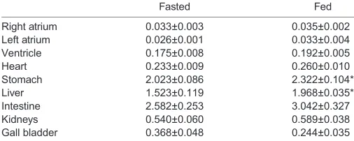
No difference in whole-heart, ventricular or atrial mass was found between the fasted and fed snakes used for the ventricular preparation experiment (Table 1). The intestine, kidney and gall bladder mass were also similar between groups. Fed snakes had larger stomachs and livers compared with fasted snakes (Table 5).
DISCUSSION
We confirmed that cardiac mass does not increase during digestion, and the isolated cardiac strips provide the first evidence that cardiac contractility per se is not affected by digestive state in pythons. Nevertheless, despite the lack of increased postprandial cardiac mass and the lack of enhanced contractile performance, we
Table 4. Activity of cytochrome oxidase (CO; ml g−1protein min−1) and
pyruvate kinase (PK;μmol g−1protein min−1) in the ventricular tissues
of fasted and fedP. regiusat 25°C
Fasted Fed
CO 1266.8±3.0 1854.1±217.1*
PK 299.5±35.8 350.0±41.3
[image:7.612.47.300.616.717.2]Values are means±s.e.m. *Significant difference from fasted animals (unpairedt-test,P<0.05).
Table 5. Relative mass of internal organs in fasting and digesting
groups ofP.regiusused for ventricular preparation studies
Fasted Fed
Right atrium 0.033±0.003 0.035±0.002
Left atrium 0.026±0.001 0.033±0.004
Ventricle 0.175±0.008 0.192±0.005
Heart 0.233±0.009 0.260±0.010
Stomach 2.023±0.086 2.322±0.104*
Liver 1.523±0.119 1.968±0.035*
Intestine 2.582±0.253 3.042±0.327
Kidneys 0.540±0.060 0.589±0.038
Gall bladder 0.368±0.048 0.244±0.035
Values are means±s.e.m. (n=5). *Significant difference from fasted snakes
(P<0.05).
Journal
of
Experimental
measured a substantial postprandial rise in VS in anaesthetized
pythons. BecauseVbdid not increase during digestion, the rise in
MCFP and the accompanying elevation of venous return seems sufficient to explain the postprandial rise in VS through an
augmented end-diastolic volume. It is also noteworthy that the maximal VS during volume loading was not elevated during
digestion, providing strong evidence that the capacity of the heart to elevateVSis not different during digestion.
We studied anaesthetized snakes to allow simultaneous measurements of the many haemodynamic variables in response to manipulations of blood volume. Pentobarbital anaesthesia does, however, mute autonomic regulation in reptiles (e.g. Crossley et al., 1998; Skals et al., 2005), and the typical withdrawal of parasympathetic tone is likely to explain the higher fH of the
anaesthetized pythons than in previous studies on fully recovered pythons (Enok et al., 2012; Skovgaard et al., 2009). The lack of body movement during anaesthesia may also affect venous return, but will do so similarly in both groups and therefore have little effect on a comparison of cardiovascular parameters. Thus, as the anaesthetized pythons exhibited the stereotypical postprandial tachycardia, it is likely that the haemodynamic state during anaesthesia sufficiently resembles that of conscious animals, and allows an identification of the mechanisms underlying the rise inVS
during digestion. Furthermore, by virtue of our manipulations of blood volume, we provide a robust and anaesthesia-independent characterization of the capacity of the python heart to increaseVS.
The Vb determined for both fasting and digesting pythons
resemble the 4–7% of body mass previously reported for other species of snakes and other terrestrial vertebrates (Hillman et al., 2010; Lillywhite and Smith, 1981; Lillywhite and Smits, 1984; Smits and Lillywhite, 1985; Thorson, 1968). We determinedVbin
awake snakes, while our haemodynamic measurements were performed with anaesthetized animals. However, given the short duration of the experimental protocol during anaesthesia and the sessile nature of the awake snakes duringVbmeasurements,Vbis
unlikely to have been altered before the initial measurements of MCFP and blood flows were concluded (Lillywhite and Smith, 1981; Lillywhite and Smits, 1984). Some studies incubate EB with native plasma before injection to ensure binding to plasma proteins and alleviate wash-out from the capillaries (Hillman et al., 2010), whereas we and others injected EB directly into the circulation (Baustian, 1988; Thorson, 1964), which could overestimate the calculatedVb. However, theVb of 3% in our study is lower than
previous measurements in other snake species (Lillywhite and Smits, 1984). The small Vb could stem from differences in the
sampling routine. A long equilibration time with infrequent sampling will tend to overestimate Vb, as the initial mixing and
equilibration within the plasma compartment is fast, followed by a prolonged equilibration with the extracellular fluid space (Nichols, 1987). The samples obtained every 5–10 min for 2 h after EB injection yielded similar dilution curves and statically similarVbin
both fasting and digesting pythons, but previous studies on other snakes relied on longer equilibration times and less frequent sampling (Lillywhite and Smith, 1981; Lillywhite and Smits, 1984; Smeller et al., 1978; Smits and Lillywhite, 1985; Thorson, 1968). In any event, there is no reason to expect that these errors would differ between fasting and digesting snakes, and our similarVb of both
groups indicate that increased Vb does not contribute to raising
cardiac filling during digestion.
BothPCVand MCFP virtually doubled during digestion, which,
in the absence of changes inVb, points to increased vascular tone.
This increased tone probably resides on the venous circulation, as
Rsyswas reduced during digestion in accordance with the rise in
metabolism. An increased vascular tone was also supported by our calculations of unstressed and stressed blood volumes. Unstressed volume is a measure of the haemodynamic inactive volume needed to fill the circulatory system without causing a positive pressure, while the remaining blood volume, the stressed blood volume, exerts a regulated pressure on the vessels (Greenway and Lautt, 1986; Samar and Coleman, 1979). The ratio between these volumes thus represents overall vascular tone. In the present study, digestion caused a shift from unstressed to stressed volume. The mobilization of unstressed volume is primarily caused by venoconstriction, as the venous circulation holds approximately two-thirds of the total blood volume (Rothe, 1993; Sandblom and Axelsson, 2007). As the veins in pythons and other vertebrates are under adrenergic control, the shift from inactive to active blood volume is likely to reflect an increased sympathetic tone, consistent with studies on rattlesnakes, various fish and mammals that report an increase in bothPCVand
MCFP because of anα-adrenergic stimulation (Lillywhite, 1987; Pang, 2001; Sandblom and Axelsson, 2007; Skals et al., 2005). In addition to a sympathetic regulation of venous tone, it is an intriguing possibility that the expansion of the stomach upon ingestion of the large prey may reduce unstressed volume by squeezing the veins.
In line with the lowerRsys,PCVtended to stabilize faster in the
digesting snakes, usually within 10–15 s compared with 20–30 s in fasted snakes, which could differentially affect the estimation of MCFP, as compensatory mechanisms are activated when Psys
declines during occlusion. These barostatic mechanisms, including vascular constriction (Pang, 2001), are rapidly activated in mammals and trout (Guyton, 1981; Sandblom and Axelsson, 2007), but seem delayed in rattlesnakes (Skals et al., 2005) and will result in overestimates of MCFP. This overestimation is likely to be most pronounced in the fasting snakes because of the longer time course for equilibration, and would therefore tend to reduce the reported difference in MCFP between fasting and digesting snakes. In any event, we do not believe this to be an important problem as the measuredPCVat 10, 20 and 30 s upon
occlusion does not differ (Fig. S1), indicating small compensatory responses.
According to Guyton’s original description (Guyton, 1955), MCFP represents the pressure in the venules and the difference between MFCP and PCV therefore provides a measure of the
pressure gradient driving venous return. Because MCFP increased relatively more thanPCVduring digestion in pythons, the venous
pressure gradient is increased. As described by Starling and Visscher (1927), this increase translates into an increasedVSby a
magnitude specified by the set point on the Starling curve (Fig. 2A). VSof fasted pythons resides on the steep part of the Starling curve,
which enhances the capacity for increases in cardiac filling to translate into increasedVS.
To evaluate the true capacities of the cardiovascular system, we volume loaded the snakes by up to 90% of the measured blood volume. At these extreme volumes,VSis no longer able to increase
with increasing pressures in either fasted or fed pythons, as the Starling capacity has been reached, and thereforeVS stagnates at
similar values, indicating equal cardiac capacities–consistent with the lack of cardiac growth. Volume loading also eliminated any influence of anaesthesia on venous return, as the system was massively distended and beyond significant nervous regulation.
The positive inotropic effects of Ca2+ and adrenaline are
consistent with previous studies on cardiac muscle from snakes and other reptiles (Shiels and Galli, 2014; Zaar et al., 2007), but
Journal
of
Experimental
more importantly, in context of the major aims of the present study, there were few effects of digestion on contractile force. Thus, although mRNA for myosin increases postprandially (Andersen et al., 2005), our study demonstrates that the expression of the contractile proteins is not translated into increased contractile force. This is consistent with the fact that reptiles are endowed with the capacity for very high ejection fractions (Burggren et al., 2013), making it unlikely that increased contractility and the associated reduction of end-systolic volume could contribute significantly to the doubling ofVSduring digestion. We did find an increase in
cytochrome oxidase activity in the cardiac muscle, consistent with the increased gene expression previously reported (Riquelme et al., 2011), identifying an increased oxidative capacity. As contractility is unchanged, this increased capacity probably supports the 50% increase in heart rate after feeding.
The increased heart rate and stroke volume of the postprandial snakes, a hallmark of the specific dynamic action response in pythons (Enok et al., 2013; Secor et al., 2000; Starck et al., 2004), clearly persisted during anaesthesia, giving credence to the experimental approach of using anaesthetized animals to unravel the physiological mechanisms underlying the rise in stroke volume during digestion. In the present study, we provide evidence supporting an unchanged contractile force during digestion, which only leaves an increased cardiac filling as the underlying cause for an increased postprandial stroke volume. We identified an increased mean circulatory filling pressure caused by an augmented venous tone, as blood volume is unchanged, sufficient to increase venous pressure gradient, which increased cardiac filling and, consequently, stroke volume.
Competing interests
The authors declare no competing or financial interests.
Author contributions
All authors were involved in conceiving and designing the study and partook in the experiments and the data analysis. S.E. collated the information and wrote the manuscript, with input from the other authors, who also approved its final version.
Funding
This study was supported by the Danish Natural Science Research Council, the CNPq and FAPESP through the INCT of Comparative Physiology.
Supplementary information
Supplementary information available online at
http://jeb.biologists.org/lookup/doi/10.1242/jeb.142729.supplemental
References
Andersen, J. B., Rourke, B. C., Caiozzo, V. J., Bennett, A. F. and Hicks, J. W.
(2005). Physiology: postprandial cardiac hypertrophy in pythons.Nature434, 37-38.
Baustian, M.(1988). The contribution of lymphatic pathways during recovery from
hemorrhage in the toadBufo marinus.Physiol. Zool.61, 555-563.
Burggren, W. W., Christoffels, V. M., Crossley, D. A., II, Enok, S., Farrell, A. P., Hedrick, M. S., Hicks, J. W., Jensen, B., Moorman, A. F. M., Mueller, C. A. et al.
(2013). Comparative cardiovascular physiology: future trends, opportunities and challenges.Acta Physiol.210, 257-276.
Chi, M. M., Hintz, C. S., Coyle, E. F., Martin, W. H., Ivy, J. L., Nemeth, P. M.,
Holloszy, J. O. and Lowry, O. H.(1983). Effects of detraining on enzymes of
energy metabolism in individual human muscle fibers. Am. J. Physiol. 244, C276-C287.
Crossley, D., Altimiras, J. and Wang, T.(1998). Hypoxia elicits an increase in
pulmonary vasculature resistance in anaesthetised turtles (Trachemys scripta).
J. Exp. Biol.201, 3367-3375.
Dawson, A. B., Evans, H. M. and Whipple, G. H.(1920). Blood volume studies.
Am. J. Physiol.51, 232-256.
Enok, S., Simonsen, L. S., Pedersen, S. V., Wang, T. and Skovgaard, N.(2012).
Humoral regulation of heart rate during digestion in pythons (Python molurusand
Python regius).Am. J. Physiol. Regul. Integr. Comp. Physiol.302, R1176-R1183.
Enok, S., Simonsen, L. S. and Wang, T.(2013). The contribution of gastric
digestion and ingestion of amino acids on the postprandial rise in oxygen consumption, heart rate and growth of visceral organs in pythons. Comp. Biochem. Physiol. A Mol. Integr. Physiol.165, 46-53.
Greenway, C. V. and Lautt, W. W.(1986). Blood volume, the venous system,
preload, and cardiac output.Can. J. Physiol. Pharmacol.64, 383-387.
Guyton, A. C.(1955). Determination of cardiac output by equating venous return
curves with cardiac response curves.Physiol. Rev.35, 123-129.
Guyton, A. C.(1981).Circulatory Physiology: Cardiac Output and its Regulation.
Philidelphia, PA: W.B. Saunders Company.
Guyton, A. C., Polizo, D. and Armstrong, D.(1954). Mean circulatory filling
pressure measured immediately after cessation of heart pumping.Am. J. Physiol.
179, 261-267.
Guyton, A. C., Lindsey, A. W. and Kaufmann, B. N.(1955). Effect of mean
circulatory filling pressure and other peripheral circulatory factors on cardiac output.Am. J. Physiol.180, 463-468.
Hansen, K., Pedersen, P. B. M., Pedersen, M. and Wang, T. (2013).
Magnetic resonance imaging volumetry for noninvasive measures of phenotypic flexibility during digestion in Burmese pythons.Physiol. Biochem. Zool.86, 149-158.
Henriksen, P. S., Enok, S., Overgaard, J. and Wang, T.(2014). Food composition
influences metabolism, heart rate and organ growth during digestion inPython regius.Comp. Biochem. Physiol. A Mol. Integr. Physiol.183C, 36-44.
Hillman, S. S., DeGrauw, E. A., Hoagland, T., Hancock, T. and Withers, P.(2010).
The role of vascular and interstitial compliance and vascular volume in the regulation of blood volume in two species of anuran.Physiol. Biochem. Zool.83, 55-67.
Jensen, B., Larsen, C. K., Nielsen, J. M., Simonsen, L. S. and Wang, T.(2011).
Change of cardiac function, but not form, in postprandial pythons. Comp. Biochem. Physiol. A Mol. Integr. Physiol.160, 35-42.
Joyce, W., Axelsson, M., Altimiras, J. and Wang, T.(2016).In situ cardiac
perfusion reveals interspecific variation of intraventricular flow separation in reptiles.J. Exp. Biol.219, 2220-2227.
Lillywhite, H. B.(1987). Circulatory adaptations of snakes to gravity.Am. Zool.27,
81-95.
Lillywhite, H. B. and Smith, L. H. (1981). Haemodynamic responses to
haemorrhage in the snake,Elaphe obsoleta obsoleta.J. Exp. Biol.94, 275-283.
Lillywhite, H. B. and Smits, A. W.(1984). Lability of blood volume in snakes and its
relation to activity and hypertension.J. Exp. Biol.110, 267-274.
McCue, M. D., Bennett, A. F. and Hicks, J. W.(2005). The effect of meal
composition on specific dynamic action in burmese pythons (Python molurus).
Physiol. Biochem. Zool.78, 182-192.
Nichols, D. J.(1987). Fluid volumes in rainbow trout,Salmo gairdneri: application
of compartmental analysis. Comp. Biochem. Physiol. A Comp. Physiol. 87, 703-709.
Olesen, M. G., Bertelsen, M. F., Perry, S. F. and Wang, T.(2008). Effects of
preoperative administration of butorphanol or meloxicam on physiologic responses to surgery in ball pythons.J. Am. Vet. Med. Assoc.233, 1883-1888.
Overgaard, J., Busk, M., Hicks, J. W., Jensen, F. B. and Wang, T.(1999).
Respiratory consequences of feeding in the snake Python molorus. Comp. Biochem. Physiol. A Mol. Integr. Physiol.124, 359-365.
Pang, C. C. Y.(2001). Autonomic control of the venous system in health and
disease: effects of drugs.Pharmacol. Ther.90, 179-230.
Riquelme, C. A., Magida, J. A., Harrison, B. C., Wall, C. E., Marr, T. G., Secor,
S. M. and Leinwand, L. A.(2011). Fatty acids identified in the Burmese python
promote beneficial cardiac growth.Science334, 528-531.
Rothe, C. F. (1993). Mean circulatory filling pressure: its meaning and
measurement.J. Appl. Physiol.74, 499-509.
Samar, R. E. and Coleman, T. G.(1979). Mean circulatory pressure and vascular
compliances in the spontaneously hypertensive rat. Am. J. Physiol. 237, H584-H589.
Sandblom, E. and Axelsson, M. (2007). The venous circulation: a piscine
perspective.Comp. Biochem. Physiol. A Mol. Integr. Physiol.148, 785-801.
Secor, S. M. and White, S. E. (2010). Prioritizing blood flow: cardiovascular
performance in response to the competing demands of locomotion and digestion for the Burmese python,Python molurus.J. Exp. Biol.213, 78-88.
Secor, S. M., Hicks, J. W. and Bennett, A. F.(2000). Ventilatory and cardiovascular
responses of a python (Python molurus) to exercise and digestion.J. Exp. Biol.
203, 2447-2454.
Shiels, H. A. and Galli, G. L. J.(2014). The sarcoplasmic reticulum and the
evolution of the vertebrate heart.Physiology29, 456-469.
Shoukas, A. A. and Sagawa, K.(1971). Total systemic vascular compliance
measured as incremental volume-pressure ratio.Circ. Res.28, 277-289.
Skals, M., Skovgaard, N., Abe, A. S. and Wang, T.(2005). Venous tone and
cardiac function in the South American rattlesnakeCrotalus durissus: mean circulatory filling pressure during adrenergic stimulation in anaesthetised and fully recovered animals.J. Exp. Biol.208, 3747-3759.
Skovgaard, N., Conlon, J. M. and Wang, T.(2007). Evidence that neurotensin
mediates postprandial intestinal hyperemia in the python, Python regius.
Am. J. Physiol. Regul. Integr. Comp. Physiol.293, R1393-R1399.
Journal
of
Experimental
Skovgaard, N., Moller, K., Gesser, H. and Wang, T.(2009). Histamine induces postprandial tachycardia through a direct effect on cardiac H-2-receptors in pythons.Am. J. Physiol. Regul. Integr. Comp. Physiol.296, R774-R785.
Slay, C. E., Enok, S., Hicks, J. W. and Wang, T.(2014). Reduction of blood oxygen
levels enhances postprandial cardiac hypertrophy in Burmese python (Python bivittatus).J. Exp. Biol.217, 1784-1789.
Smeller, J. M., Bush, M. and Seal, U. S.(1978). Blood volume measurements in
gopher snakes, using autologous 51Cr-labeled red blood cells.Am. J. Vet. Res.
39, 355-356.
Smits, A. W. and Lillywhite, H. B.(1985). Maintenance of blood volume in snakes:
transcapillary shifts of extravascular fluids during acute hemorrhage.J. Comp. Physiol. B Environ. Physiol.155, 305-310.
Starck, J. M., Moser, P., Werner, R. A. and Linke, P. (2004). Pythons
metabolize prey to fuel the response to feeding.Proc. R. Soc. Lond. B Biol. Sci.
271, 903-908.
Starling, E. H. and Visscher, M. B.(1927). The regulation of the energy output of
the heart.J. Physiol.62, 243-261.
Thorson, T. B.(1964). The partitioning of body water in amphibia.Physiol. Zool.37,
395-399.
Thorson, T. B.(1968). Body fluid partitioning in reptilia.Copeia1968, 592.
Trippodo, N. C. (1981). Total circulatory capacity in the rat. Effects of
epinephrine and vasopressin on compliance and unstressed volume.Circ. Res.
49, 923-931.
Wamberg, S., Sandgaard, N. C. F. and Bie, P.(2002). Simultaneous determination
of total body water and plasma volume in conscious dogs by the indicator dilution principle.J. Nutr.132, 1711S-1713S.
Wang, T., Axelsson, M., Jensen, J. and Conlon, J.(2000). Cardiovascular actions
of python bradykinin and substance P in the anesthetized python,Python regius.
Am. J. Physiol. Regul. Integr. Comp. Physiol.279, R531-R538.
Wang, T., Zaar, M., Arvedsen, S., Vedel-Smith, C. and Overgaard, J.(2002).
Effects of temperature on the metabolic response to feeding inPython molurus.
Comp. Biochem. Physiol. A Mol. Integr. Physiol.133, 519-527.
Zaar, M., Overgaard, J., Gesser, H. and Wang, T.(2007). Contractile properties of
the functionally divided python heart: two sides of the same matter.Comp. Biochem. Physiol. A Mol. Integr. Physiol.146, 163-173.
Zerbe, P., Glaus, T., Clauss, M., Hatt, J.-M. and Steinmetz, H. W. (2011).
Ultrasonographic evaluation of postprandial heart variation in juvenile Paraguay anacondas (Eunectes notaeus).Am. J. Vet. Res.72, 1253-1258.
Journal
of
Experimental