Original Article
Clinical features of MELAS and its
relation with A3243G gene point mutation
Jin Zhang1, Junhong Guo1, Wanghui Fang1, Qili Jun1, Kaili Shi2
1Department of Internal Medicine-Neurology, The First Hospital of Shanxi Medical University, Taiyuan 030001, Shanxi, China; 2Department of Neurology, Shanxi Province Children’s Hospital, Taiyuan 030001, Shanxi, China Received June 28, 2015; Accepted July 29, 2015; Epub October 1, 2015; Published October 15, 2015
Abstract: Mitochondrial encephalopathy with lactic acidosis and stroke-like episodes (MELAS) mostly occur in chil-dren. The point mutation A3243G of mitochondrial DNA (mtDNA) may work as a specific bio-marker for mitochon -drial disorders. The related clinical features, however, may vary among individuals. This study therefore investigated the relation between MELAS clinical features and point mutation A3243G of mtDNA, in an attempt to provide further evidences for genetic diagnosis of MELAS. Children with MELAS-like syndromes were tested for both blood lactate level and point mutation A3243G of mtDNA. Further family study was performed by mtDNA mutation screening at the same loci for those who had positive gene mutation at A3243G loci. Those who were negative for A3243G point mutation were examined by muscle biopsy and genetic screening. Both clinical and genetic features were analyzed. In all 40 cases with positive A3243G mutation, 36 children fitted clinical diagnosis of MELAS. In other 484 cases with negative mutation, only 8 children were clinically diagnosed with MELAS. Blood lactate levels in both groups were all elevated (P>0.05). In a further genetic screening of 28 families, 10 biological mothers and 8 silbings of MELAS children had positive A3243G point mutations but without any clinical symptoms. Certain difference existed in the clinical manifestations between children who were positive and negative for A3243G mutation of mtDNA but without statistical significance. MELAS showed maternal inheritance under most circumstances.
Keywords: MELAS, point mutation, mitochondrial DNA, maternal inheritance
Introduction
As one important sub-type of mitochondrial encephalopathy, mitochondrial encephalopa-thy with lactic acidosis and stroke-like episodes (MELAS) mostly occur in childhood but without clear illustration of pathogenesis. Common clinical symptoms of MELAS vary from child to child, including coma, dementia, epilepsy, aphasia, vomiting, fever, weakness, headache, ataxia, external ophthalmoplegia, hemiparaly-sis, periodic encephalopathy, audiovisual
disor-der, hypothyroidism, hirsutism and dwarfism [1, 2]. Due to the lack of specific laboratory indica
-tor, it has been difficult for making a definitive
diagnosis of MELAS, although the occurrence
of fragile red edged fibers in muscular patho
-logical examination has been postulated [3].
With the advancement of genetic studies, it has been reported that the mutation at loci A3243G of mitochondrial DNA (mtDNA) played an impor-tant role in the occurrence of mitochondrial
encephalopathy. Due to the unique cytoplasmic distribution, mtDNA has certain differences regarding its inheritance mechanism compared to nuclear DNA, as various phenomena includ-ing higher mutation rates, maternal inheritance, heterozygous, genetic drift may occur more
fre-quently in mtDNA [4]. Novel mutation loci for MELAS have been identified, such as A3252G,
T3271C, A3260G, T7512C, G583A, G1642A
and T3291C [5]. Epidemiological surveys have
contributed more than 80% of MELAS cases into point mutation A3252G, which was the most prevalent point mutation in Chinese Han
people with MELAS [6]. When comparing
mutation patterns, in order to analyze the genetic feature of the disease.
Materials and methods
Patients and inclusive/exclusive criteria
We adopted the primary diagnostic criteria of
MELAS as previously established [7], including:
(1) Brain stroke before 40-years old; (2) Manifested with progressive encephalopathy with dementia and seizure; (3) Multiple systems including central and peripheral organs affect-ed; (4) Hyperlactacidemia; (5) Cerebral infarc-tion/atrophy and basal ganglia affected by head CT; (6) No other encephalopathy or
meta-bolic disorders. In general, individuals who fit -ted (4), (5) and (6), plus any one of (1), (2) or (3) were diagnosed with MELAS-like cases.
Further confirmed diagnosis was made as pre
-viously reported [8]. In brief, a confirmed diag -nosis was made based on the primary screen-ing as abovementioned plus: (1) atypical red
edged fiber (RRF) in muscle biopsy by succinate
dehydrogenase (SDH) staining; (2) Negative for sub-sarcolemma under cytochrome-c (COX) staining; and (3) positive point mutation for A3243G in genetic screening.
Those children who were positive for A3243G
point mutation and fitted MELAS clinical symp
-toms were classified as mutation-positive group. Those who fitted clinical symptoms of
MELAS but had negative A3243G mutation and
negative for COX or RRF staining were classified
in mutation-negative group. This study has been pre-approved by the ethical committee of our hospital and has obtained written consents from all children’s guardians.
Genetic screening for point mutation
We adopted PCR-RFLP technique for screening of A3243G point mutation of mtDNA. In brief, peripheral blood samples were collected from patients and extracted for genomic DNA. The
PCR utilized specific primers for A3243G muta -tion (Forward, 5’-CCTCC CTGTA CGAAA GGACA-3’; Reverse, 5’-CACCC TGATC AGAGG ATTGA G-3’) under the following condition: 95°C pre-denature for 5 min, followed by 30 cycles each containing 94°C denature for 30 sec, 60°C annealing for 30 sec and 72°C elongation for 60 sec. The reaction ended after a further 72°C elongation for 7 min. PCR products were then cut by ApaI restriction enzyme, which can cut the mutated gene into two fragments (130 and 423 bp) while leaving the wild type gene intact
(553 bp). Digestion fragments were finally sep -arated under agarose gel electrophoresis. The gel image was captured and analyzed by Quantity One software (BioRad, US).
Statistical analysis
All collected data were analyzed by SPSS 21.0 software package. The correlation between MELAS clinical symptoms and A3243G point mutation was processed by one-factor
correla-tion analysis. A statistical significance was defined when P<0.05.
Results
General information of patients
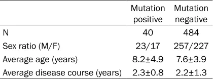
As shown in Table 1, among all 524 cases with MELAS-like symptoms, 40 of them were posi-tive for mutation while 484 cases were nega-tive for the point mutation. General information including onset age, disease course and sex
ratio had no significant difference between
those two groups (P>0.05). Blood lactate level assay
Serum assays showed that the blood lactic acid level was between 4.2 and 9.9 mM in mutation positive group, and was between 2.3 and 10.8 mM in mutation negative group. Both groups showed higher lactate level than normal people (0.5 to 2.0 mM) but without any statistically
sig-nificant difference (P>0.05).
Gene screening for A3243G mutation
[image:2.612.90.302.84.163.2]Figure 1 showed the RFLP pattern of both mutation positive (Lane 3) and mutation nega-tive (Lane 4) individuals. Mutant gene form had two restriction bands of 423 and 130 bp while wild type form had one single band at 553 bp after restriction digestion.
Table 1. General information of patients
Mutation
positive Mutation negative
N 40 484
Clinical features and its relation with A3243G point mutation
In mutation positive group, patients were
main-ly manifested as dwarfism and hirsutism, along
with other features including weakness, sei-zure, fever, aphasia, omitting, progressive dementia and vision loss. In some patients, there were also headache, coma, cerebral stroke and auditory dysfunctions. A total of 26 cases were complicated with epilepsy. In muta-tion negative group, patients also showed sei-zure, weakness, dementia and vision loss but
no hirsutism or dwarfism (but not in one case).
A tabulated description of all major clinical tures was shown in Table 2. Some of those
fea-tures showed significant difference between
two groups (P<0.05).
Family survey of MELAS patients
In mutation positive group, four children had
definitive family history, as two cousins of
patients had been diagnosed with MELAS by muscular biop-sy when alive. One patient’s sister has been misdiagnosed with renal tubular acidosis since two years old, and mani-fested with progressive dementia and ptosis at 10 years old. The patient’s moth-er showed genmoth-eral weakness and lower body weight but without A3243G mutation. In mutation negative group, one child’s mother had psychiatric disorders. One sibling of this patient also died without
definitive reason. No family
history has been discovered in any patient in the mutation negative group.
Family screening of A3243G mutation
[image:3.612.89.373.73.143.2]We also performed the genet-ic screening targeting A3243G mutation in a total of 74 family members in 28 families with mutation positive MELAS child. A total of 10 biological mothers were positive for A3243G mutation, along with
Figure 1. RFLP patterns of A3243G mutation. Lane 1, negative control; Lane 2, positive control; Lane 3, mutation negative; Lane 4, mutation positive.
8 siblings. All these carriers had no clinical symptoms. Other family members had no genetic mutation at the given locus.
Discussion
Mitochondrial encephalopathy was a multi-sys-tem heterogeneous disease that has both genetic and acquired factors. As the organelle for energy transformation inside eukaryotic cells, mitochondria is unique with other cellular compartments as it has it self-organized DNA and unique genetic mechanism. With the advancement of genetic diagnosis, the muta-tion of mtDNA has been recognized to be relat-ed with various mitochondria diseases
includ-ing MELAS [9, 10]. Studies have revealed the
A3243G mutation as the most commonly occurred pathogenic mutation locus of this dis-ease. The location of this mutation is within UUR coding region of tRNA. The A to G substitu-tion lead to per-terminasubstitu-tion of transcripsubstitu-tion, impeding expression of normal rRNA, thus Table 2. Clinical features of MELAS
Symptom Mutation positive (N=36) Mutation negative (N=8) χ2 P
Weakness 30 5 0.215 0.764 Dementia 28 7 0.042 1.000 Seizure 35 6 0.194 0.773 Omitting 17 0 3.557 0.092
Fever 16 0 3.357 0.095
Hirsutism 30 0 6.116 0.018
Dwarfism 35 1 4.706 0.037
Vision loss 22 5 0.001 1.000
Fever 7 2 0.080 1.000
Vomiting 5 1 0.008 1.000 Aphasia 8 2 0.018 1.000
Coma 9 3 0.278 0.686
[image:3.612.91.375.210.434.2]compromising mitochondrial protein synthesis,
ATP production and body metabolism [11, 12].
MELAS may occur in all age groups but predom-inantly during childhood with various clinical manifestations including periodic headache, seizure, vomiting, vision loss and weakness. Currently, diagnosis is mainly dependent on the
occurrence of raged red fiber and negative
cytochrome C staining in muscular biopsy. Atypical features including various central ner-vous injury and progressive extraocular paraly-sis but without cerebral stoke may also occur in children who also had growth retard, seizure and cognitive disorder but perhaps no raged
red fiber in biopsy [13-15]. This study observed
the correlation between A3243G mutation in MELAS children and their clinical features, thus providing references for genetic diagnosis. In clinical practice, similar manifestations exist between MELAS patients who were positive for mtDNA point mutation and for those who were negative for point mutation, further complicat-ing the sub-typcomplicat-ing of patients. As negative results in pathological examination may not completely indicate the clinical sub-type, fur-ther genetic screening can work as one labora-tory index. The correlation between mutation and clinical features, however, remained poor
understood [16-18]. This study performed a
correlation analysis between the clinical fea-tures and MELAS patients who were positive or negative for mtDNA point mutation, by the means of one-factor or multi-factor correlation analysis. Our results showed mutation positive patients had disease-related features including weakness, dementia, seizure, vomiting, fever,
hirsutism and dwarfism, while mutation nega -tive patients had other disease-related mani-festations including weakness, seizure, demen-tia and vision loss. By a multi-factor regression analysis, the most commonly occurred clinical symptoms in mutation positive ones were (in descending order) weakness, hirsutism, dementia and seizure; whilst mutation negative patients most frequently had seizure, demen-tia, vision loss and weakness.
Meanwhile we also performed family survey on families of MELAS patients, including mutation positive and negative ones. In a genetic screen-ing for mtDNA A3243G mutation, we found 10 mothers and 8 siblings of mutation positive children also carried such point mutations. In mutation negative group, however, no A3243G
genotype has been detected. These results support the maternal inheritance pattern of A3243G mutation, consistent with previous
reports [19, 20].
In summary, certain but not significant differ -ences exist in clinical features between MELAS patients with positive and negative A323G mutation, which follow a maternal inheritance
pattern. Our study for the first time provides
correlation between clinical features and mtDNA mutation of MELAS, although further independent multi-centered survey is required for the substantiation.
Acknowledgements
Department of science research project in Shanxi Province (NO. 20130313022), Yuan
funds The first hospital of ShanXi medical uni -versity (YJ1402).
Disclosure of conflict of interest
None.
Address correspondence to: Dr. Jin Zhang, Depart- ment of Internal Medicine-Neurology, The First Hospital of Shanxi Medical University, 85 Jiefang South Road, Taiyuan 030001, Shanxi, China. Tel: +86-351-4867014; Fax: +86-351-4867014; E-mail: [email protected]
References
[1] Abeliovich A. Parkinson’s disease: Mitochon- drial damage control. Nature 2010; 463: 744-5.
[2] McFarland R, Taylor RW and Turnbull DM. A neurological perspective on mitochondrial dis-ease. Lancet Neurol 2010; 9: 829-40.
[3] Calvo SE and Mootha VK. The mitochondrial proteome and human disease. Annu Rev Genomics Hum Genet 2010; 11: 25-44. [4] Schaefer AM, McFarland R, Blakely EL, He L,
Whittaker RG, Taylor RW, Chinnery PF, Turnbull DM. Prevalence of mitochondrial DNA disease in adults. Ann Neurol 2008; 63: 35-9.
[5] Wang K, Takahashi Y, Gao ZL, Wang GX, Chen XW, Goto J, Lou JN, Tsuji S. Mitochondrial ND3 as the novel causative gene for Leber he- reditary optic neuropathy and dystonia. Neurogenetics 2009; 10: 337-45.
[7] Tschampa HJ, Urbach H, Greschus S, Kunz WS, Kornblum C. Neuroimaging characteristics in mitochondrial encephalopathies associated with the m.3243A>G MTTL1 mutation. J Neurol 2013; 260: 1071-80.
[8] Chinnery PF, Elliott HR, Hudson G, Samuels DC, Relton CL. Epigenetics, epidemiology and mitochondrial DNA diseases. Int J Epidemiol 2012; 41: 177-87.
[9] Clark J, Reddy S, Zheng K, Betensky RA, Simon DK. Association of PGC-1alpha polymorphisms with age of onset and risk of Parkinson’s dis-ease. BMC Med Genet 2011; 12: 69.
[10] Prasad M, Narayan B, Prasad AN, Rupar CA, Levin S, Kronick J, Ramsay D, Tay KY, Prasad C. MELAS: A Multigenerational Impact of the MTTL1 A3243G MELAS Mutation. Can J Neurol Sci 2014; 41: 210-9.
[11] Zhang YM, Ji YC, Liu XL, Zhou XT, Zhao FX, Sun YH, Wei QP, Zhang JJ, Liu Y, Qu J, Guan MX. [Leber’s hereditary optic neuropathy may be associated with the mitochondrial tRNAGlu A14693G mutation in three Chinese families]. Yi Chuan 2010; 32: 353-9.
[12] Suomalainen A. Biomarkers for mitochondrial respiratory chain disorders. J Inherit Metab Dis 2011; 34: 277-82.
[13] Ito H, Mori K and Kagami S. Neuroimaging of stroke-like episodes in MELAS. Brain Dev 2011; 33: 283-8.
[14] Cwerman-Thibault H, Sahel JA and Corral-Debrinski M. Mitochondrial medicine: to a new era of gene therapy for mitochondrial DNA mu-tations. J Inherit Metab Dis 2011; 34: 327-44. [15] Lim ST, Esfahani K, Avdoshina V, Mocchetti I.
Exogenous gangliosides increase the release of brain-derived neurotrophic factor. Neuro- pharmacology 2011; 60: 1160-7.
[16] Kim HK, Ha SH and Han J. Potential therapeu-tic applications of tetrahydrobiopterin: from in-herited hyperphenylalaninemia to mitochon-drial diseases. Ann N Y Acad Sci 2010; 1201: 177-82.
[17] Halter J, Schüpbach WM, Casali C, Elhasid R, Fay K, Hammans S, Illa I, Kappeler L, Krähenbühl S, Lehmann T, Mandel H, Marti R, Mattle H, Orchard K, Savage D, Sue CM, Valcarcel D, Gratwohl A, Hirano M. Allogeneic hematopoietic SCT as treatment option for pa-tients with mitochondrial neurogastrointesti-nal encephalomyopathy (MNGIE): a consensus conference proposal for a standardized ap-proach. Bone Marrow Transplant 2011; 46: 330-7.
[18] de Laat P, Koene S, van den Heuvel LP, Rodenburg RJ, Janssen MC, Smeitink JA. Clinical features and heteroplasmy in blood, urine and saliva in 34 Dutch families carrying the m.3243A > G mutation. J Inherit Metab Dis 2012; 35: 1059-69.
[19] Lembke A, Gomez R, Tenakoon L, Keller J, Cohen G, Williams GH, Kraemer FB, Schatzberg AF. The mineralocorticoid receptor agonist, fludrocortisone, differentially inhibits pituitary-adrenal activity in humans with psychotic ma-jor depression. Psychoneuroendocrinology 2013; 38: 115-21.