Introduction
Benign lesions of the duodenum include het-erotopic pancreas, hethet-erotopic gastric mucosa, duplication, atresia, diverticulum, Celiac dis-ease, tropical sprue, Whipples disdis-ease, amyloi-dosis, parasite infestation, duodenal ulcer, duo-denitis, AIDS-related inflammatory disease, fun-gal infection, cytomefun-galovirus infection, radia-tion duodenitis, Brunner’s glands hyperplasia, Brunner’s glands adenoma, adenoma, hamar-tomatous polyp, endometriosis, inflammatory fibroid polyp, lipoma, hemangioma, lymphan-gioma, telangiectasia, neurofibroma, gan-glioneurofibroma, and congenital fibromatosis [1]. In the present study, 567 benign duodenal lesions were described.
Materials and methods
specimens in the last 10 years of our pathology laboratory. Review of the histological slides was done when appropriate. The duodenal speci-mens were composed of 567 benign lesions and 48 malignant lesions. Computer search of clinical records were also reviewed. The pa-tients ranged from 25 years to 95 years with a mean of 53 years. Male to female ratio was 321:294. In appropriate cases, an immunohis-tochemical analysis had been performed with the use of Dako Envision method (Dako), as previously described [2-6].
Results
The duodenal specimens were composed of 567 benign lesions and 48 malignant lesions. In this report, the benign duodenal lesions were described. The 567 benign lesions were com-posed of chronic non-specific duodenitis in 334
Original Article
Pathologic observations of the duodenum in 615
consecutive duodenal specimens: I. benign lesions
Tadashi Terada
Department of Pathology, Shizuoka City Shimizu Hospital, Shizuoka, Japan
Received November 18, 2011; accepted December 14, 2011; Epub January 1, 2012; Published January 15, 2012
Abstract: The author investigated histopathology of 615 consecutive duodenal specimens in our pathology laboratory. Computer search of the duodenal lesions was performed. Review of histological slides was done, when appropriate. The duodenal specimens were composed of 567 benign lesions and 48 malignant lesions. The 567 benign lesions were composed of chronic non-specific duodenitis in 334 cases (60.0%), duodenal ulcer in 101 cases (17,8%), het-erotopic gastric mucosa in 81 cases (14.3%), hyperplastic polyp in 16 cases (2.8%), Brunner’s gland hyperplasia in 14 cases (2.5%), Brunner’s gland adenoma in 8 cases (1.4%), lymphoid polyp in 5 cases (0.8%), tubular adenoma in 4 cases (0.7%), lymphangioma in 2 cases (0.4%), endocrine nests in 1 case (0.2%), and amyloidosis in 1 case (0.2%). The chronic non-specific duodenitis was characterized by edema and lymphocytic infiltration. The duodenal ulcer was characterized by exudate, necrosis, granulation tissue and regenerative epithelium. The heterotopic gastric mucosa consisted of two types: one was composed of only foveolar epithelium (n=21) and another foveolar epithelium and fundic glands (n=60). Hyperplastic polyp was characterized by proliferation of gastric foveolar-like epithelium. The Brunner’s gland hyperplasia was characterized by hyperplastic proliferation of the gland. The Brunner gland adenoma was characterized by neoplastic proliferation of the gland. The lymphoid polyp was characterized by large lymph folli-cles with large germinal centers. The tubular adenoma was characterized by adenomatous proliferation of intestinal epithelium, similar to colon adenoma. The lymphangioma was characterized by submucosal cavernous proliferation of lymphatics. The endocrine cell nests were characterized by non-neoplasmic proliferation of neuroendocrine cells. The amyloidosis was characterized by deposition of amorphous materials positive with Congo-red stain.
cases (14.3%), hyperplastic polyp in 16 cases (2.8%), Brunner’s gland hyperplasia in 14 cases (2.5%), Brunner’s gland adenoma in 8 cases (1.4%), lymphoid polyp in 5 cases (0.8%), tubu-lar adenoma in 4 cases (0.7%), lymphagioma in 2 cases (0.4%), endocrine cell micronests in 1 case (0.2%), and amyloidosis in 1 case (0.2%).
The chronic non-specific duodenitis (n=334) was characterized by edema and lymphocytic infiltration (Figure 1). This condition was very frequently recognized. Almost all duodenal specimens showed more or less lymphocytic infiltration.
The duodenal ulcer (n=101) was characterized
by exudate, necrosis, granulation tissue and regenerative epithelium (Figure 2). The regen-erative epithelium infrequently mimicked ade-nocarcinoma. No evidence for viral or fungal infection was noted in the present series. The location was fist portion in 87 cases, and sec-ond portion in 14 cases. Perforation of the ulcer was recognized in two cases, which needed emergency operations.
[image:2.612.324.531.81.397.2]The hererotopic gastric mucosa (n=81) was lo-cated in the first portion in 42 cases, second portion in 32 cases, and third portion in 7 cases. Endoscopically, it was recognized as slight elevated or discolored lesion. Heterotopic gastric mucosa consisted of the following two types: one was composed of only foveolar epi-thelium (n=21) (Figure 3A) and another foveolar epithelium and fundic glands (n=60) (Figure 3B). The foveolar epithelium occasionally Figure 1. Chronic duodenitis. Much lymphocytes
[image:2.612.80.291.85.244.2]infil-tration is seen. HE, x200.
Figure 2. Duodenal ulcer. Necrosis, exudates, infiltra-tion of neutrophils and lymphocytes are recognized. HE, x200.
[image:2.612.80.292.301.460.2]showed hyperplastic changes (Figure 3A).
The hyperplastic polyp (n=16) was located in the first portion in 9 cases, in the second por-tion in 5 cases and in the third porpor-tion in 2 cases. Endoscopically, it was detected as duo-denal polyp. Histologically, it was composed of hyperplastic columnar epithelium with mucins, and resembled to hyperplastic polyp of the stomach (Figure 4).
The Brunner’s gland hyperplasia (n=14) was located in the first portion in 9 cases and the second portion in 5 cases. Endoscopically, it was recognized as an elevated or polyp lesion. It was histologically characterized by hyperplastic proliferation of the gland. However, in biopsy specimens, differentiation from Brunner’s gland adenoma was occasionally difficult.
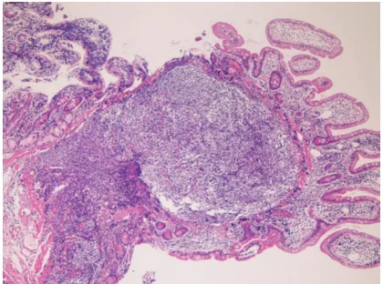
The Brunner gland adenoma (n=8) was situated in the first portion in 3 cases and the second portion in 5 cases. Endoscopically, it was recog-nized as a polypoid elevation or polyp lesion. It was histologically characterized by neoplastic proliferation of the gland (Figure 5).
The lymphoid polyp (n=5) was present in the first portion in 2, second portion in 2, and third portion in 1 cases. Endoscopically, it was recog-nized as a polyp. It was histologically character-ized by a large lymph follicle with a large germi-nal center (Figure 6). The histology and imuno-histochemical study demonstrated that it was different from follicular lymphoma and other
types of lymphoma.
The tubular adenoma (n=4) was located in the first portion in 1 case, second portion in 2 cases, and third portion in 1 case. Endoscopi-cally, it was recognized as flat or elevated le-sions. It was histologically characterized by ade-nomatous proliferation of intestinal epithelium (Figure 7), similar to colon adenoma. Immuno-histochemically, the tubular adenoma was nega-tive for p53 protein, and Ki-67 labeling was low (mean Ki-67 labeling = 8%).
[image:3.612.323.533.84.242.2]The lymphangioma (n=2) was present in the second portion in all the two cases. Endoscopi-cally, it was recognized as polyp or submucosal Figure 4. Hyperplastic polyp of the duodenum.
Hyper-plasia of gastric foveolar-like cells is recognized. HE, x100.
[image:3.612.79.290.85.244.2]Figure 5. Brunner’s gland adenoma. Neoplastic Brunner’s gland is noted. No atypia is seen. HE, x100
[image:3.612.323.533.311.467.2]tumor. Pathologically, it was characterized by submucosal cavernous proliferation of lymphat-ics free of red blood cells (Figure 8). No atypia was recognized.
The endocrine cell micronests (n=1) was lo-cated in the second portion. Endoscopically, it was recognized as a flat discolored lesion. It was pathologically characterized by non-neoplasmic proliferation of neuroendocrine cells positive for synaptophysin, neuro- specific enolase, and CD56.
In amyloidosis (n=1), biopsy was taken from the bulb. Endoscopically, it was recognized as a
polyp. Pathologically, it was characterized by deposition of amorphous materials (Figure 9A) positive with Congo-red stain (Figure 9B). Later the patient (76-year-old man) was found to have multiple myeloma.
Discussion
In the present series, chronic non-specific duo-denitis was the most common benign condition. Almost all patients in the present study showed more or less lymphocytic infiltration of the duo-denal mucosa, similar to the stomach. The lym-phocytes may play an important role of local immunity.
[image:4.612.80.291.85.244.2]The duodenal ulcer was the second common benign condition next to duodenitis. It was a simple ulcer. However, penetration and perfora-tion may occur, causing serious problems, as seen in the present study. Pathologically, the Figure 7. Tubular adenoma of the duodenum.
[image:4.612.80.291.318.476.2]Adeno-matous proliferation of intestinal epithelium is recog-nized. The appearances are similar to colonic ade-noma. HE, x200.
Figure 8. Lymphagioma of the duodenum. Neoplastifc proliferation of lymphatics is recognized. HE, x100.
[image:4.612.325.532.344.657.2]ulcer margin regenerative epithelium must be differentiated from adenocarcinoma.
Heterotopic gastric mucosa is well recognized entity, and several case report or small case series have been published [7-10]. However, the frequency and common site have been un-clear. In the present series, the frequency was 14.3%. In the present study, it was located in the first portion in 42 cases, second portion in 32 cases, and third portion in 7 cases. There-fore, the common location is the first and sec-ond portions of the dusec-ondenum. In the present study, heterotpic gastric mucosa consisted of the following two types: one was composed of only foveolar epithelium (n=21) and another foveolar epithelium and fundic glands (n=60). The author speculates that the latter is a genital maliformation while the former is a con-genital or acquired lesion (gastric foveolar meta-plasia). Of importance, adenoma and adenocar-cinoma very infrequently arise in heterotopic gastric mucosa [11, 12]. The foveolar epithe-lium occasionally showed hyperplastic changes in the present series.
Hyperplastic polyp was recognized in 2.8% in the present study. Its common locations are the first and second portions of the duodenum. His-tologically, it was composed of hyperplastic co-lumnar epithelium with mucins, and resembled to hyperplastic polyp of the stomach. The author speculates that the hyperplastic polyp is in fact hyperplastic changes of ectopic foveolar gastric mucosa. It should be kept in mild that hyper-plastic polyp may undergo malignant transfor-mation [13-15].
In the present study, Brunner’s gland hyperpla-sia was seen in 2.5%. Its preferential locations were the first portion and the second portion of the duodenum. In biopsy specimens, differentia-tion from Brunner’s gland adenoma was occa-sionally difficult. A few case studies of Brunner’s gland hyperplasia have been reported [16, 17]. It is important to recognize that carcinoma may arise in Brunner’s gland hyperplasia [17].
Brunner gland adenoma was recognized in 1.4% in the present series. The preferential lo-cation was the second portion of the duode-num. A few case reports of this neoplasm have been reported [18, 19]. It was noteworthy that carcinoma may arise from Brunner’s gland ade-noma [19].
The lymphoid polyp was noted in 5 cases in the
present study. In the English literature, no re-ports of lymphoid polyp of the duodenum are present. It has been rarely reported in the rec-tum [20]. The important point is a differential diagnosis from malignant lymphoma, follicular lymphoma, and lymphomatoid hyperplasia. In the present study, lymphoma was denied from various immunohistochemical stainings. Other-wise, it has little clinical relevance.
In the present study, tubular adenoma of the duodenum was recognized in 4 patients. Endo-scopically, it was recognized as flat or elevated lesions. It was histologically characterized by adenomatous proliferation of intestinal epithe-lium, similar to colon adenoma. Adenoma of the duodenum is extremely rare [22]. In the present study, the neoplasm is not adenocarcinoma in histology as well as immunohistochemistry.
Lymphangioma of the duodenum was recog-nized in 2 cases in the present series. Lymphan-gioma of the duodenum is very rare, and a few case reports are present in the English literature [23, 24]. Lymphangioma of the duodenum was characterized by submucosal cavernous prolif-eration of lymphatics free of red blood cells in the present study. No atypia was recognized. It has no clinical relevance.
Endocrine cell micronests were noted in 1 case. This is a rare condition, frequently associated with duodenal carcinoid tumors [25, 26]. The micronests were pathologically characterized by non-neoplasmic proliferation of neuroendocrine cells positive for synaptophysin, neuron-specific enolase, and CD56. In the present series, no association with carcinoids was noted.
In the present study, amyloidosis was nized in 1 case. Endoscopically, it was recog-nized as a polyp. Pathologically, it was charac-terized by deposition of amorphous materials positive with Congo-red stain. The patient (76-year-old man) was found to have multiple mye-loma later, suggesting that systemic amyloidosis may present initially as duodenal polyp.
In summary, the present study reported the histopathology of various benign lesions of the duodenum.
References
[1] Rosai J. Small intestine. In Rosai and Acker-man’s Surgical Pathology. Ninth edition Mosby 2004; pp: 712-756.
[2] Terada T, Kawaguchi M, Furukawa K, Sekido Y, Osamura Y. Minute mixed ductal-endocrine carcinoma of the pancreas with predominant intraductal growth. Pathol Int 2002; 52: 740-746.
[3] Terada T, Tanigichi M. Intraductal oncocytic papillary neoplasm of the liver. Pathol Int 2004; 54: 116-123.
[4] Terada T. Primary multiple extragastrointestinal stromal tumors of the omentum with different mutations of c-kit gene. World J Gastroenterol 2008; 14: 7256-7259.
[5] Terada T. Gastrointestinal stromal tumor of the uterus: A case report with genetic analyses of c-kit and PDGFRA genes. Int J Gynecol Pathol 2009; 28: 29-34.
[6] Terada T. Large endocervical polyp with carti-laginous and osseous metaplasia: a hitherto unreported entity. Int J Gynecol Pathol 2009; 28: 98-100.
[7] Spiller RC, Shousa S, Barrison IG. Heterotopic gastric mucosa in the duodenum: a report of eight cases. Dig Dis Sci 1982; 27: 880-883. [8] Lessells AM, Martin DF. Heterotopic gastric
mucosa in the duodenum. J Clin Pathol 1982; 35: 591-595.
[9] Franzin G, Musola R, Negri A, Mencarelli R, Fratton A. Heterotopic gastric (fundic) mucosa in the duodenum. Endoscopy 1982; 14: 166-167.
[10] Mann NS, Mann SK, Rachut E. Heterotopic gastric mucosa in the duodenal bulb. J Clin Gastroenterol 2000; 30: 303-306.
[11] Russin V, Krevsky B, Caroline DF, Tang CK, Ming SC. Mixed hyperplastic and adenomatous polyp arising from ectopic gastric mucosa of the duodenum. Arch Pathol Lab Med 1986; 110: 556-558.
[12] Abe T, Hosokawa M, Kusami T, Kusano M, Ho-kari K, Kagaya H, Watanabe A, Fujita M, Sasaki S. Adenocarcinoma arising from ectopic gastric mucosa in the cervical esophagus. Am J Clin Pathol 2004; 27: 644-645.
[13] Daibo M, Itabashi M, Hirota T. Malignant trans-formation of gastric hyperplastic polyp. Am J Gastroenterol 1987; 82: 1016-1025.
[14] Oriowska J, Jarosz D, Pachlewski J, Butruk E. Malignant transformation of benign epithelial gastric polyps. Am J Gastroenterol 1995; 90: 2152-2159.
[15] Zea-Iriarte WL, Sekine I, Itsuno M, Makiyama K, Naito S, Nakayama T, Nishizawa-Takano JE, Hattori T. Carcinoma in gastric hyperplastic polyps: a phenotypic study. Dig Dis Sci 1996; 41: 377-386.
[16] Ueno N, Tomiyama T, Tano S, Aizawa T,
Na-gamine N, Kihara K, Kumakura Y, Ishino Y, Kimura K. A case of Brunner’s gland hyperpla-sia: endoscopic color Doppler ultrasonic find-ings. Endoscopy 1997; 29: 51.
[17] Sakirai T, Sakashita H, Honjo G, Kasyu I, Ma-nabe T. Gastric foveolar metaplasia with dys-plastic changes in Brunner gland hyperplasia: possible precursor lesions for Brunner gland adenocarcinoma. Am J Surg Pathol 2005; 29: 1442-1448.
[18] Gao YP, Zhu JS, Zheng WJ. Brunner’s gland adenoma: a case report and literature review. World J gastroenterol 2004; 10: 2616-2617. [19] Faller G, Kirchner T. Low-grade intraepithelial
neoplasm of Brunner’s gland. Histopathology 2005; 47: 118-119.
[20] Rutsch F, Henker J, Fischer R, Gobel P. Gastro-intestinal lymphonodular polyp and lymphoid polyps of the rectum: a rare condition. Z Gastro-enterol 1997; 35: 271-275.
[21] Reddy RR, Schuman BM, Priest RJ. Duodenal polyps: diagnosis and management. J Clin Gas-troenterol 1981; 3: 139-147.
[22] Seifert E, Schulte F, Stolte M. Adenoma and carcinoma of the duodenum and papilla of Vater: a clinicopathologic study. Am J Gastroen-terol 1992; 87: 37-42.
[23] Salta HH, Mercader J, Navorro A, Cortes JM, Gonzales-Campos C: Lymphangioma of the duodenum. Endoscopy 1984; 16: 30-32. [24] Aneiros J, Pleguezuelos J, Garcia del Moral R,
Cabollero T, Rodrigo M, Salido E. Lymphan-gioma of the duodenum: an ultrastructural study. Endoscopy 1986; 18: 245-248.
[25] Abe H, Kubota K, Oka T, Kobayashi T, Makuuchi M. A rare case of multiple carcinoids and endo-crine cell micronests in a patient with chronic duodenitis. Cancer 2000; 89: 963-969. [26] Noda Y, Watanabe H, Iwafuchi M, Furuta K,