Original Article
B7-H6 expression in non-small cell lung cancers
Xiuqin Zhang1,3, Guangbo Zhang2, Yan Qin3, Ruizhen Bai3, Jianan Huang1,2
1Department of Respiratory Medicine, The First Affiliated Hospital of Soochow University, 188 Shizi Street, Suzhou 215006, China; 2Clinical Immunology Laboratory of Jiangsu Province, 708 Renmin Road, Suzhou 215006, China; 3The First Affiliated Hospital of Jiangnan University, 200 Huihe street, Binhu District, Wuxi 214000, China
Received August 19, 2014; Accepted September 15, 2014; Epub September 15, 2014; Published October 1, 2014
Abstract: B7 family has been known to be a negative regulator of immunity response in patients with lung cancer. B7-H6 as a novel identified member of B7 family is found to trigger natural killer (NK) cell cytotoxicity and cytokine secretion by binding natural cytotoxicity receptor NKp30. Up until now, no investigations have been made about B7-H6 expression in lung cancer. We present the result of the B7-H6 prognostic value in 65 non-small cell lung cancer (NSCLC) tissues and 65 matched adjacent non-tumor tissues by Immunohistochemistry (IHC). Meanwhile, fluorescence activated cell sorter (FACS) analysis was used to detect B7-H6 receptor NKp30 expression in 7 non-small cell lung cancer tissues and 7 adjacent non-tumor tissues. Here, the result showed B7-H6 immunoreactivity in 6/65 (9.23%) lung cancer patients and 4/65 (6.15%) in adjacent non-tumor tissues. No relationship was found between B7-H6 expression and clinic pathological features. Similarly, no relevance was found for NKp30 expression in lung cancer tissues and non-tumor tissues. However, B7-H6 positive carcinomas were significantly correlated with degree of differentiation (P= 0.044). Three year survival rate after operation did not show the prognostic value for B7-H6 expression. Our study suggests that B7-H6 has a limited value as a prognostic marker in the patients of lung cancer.
Keywords: B7-H6, NKp30, lung cancer, immunohistochemistry
Introduction
Worldwide, lung cancer is a leading cause of cancer death in men, and it is the second cause of cancer death for women. Roughly 85% patients are non-small cell lung cancer, over half of lung cancer is diagnosed at an advanced stage, 16% are diagnosed at the early stage. In spite of many available adjuvant therapies are available, however, the overall five-year survival rate remain low for lung cancer patients, a major reason is diagnosis of lung cancer at advanced stage [1, 2]. Therefore, we would make great efforts to explore novel markers and contribute to early diagnosis of lung cancer.
The B7 family members control T cell mediated immune response by binding their CD28 recep-tors on activated T cells, the inhibitory mem-bers of B7 family can yield regulatory signals to terminate or weaken functions of activated T cells in tumor environment [3-5]. At present,
several B7 family numbers have been found in lung cancer. B7-H6 known as NGR3LG1 is a newly identified member in the B7 family. B7-H6 mRNA and protein expression are not detected in normal tissues, and expressed mainly on the cell surface of various tumor cells such as hematological malignancies, it seems that its expression maybe associate with tumor prog-nosis in a large number of tumor patients [6, 7]. B7-H6 triggers antitumor of natural killer cell cytoxicity and cytokine secretion by binding NKp30 receptor which is a natural cytotoxicity receptor expressed mainly on the surface of natural killer cells [6-8]. However, there are no data about the clinical significance of B7-H6 expression in patients of lung cancer.
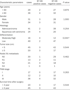
Table 2. Relationship between B7-H6 expression and clinicopathologi-cal parameters in lung carcinoma patients
Characteristic parameters cases positive cases B7-H6 negative casesB7-H6 P value Age (years)
< 60 29 2 27 0.879
≥ 60 36 4 32
Gender
Male 31 3 28 1.000
Female 34 3 31
Histology
Adenocarcinoma 41 2 39 0.254
Squamous cell carcinoma 24 4 20
Differentiation
Moderate/High 16 4 12 0.044*
Low 49 2 47
Tumor size (cm)
< 5 45 3 42 0.544
≥ 5 20 3 17
Nodal (N) metastasis
N0 30 1 29 0.462
N1 13 2 11
N2 21 3 18
N3 1 0 1
TNM stage
I 17 0 17 0.263
II 12 2 10
III 36 4 32
Survival time after surgery
< 3 year 20 3 17 0.544
≥ 3 year 45 3 42
*P < 0.05.
investigate whether B7-H6 acts as a novel iden-tified prognostic marker in lung cancer patients.
Materials and methods
Patients and tissue specimens
Formalin-fixed, paraffin-embedded tumor tis -sues and adjacent non-tumor tis-sues were selected retrospectively from the First Affiliated
utes in a citrate buffer (10 nmol/L; pH 6.0), then endogenous peroxidase activity was blocked with 0.3% hydrogen peroxide solution for 30 minutes. After washing three times with phosphate buffer saline (PBS) for 5 minutes each, sections were incubated with rabbit anti-human B7-H6 polyclonal antibody (1:50 dilu-tions, Abcam) at 4°C overnight, a negative con-trol was carried out by replacing the primary antibody with PBS, then sections were incubat-Table 1. The percent of B7-H6 expression in lung cancer tissues and
non-tumor tissues
B7-H6 expression Tumor (N ratios [%]) Non-tumor (N ratios [%])
Positive (n = 10) 6/9.23 4/6.15
Negative (n = 120) 59/90.77 61/91.85
Hospital of Jiangnan Uni- versity (the Fourth People’s Hospital, Wu Xi, China). All of 130 cases including 65 tumor tissues and 65 adja-cent non-tumor tissues underwent surgical resec-tion between January 2008 and December 2009. None of the patients had received radiotherapy and chemo-therapy before operation. According to the American Joint Committee on cancer staging system (AJCC) (7th, edition), these patients we- re classified as 17 cases (stage I); 12 cases (stage II); 36 cases (stage III), and include 31 males and 34 females. These patients were followed-up for 3 years. The median age at diagnosis was 59.94 (ran- ge: 28-78) years. The proto-col in this study was approved by the ethics committees of the First Affiliated Hospital of Jiang-nan University.
Immunohistochemistry
[image:2.612.93.391.189.569.2]min-ed with horseradish peroxide-labelmin-ed goat anti-Rabbit second antibody (1:1000 dilutions, Merck & Millipore).
Evaluation of B7-H6 staining
The sections were examined and evaluated independently by two pathologists. B7-H6 pro-tein was quantified using a visual grading according to the extent of staining: 0 (< 5%); 1 (6%-25%); 2 (26%-50%); 3 (51%-75%); 4 (> 75%), and intensity of staining: 0 (no staining); 1 (weak staining, light yellow); 2 (moderate staining, yellowish brown); 3 (strong staining, brown). The sum of score was determined as followed: 0 (negative); 1-4 (weakly positive); 5-8 (moderately positive); 9-12 (strongly positive) [9].
Flow cytometry for NKp30
The specimens dissected from 7 matched patients of lung cancer and adjacent non tumor tissues were cut into 2 mm fragments, these fragments were washed in PBS, and trans-ferred to a conical tube containing 0.05% col-lagenase at 37°C for 20 minutes, then these specimens were passed with a mesh to provide a single cell suspension. The cell suspension from lung cancer and adjacent non tumor was washed with PBS for 2 times, stained by phyco-erythrin-cyanine 5 (PC5) conjugated anti-CD45, fluorescein isothiocyanate (FITC) conjugated anti-CD56 and phycoerythrin (PE) conjugated-NKp30 antibody (Biolegend) in 4°C incubation for 30 minutes. The staining with anti-CD45 PC5 and anti-CD56 FITC was marked as natural kill cells in lung carcinomas and non-tumor tis-sues; PE-conjugated-NKp30 antibody was used to detect B7-H6 receptor NKP30 expression on the surface of CD45+ and CD56+ cells, then cells were washed by PBS and detected imme-diately by flow cytometry on fluorescence acti -vated cell sorter (FACS Calibur cytometer, Becton Dickinson, Heidelberg, Germany).
Statistical analysis
Statistical analysis was performed with SPSS 17.0 software. The association between B7-H6 expression and clinic pathologic features was analyzed by using chi-square test or Fisher’s exact test. NKp30 expression was analyzed with rank sum test, P-values less than 0.05
were considered as being statistically sig- nificant.
Results
B7-H6 expression in tumor tissues and non-tumor tissues
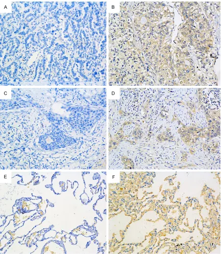
In this study, B7-H6 expression was found in the cytoplasm of cells. B7-H6 protein expres-sion was detected in 6/65 (9.23%) lung carci-nomas and 4/65 (6.15%) non tumor tissues (Table 1). Lung cancer samples were all moder-ate staining (6/6), strong staining was not detected in all lung cancer specimens. However, adjacent non-tumor tissues were all weak stain-ing (4/4) in cytoplasm of lung tissues, moderate and strong staining were not found in all lung tissues (Figure 1).
B7-H6 expression and clinic pathological
char-acteristics and prognosis
B7-H6 expression presented no significant dif -ferences between lung cancer tissues and non-tumor tissues. No matter lung carcinomas or adjacent non-tumor tissues, B7-H6 expression difference was not found with regard to patients’ age, sex distribution, histological clas -sification, tumor size, distant metastasis. However, B7-H6 immunoreactivity was associ-ated with degree of differentiation (P = 0.044) (Table 2). Three year survival rate of B7-H6 positive and B7-H6 negative cases provided no significant difference in lung carcinomas and non-tumor tissues (the data of non tumor tis-sues was not shown).
NKp30 expression insurgical resected
speci-mens
Discussion
The B7 family has been studied in human malignancies. The inhibitory B7 family mem-bers interact with their receptors CD28 family to inhibit cytokine secretion, cytotoxicity deve- lopment and activation of T cells [10, 11]. Many inhibitory B7 family members are expressed in
[image:4.612.90.525.69.566.2]lung cancer and associated with adverse prog-nosis. The inhibitory B7 family include B7-H1 (PD-L1, CD274), B7-DC (PD-L2, CD273), B7-H2 (ICOSL, CD275), B7-H3 (CD276, B7RP-2), B7-H4 (VTCN1, B7X, B7S1), and newly identified B7-H6 [12]. B7-H1 expression was found in 53.2% of lung cancer tissues, the patients with B7-H1 high expression presented disadvantageous
Figure 2. A. NKp30 expression in CD45+ and CD56+ cells of tumor and non-tumor. CD45+ and CD56+ cells were regarded as nature kill cells in tumor and non-tumor by FACS. B. The comparison of NKp30 expression on the sur-face of nature kill cells between tumor and non-tumor. (P = 0.750, P > 0.05).
clinical outcome with low infiltration of tumor infiltrating lymphocyte (TIL) [13, 14]. However, the clinical significance of B7-H2 is unclear in lung cancer. B7-H3 and B7-H4 inhibitory mole-cules are found on the surface and in the cyto-plasm of lung cancer, and have significant rele -vance with lymph node metastasis [15]. The soluble B7-H3 level was significantly associated with tumor size, TNM stage, lymph node
expression had short median overall survival time and adverse distant metastasis [9]. However, there are no data about clinical sig-nificance of B7-H6 expression in lung cancer.
In this study, we provide the first investigation about the relationship of prognostic and clinical value of B7-H6 protein in lung carcinomas and adjacent non tumor tissues. All of lung carcino-mas and adjacent non-tumor tissues were stained for B7-H6. The B7-H6 expression had no obvious difference between tumor tissues and non-tumor tissues. Meanwhile, the B7-H6 expression did not reveal significant relevance with clinical pathological features, such as age, sex, tumor size, histological classification, lym -phocyte node metastasis, TNM stage and dis-tant metastasis in lung cancer patients, but B7-H6 positive expression significantly corre -lated with degree of differentiation (P= 0.044). This result is similar with study in gastric carci-noma [19]. Three year survival rate after opera-tion presented no significant difference of B7-H6 expression in lung carcinomas. Similarly, the B7-H6 receptor NKp30 expression did not reveal any significant difference between lung carcinomas and adjacent non tumor tissues. Above, it seems that B7-H6 and its receptor NKp30 have no essential clinical meanings in lung carcinomas patients.
Altogether, our investigations suggest that B7-H6 has a limited clinical value as prognostic marker for lung cancer. Although the number of lung cancer patients enrolled is small, the results of B7-H6 expression might be different in many other tumors. So, the B7-H6 expres-sion of other tumors would need to be investi-gated in a great deal of patients.
Acknowledgements
This work was supported by graduate innova-tion project of Jiangsu Province, Soochow University (No. CXZZ13_0831), a project fund-ed by National Clinical key specialty construc-tion projects, Naconstruc-tional Natural Science Foun-dation of China (No. K112211412), Natural Science Foundation of Jiangsu Province (BK20-12606) and supported by Science and Tech- nology Development Fund of Wuxi (scientific and technological support social development-Demonstration) (No. CSE31N1317), China.
Disclosure of conflict of interest
None.
Address correspondence to: Dr. Jianan Huang, De- partment of Respiratory Medicine, The First Affiliated Hospital of Soochow University, 188 Shizi Street, Suzhou 215006, China. Tel: 8613506218900; E-mail: [email protected]
References
[1] Jemal A, Bray F, Center MM, Ferlay J, Ward E, Forman D. Global cancer statistics. CA Cancer J Clin 2011; 61: 69-90.
[2] Jeremic B, Milicic B, Milisavljevic S. Toxicity of concurrent hyperfractionated radiation thera-py and chemotherathera-py in locally advanced (stage III) non-small cell lung cancer (NSCLC): single institution experience in 600 patients. Clin Transl Oncol 2012; 14: 613-8.
[3] Greaves P, Gribben JG. The role of B7 family molecules in hematologic malignancy. Blood 2013; 121: 734-44.
[4] Zang X, Loke P, Kim J, Murphy K, Waitz R, Allison JP. B7x: a widely expressed B7 family member that inhibits T cell activation. Proc Natl Acad Sci U S A 2003; 100: 10388-92. [5] Seliger B, Quandt D. The expression, function,
and clinical relevance of B7 family members in cancer. Cancer Immunol Immunother 2012; 61: 1327-41.
[6] Kaifu T, Escaliere B, Gastinel LN, Vivier E, Baratin M. B7-H6/NKp30 interaction: a mech-anism of alerting NK cells against tumors. Cell Mol Life Sci 2011; 68: 3531-9.
[7] Brandt CS, Baratin M, Yi EC, Kennedy J, Gao Z, Fox B, Haldeman B, Ostrander CD, Kaifu T, Chabannon C, Moretta A, West R, Xu W, Vivier E, Levin SD. The B7 family member B7-H6 is a tumor cell ligand for the activating natural kill-er cell receptor NKp30 in humans. J Exp Medicine. 2009; 206: 1495-503.
[8] Li Y, Wang Q, Mariuzza RA. Structure of the hu-man activating natural cytotoxicity receptor NKp30 bound to its tumor cell ligand B7-H6. J Exp Med 2011; 208: 703-14.
[9] Li ZY, Zhang XH, Chen Y, Guo JG, Sai K, Yang QY, Chen ZP, Mou YG. Clinical significance of B7-H4 expression in matched non-small cell lung cancer brain metastases and primary tu-mors. Onco Targets Ther 2013; 6: 869-75. [10] Wang S, Chen L. Co-signaling molecules of the
B7-CD28 family in positive and negative regu-lation of T lymphocyte responses. Microbes Infect 2004; 6: 759-66.
[11] Leitner J, Grabmeier-Pfistershammer K, Steinberger P. Receptors and ligands implicat-ed in human T cell costimulatory processes. Immunol Lett 2010; 128: 89-97.
and disease. Trends Immunol 2013; 34: 556-63.
[13] Mu CY, Huang JA, Chen Y, Chen C, Zhang XG. High expression of PD-L1 in lung cancer may contribute to poor prognosis and tumor cells immune escape through suppressing tumor infiltrating dendritic cells maturation. Med Oncol 2011; 28: 682-8.
[14] Konishi J, Yamazaki K, Azuma M, Kinoshita I, Dosaka-Akita H, Nishimura M. B7-H1 expres-sion on non-small cell lung cancer cells and its relationship with tumor-infiltrating lympho -cytes and their PD-1 expression. Clin Cancer Res 2004; 10: 5094-100.
[15] Sun Y, Wang Y, Zhao J, Gu M, Giscombe R, Lefvert AK, Wang X. B7-H3 and B7-H4 expres-sion in non-small-cell lung cancer. Lung Cancer 2006; 53: 143-51.
[16] Zhang G, Xu Y, Lu X, Huang H, Zhou Y, Lu B, Zhang X. Diagnosis value of serum B7-H3 ex-pression in non-small cell lung cancer. Lung Cancer 2009; 66: 245-9.
[17] Xu YH, Zhang GB, Wang JM, Hu HC. B7-H3 and CD133 expression in non-small cell lung can-cer and correlation with clinicopathologic fac-tors and prognosis. Saudi Med J 2010; 31: 980-6.
[18] Chen C, Zhu YB, Shen Y, Zhu YH, Zhang XG, Huang JA. Increase of circulating B7-H4-expressing CD68+ macrophage correlated with clinical stage of lung carcinomas. J Immunother 2012; 35: 354-8.