A STUDY ON SURGICAL MANAGEMENT OF PARAUMBILICAL HERNIA
*Arun Prasath Sinraj
ESIC
ARTICLE INFO ABSTRACT
Paraumbilical hernia is
be associated with high morbidity and mortality in comparison with inguinal hernia because of the higher risk of incarceration and strangulation, there still appears to be a certain d
its importance and the attention it has received in the literature. Hence PUH needs to be studied further.
Objectives of the study:
Methods
PGIMSR from January 2015 to June 2016. Ninenty cases have bee
port site hernia, with previous mesh implantation, pregnant women and those requiring emergency surgery have been excluded. Twenty four patients underwent Anatomical repair and 66 patients underwent Mesh repair. Follow up
Results
predisposing factors were multiparity and obesity. Postoperative complications like seroma, haematoma, infection were similar in both procedures (Anatomical repair and Mesh repair). There were no recurrence following Anatomical repair. There was one recurrence following Mesh repair
Conclusion:
decade of life with female: male ratio of 1.4:1. Most
located adjacent to the umbilicus which is readily reducible. Multiparity was the most common precipitating factor in females an
Percentage of recurrence following Anatomical repair was 0% and following mesh repair was 0.015%.
Copyright©2017, Arun Prasath Sinraj et al. This is an open access article distributed under the unrestricted use, distribution, and reproduction in any medium, provided the original work is properly cited.
INTRODUCTION
Umbilical hernia occurs when the umbilical scar closes incompletely in the child or fails and stretches in later years in the adult patient. Midline hernias from 3 cm above to 3 cm below the umbilicus constitute “Paraumbilical hernia majority of congenital paediatricparaumbilical hernias are known to close over time, as the infant becomes a child. Infant and children umbilical hernias are rarely the sites of obstruction and strangulation. A hernia that persists after 3 years and having diameter of more than 2 cm should be treated with surgery. The hernia in the adult is often symptomatic and does not show a tendency to close spontaneously and there is increased risk of strangulation. Therefore, adult paraumbilical hernias should be repaired at the earliest.
2013)The management of paraumbilical hernia remains
*Corresponding author: Arun Prasath, S.
ESIC MC & PGIMSR, Bangalore, India.
ISSN: 0975-833X
Article History:
Received 23rd January, 2017
Received in revised form 04th February, 2017
Accepted 30th March, 2017
Published online 20th April,2017
Key words:
Paraumbilical hernia, Multiparity, Obesity, Anatomical repair, Mesh repair.
Citation: Arun Prasath Sinraj, Nagaraja Anekal, L. and Surag Kajoor Rathnakar,
International Journal of Current Research, 9, (04), 48827
RESEARCH ARTICLE
A STUDY ON SURGICAL MANAGEMENT OF PARAUMBILICAL HERNIA
Arun Prasath Sinraj, Nagaraja Anekal, L. and Surag Kajoor Rathnakar
ESIC MC & PGIMSR, Bangalore, India
ABSTRACT
Paraumbilical hernia is a common condition encountered by surgeons. Though PUH has a tendency to be associated with high morbidity and mortality in comparison with inguinal hernia because of the higher risk of incarceration and strangulation, there still appears to be a certain d
its importance and the attention it has received in the literature. Hence PUH needs to be studied further.
Objectives of the study:
To study the different surgical Management of paraumbilical hernia. Immediate Postoperative complications.
Methods: This is a prospective study conducted in ESI Model Hospital attached to ESIC MC & PGIMSR from January 2015 to June 2016. Ninenty cases have bee
port site hernia, with previous mesh implantation, pregnant women and those requiring emergency surgery have been excluded. Twenty four patients underwent Anatomical repair and 66 patients underwent Mesh repair. Follow up was done in 1week, 4 weeks, 6 months, 12 months and 18 months.
Results: Paraumbilical hernia was more common in middle-aged patients and in females. Commonest predisposing factors were multiparity and obesity. Postoperative complications like seroma, ematoma, infection were similar in both procedures (Anatomical repair and Mesh repair). There were no recurrence following Anatomical repair. There was one recurrence following Mesh repair
Conclusion: In our study, paraumbilical hernia was found more com
decade of life with female: male ratio of 1.4:1. Most common presenting symptom was
located adjacent to the umbilicus which is readily reducible. Multiparity was the most common precipitating factor in females and smoking was most common precipitating factor in males. Percentage of recurrence following Anatomical repair was 0% and following mesh repair was 0.015%.
is an open access article distributed under the Creative Commons Att use, distribution, and reproduction in any medium, provided the original work is properly cited.
Umbilical hernia occurs when the umbilical scar closes in the child or fails and stretches in later years in the adult patient. Midline hernias from 3 cm above to 3 cm araumbilical hernia”. The majority of congenital paediatricparaumbilical hernias are as the infant becomes a child. Infant and children umbilical hernias are rarely the sites of obstruction and strangulation. A hernia that persists after 3-4 years and having diameter of more than 2 cm should be treated t is often symptomatic and does not show a tendency to close spontaneously and there is increased risk of strangulation. Therefore, adult paraumbilical (Williams et al., The management of paraumbilical hernia remains
surgical and the choice of the appropriate surgical procedure is preperitoneal mesh repair for defects larger than 2cm.
defects up to 2 cm in diameter may be sutured primarily with minimal tension. But large paraumbilical hernias are difficult to manage by anatomical repair, which if done will result in early recurrence due to undue tension resulting in tissue necrosis. Such hernias should be treated with prosthetic mesh repair. (Williams et al., 2013; Brunicardi
Objectives of the study
To study the surgical Management of paraumbilical
hernia.
Immediate Postoperative complications and its
management.
MATERIALS AND METHODS
This study included 90 patients who were admitted in ESIC MC & PGIMSR with diagnosis of paraumbilical hernia from
Available online at http://www.journalcra.com
International Journal of Current Research Vol. 9, Issue, 04, pp.48827-48833, April, 2017
INTERNATIONAL
OF CURRENT RESEARCH
Arun Prasath Sinraj, Nagaraja Anekal, L. and Surag Kajoor Rathnakar, 2017. “A study on surgical management of Paraumbilical hernia”,
48827-48833.
z
A STUDY ON SURGICAL MANAGEMENT OF PARAUMBILICAL HERNIA
Surag Kajoor Rathnakar
a common condition encountered by surgeons. Though PUH has a tendency to be associated with high morbidity and mortality in comparison with inguinal hernia because of the higher risk of incarceration and strangulation, there still appears to be a certain discrepancy between its importance and the attention it has received in the literature. Hence PUH needs to be studied
paraumbilical hernia.
This is a prospective study conducted in ESI Model Hospital attached to ESIC MC & PGIMSR from January 2015 to June 2016. Ninenty cases have been studied. Patients with umbilical port site hernia, with previous mesh implantation, pregnant women and those requiring emergency surgery have been excluded. Twenty four patients underwent Anatomical repair and 66 patients was done in 1week, 4 weeks, 6 months, 12 months and 18 months.
aged patients and in females. Commonest predisposing factors were multiparity and obesity. Postoperative complications like seroma, ematoma, infection were similar in both procedures (Anatomical repair and Mesh repair). There were no recurrence following Anatomical repair. There was one recurrence following Mesh repair.
In our study, paraumbilical hernia was found more commonly between fourth and fifth common presenting symptom was a soft bulge located adjacent to the umbilicus which is readily reducible. Multiparity was the most common d smoking was most common precipitating factor in males. Percentage of recurrence following Anatomical repair was 0% and following mesh repair was
Creative Commons Attribution License, which permits
surgical and the choice of the appropriate surgical procedure is preperitoneal mesh repair for defects larger than 2cm. Smaller defects up to 2 cm in diameter may be sutured primarily with minimal tension. But large paraumbilical hernias are difficult to manage by anatomical repair, which if done will result in early recurrence due to undue tension resulting in tissue ch hernias should be treated with prosthetic mesh
Brunicardi, 2015)
To study the surgical Management of paraumbilical
Immediate Postoperative complications and its
METHODS
This study included 90 patients who were admitted in ESIC MC & PGIMSR with diagnosis of paraumbilical hernia from
INTERNATIONAL JOURNAL OF CURRENT RESEARCH
January 2015 to June 2016. The study constitutes PUH patients who were treated by either Anatomical or mesh repair. All the patients underwent USG abdomen the day prior to surgery and patients with abdominal wall defect less than 2cm underwent anatomical repair. Patients with defect larger than 2cm underwent mesh repair; the plane at which mesh was to be placed was decided intraoperatively – preperitoneal and retrorectus plane was preferred when the dissection was feasible and plane could be created. In other patients mesh was placed either over anterior rectus sheath or intraperitoneally.
Patients with severe comorbid condition
cardiopulmonary disease, uncontrolled ascites) and patients undergoing emergency surgery were excluded. All patients included in the study underwent surgery following routine preoperative investigations in the form of Complete Hemogram, Bleeding time, Clotting Time, Fasting & Postprandial Blood Sugar, Blood urea, serum creatinine, Urine for albumin, sugar and microscopy, Electrocardiogram, chest X ray, USG abdomen to look for size of the defect. Cases were prepared for surgery after preoperative optimisation of anemia, hypertension, obesity, diabetes and local skin conditions. All patients underwent surgical procedure after following preoperative preparation.
•
Informed and written consent was obtained after explainingthe pros and cons of each surgical procedure.
•
Nil by mouth after 10:00 pm on the previous night of surgery.•
Injection tetanus toxoid 0.5 ml IM.•
Injection xylocaine test dose.•
Preparation of the parts by shaving.All patients received one dose of preoperative antibiotic (1 gram of third generation cephalosporin) during induction of anaesthesia. Patients were operated either under spinal anaesthesia or general anaesthesia. On operating table betadine scrub was given to the anterior abdominal wall. Surgical procedures done were anatomical closure and prosthetic mesh repair. Patients with a defect of less than 2cm underwent anatomical repair and those with defect size larger than underwent mesh repair although they were selected for particular type of mesh repair intraoperatively and consideration was given to size of defec
abdominal wall.
Number of patients who underwent anatomical closure
Number of patients who underwent polypropylene mesh repair Onlay repair - 32
Preperitoneal repair - 14 Intraperitoneal repair - 16 Retrorectus repair - 04
RESULTS
Study Design
A Prospective study consisting of 90 PUH patients was taken up for investigating the etiology, clinical features and the factors associated with the development of paraumbilical hernia, to discuss the methods of treatment of paraumbilical hernia and to study the morbidity and postoperative complications.
48828 Arun Prasath Sinraj et al.
The study constitutes PUH patients who were treated by either Anatomical or mesh repair. All the patients underwent USG abdomen the day prior to surgery and patients with abdominal wall defect less than 2cm underwent anatomical repair. Patients with defect larger than 2cm underwent mesh repair; the plane at which mesh was to be preperitoneal and retrorectus plane was preferred when the dissection was feasible and plane could be created. In other patients mesh was placed either over anterior rectus sheath or intraperitoneally.
Patients with severe comorbid conditions (severe
cardiopulmonary disease, uncontrolled ascites) and patients undergoing emergency surgery were excluded. All patients included in the study underwent surgery following routine n the form of Complete time, Clotting Time, Fasting & Postprandial Blood Sugar, Blood urea, serum creatinine, Urine for albumin, sugar and microscopy, Electrocardiogram, chest X ray, USG abdomen to look for size of the defect. Cases were ptimisation of anemia, hypertension, obesity, diabetes and local skin conditions. All patients underwent surgical procedure after following
Informed and written consent was obtained after explaining the pros and cons of each surgical procedure.
Nil by mouth after 10:00 pm on the previous night of
All patients received one dose of preoperative antibiotic (1 gram of third generation cephalosporin) during induction of anaesthesia. Patients were operated either under spinal perating table betadine scrub was given to the anterior abdominal wall. Surgical procedures done were anatomical closure and prosthetic mesh repair. Patients with a defect of less than 2cm underwent anatomical repair and those with defect size larger than 2cm underwent mesh repair although they were selected for particular type of mesh repair intraoperatively and consideration was given to size of defect, tone of the
Number of patients who underwent anatomical closure – 24.
ients who underwent polypropylene mesh repair - 66
[image:2.595.309.557.74.155.2]A Prospective study consisting of 90 PUH patients was taken up for investigating the etiology, clinical features and the factors associated with the development of paraumbilical hernia, to discuss the methods of treatment of paraumbilical dy the morbidity and postoperative
Table 1. Occurence of Paraumbilical Hernia
Total hernias Operated from Jan 2015 to June 2016
Inguinal Hernia Paraumbilical Hernia Incisional Hernia Epigastric Hernia Femoral Hernia
[image:2.595.330.534.239.320.2]Total number of hernias operated were 621 from January 2015 to June 2016 in General Surgery Department in ESI Model Hospital of which paraumbilical hernia accounts for 14.8% of cases.
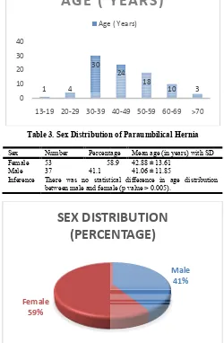
Table 2. Age distribution of Paraumbilical Hernia
Age (in years) Number 13 –19 1 20 - 29 4 30 – 39 30 40 – 49 24 50 – 59 18 60 – 69 10 > 70 years 3
[image:2.595.307.557.402.784.2]This table shows that majority of the patients are in the age group of 30-50 years. Only 13 patients were above 60 years. Youngest patient in this group was 16 years old and eldest patient was 76 years old.
Table 3. Sex Distribution of Paraumbilical
Sex Number Percentage Female 53 58.9 Male 37 41.1
Inference There was no statistical difference in age distribution between male and female (p value > 0.005).
1 4 30 0 10 20 30 40
13-19 20-29 30-39
AGE ( YEARS)
Age ( Years)
Female 59%
SEX DISTRIBUTION
(PERCENTAGE)
n Prasath Sinraj et al. A study on surgical management of Paraumbilical herniaOccurence of Paraumbilical Hernia
Number Occurence Rate Total hernias Operated from Jan 621 100%
418 67.3% 90 14.8% 79 12.7% 30 4.8% 02 0.3%
Total number of hernias operated were 621 from January 2015 to June 2016 in General Surgery Department in ESI Model Hospital of which paraumbilical hernia accounts for 14.8% of
Age distribution of Paraumbilical Hernia
mber Percentage 1.1% 4.4 % 33.3 % 26.6 % 20.0 % 11.1 % 3.3%
This table shows that majority of the patients are in the age 50 years. Only 13 patients were above 60 years. Youngest patient in this group was 16 years old and eldest
Sex Distribution of Paraumbilical Hernia
Percentage Mean age (in years) with SD 58.9 42.88 ± 13.61
41.06 ± 11.85
There was no statistical difference in age distribution between male and female (p value > 0.005).
24 18
10 3
40-49 50-59 60-69 >70
AGE ( YEARS)
Age ( Years)
Male 41%
SEX DISTRIBUTION
This table shows that 53 patients (58.9%) were female and 37 patients (41.1%) were male. Mean age distribution in females was 42.88±13.61 years; Mean age distribution in males was 41.06±11.85 years.
Table 4. Presentation of Symptoms
Symptoms Number
Swelling around Umbilicus 90 Pain in the Swelling or Pain Abdomen 51
[image:3.595.34.292.222.425.2]In our study most common presenting symptom was swelling around the umbilicus. 56.7% of patients had associated dragging type of pain in the abdomen.
Table 5. Duration of Symptoms
Duration Number
Since Childhood 1 0-6 months 16 7-12 months 27 1 year –3 years 35 3 years –6 years 7 6 years –10 years 2 More than 10 years 1
This table shows that 39% of patients had swelling around the umbilicus for 1-3 years before presenting to hospital. 30% of patients had swelling for 7-12 months;
18% of patients had recent onset swelling.
1 16
27 35
7
0 10 20 30 40
Duration of Symptoms
Duration of Symptoms0 20 40 60 80 100
Swelling around Umblicus Pain in Swelling/Abdomen
Presenting Complaints
Presenting Complaints
48829 International Journal of Current Research,
patients (58.9%) were female and 37 patients (41.1%) were male. Mean age distribution in females 13.61 years; Mean age distribution in males was
Presentation of Symptoms
Number Percentages 100.0 %
56.7 %
In our study most common presenting symptom was swelling around the umbilicus. 56.7% of patients had associated
Duration of Symptoms
Percentage 1.1 % 17.8 % 30 % 38.9 % 7.8 % 2.2 % 1.1 %
This table shows that 39% of patients had swelling around the 3 years before presenting to hospital. 30% of
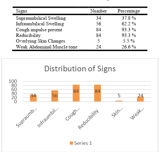
Table 6. Distribution of signs
Signs
Supraumbilical Swelling Infraumbilical Swelling Cough impulse present Reducibility
Overlying Skin Changes Weak Abdominal Muscle tone
[image:3.595.323.542.408.460.2]In our study, infraumbilical swelling was present in 62% of patients; supraumbilical swelling was present in 38% of patients. Hernia was reducible with cough impulse present in 93% of patients. Tone of abdominal muscle was poor in 27% of patients.
Table 7. Precipitating factors in female sex
Precipitating factors Multiparity (≥ 2 children) Obesity
Chronic Cough Constipation
[image:3.595.45.281.449.528.2]In females most common precipitating factor of paraumbilical hernia was multiparity (83%) followed by obesity (36%).
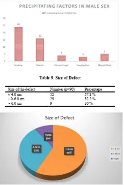
Table 8. Precipitating factors in male sex
Precipitating factors (Males) Smoking
Obesity Chronic Cough Constipation Manual Work
2 1
Duration of Symptoms
34 56 84
0 20 40 60 80 100
Distribution of Signs
Series 1
0 10 20 30 40 50
44
19
Precipitating factors in
Female Sex
Column1 Pain in Swelling/Abdomen
Presenting Complaints
International Journal of Current Research, Vol. 9, Issue, 04, pp.48827-48833, April, 2017
Distribution of signs
Number Percentage 34 37.8 % 56 62.2 % 84 93.3 % 84 93.3 % 5 5.5 % Weak Abdominal Muscle tone 24 26.6 %
In our study, infraumbilical swelling was present in 62% of patients; supraumbilical swelling was present in 38% of patients. Hernia was reducible with cough impulse present in 93% of patients. Tone of abdominal muscle was poor in 27%
Precipitating factors in female sex
Number (n=53) Percentage
44 83 %
19 35.8 % 2 3.8 % 3 5.7 %
In females most common precipitating factor of paraumbilical hernia was multiparity (83%) followed by obesity (36%).
Precipitating factors in male sex
Number (n=37) Percentage
24 64.8 %
16 43.2 %
4 10.8 %
3 8.1 %
5 13.5 %
84 84
5 24
Distribution of Signs
Series 1
2 3
Precipitating factors in
Female Sex
Column1
[image:3.595.303.562.511.711.2]In males most common precipitating factor was smoking (65%) followed by obesity (43%).
Table 9. Size of Defect
Size of the defect Number (n=90) Percentage < 4.0 cm 52 57.8 % 4.0-6.0 cm 29 32.2 % > 6.0 cm 9 10 %
This table shows that size of the defect was < 4 cm in 52 patients (57.8%), between 4-6 cm in 29 patients (32.2%) and > 6 cm in 9 patients (10%).
Table 10. Co-morbidities
Associated Diseases Number (n=90) Percentage Hyper tension 19 21.1 Diabetes Mellitus 14 15.5
Anemia 3 3.3
Others 6 6.7
[image:4.595.307.553.113.329.2]Out of 90 patients in our study, 42 had comorbid illness of which hypertension was the most common seen in 19 patients followed by diabetes mellitus seen in 14 patients.
Table 11. Surgical procedures
Procedures Number (n=90) Percentage Anatomical Repair 24 26.7 % Prosthetic Mesh Repair 66 73.3 %
On Lay 32 35.5 %
Preperitoneal 14 15.5 % Intraperitoneal 16 17.8 %
Retrorectus 4 4.4 %
Table 12. Postoperative complications
Postoperative Complications
Anatomical Repair (n=24)
Onlay Mesh Repair (n=32)
PreperitonealMesh Repair (n=14)
Intraperitoneal Mesh Repair (n=16)
Retrorectus mesh repair (n=4)
Seroma 1 2 2 1 -
Haematoma - - 1 1 1
Wound Infection - 1 2 2 -
Wound Dehiscence - - - - -
Sinus - - - 1 -
[image:4.595.317.547.400.472.2]Duration of surgery (min) 30+4 37+6 55+5 34+4 72+6
Table 13. Duration of Postoperative hospital Stay
Procedures Mean Hospital Stay (days) SD Anatomical Repair (n=24) 3.1 0.6 Onlay Mesh Repair (n=32) 4.4 0.4 Preperitoneal Mesh Repair (n=14) 5.2 0.5 Intraperitoneal Mesh Repair (n=16) 4.6 0.3 Retrorectus Mesh Repair (n=4) 5.1 0.4
Table 14. Recurrence of the Disease
Procedures Number Percentage Anatomical Repair - - Onlay Mesh Repair 2 6.2 % Preperitoneal Mesh Repair - - Intraperitoneal Mesh Repair - - Retromuscular Mesh Repair - -
[image:4.595.58.538.508.589.2] [image:4.595.177.420.620.681.2]In our study, 24 patients (26%) underwent anatomical repair and rest of the patients (66) underwent prosthetic mesh repair of which 32 were onlay mesh repair, 14 were preperitoneal, 16 intraperitoneal and 4 were retromuscular mesh repair.
This table shows incidence of postoperative complication in different surgical procedures. Seroma was present in 6 patients, hematoma in 3 patients and wound infection in 5 patients in both groups (Anatomical repair and prosthetic mesh repair). The duration of surgery is more in patients in whom mesh repair was done and among these, longest time were consumed in retromuscular mesh repair and prepritoneal mesh repair groups. The duration of surgery in our study is not significant statistically. (p value-0.106)
Again the duration if hospital stay is not statistically significant (p value-0.284).
DISCUSSION
In this study, 90 patients with paraumbilical hernia were admitted and treated with different surgical procedures from January 2015 to June 2016. The same group of patients were studied for ocurrence, risk factors, clinical features, treatment and postoperative complications pertaining to disease. Discussion is mainly concentrated on Anatomical repair and Mesh repair as they comprise major bulk of the sample. Occurence of different types of hernia operated in our hospital is as follows: Inguinal hernia – 67.3%, paraumbilical hernia-14.8%, incisional hernia-12.7%, epigastric hernia-4.8% and
femoral hernia-0.3%. Although exact incidence of
[image:5.595.51.553.82.233.2]paraumbilical hernia is not mentioned in available literature, it is considered as one of the common hernias apart from inguinal hernia. Paraumbilical hernia is more common in patients aged between 30-50 years (59%) in our study.
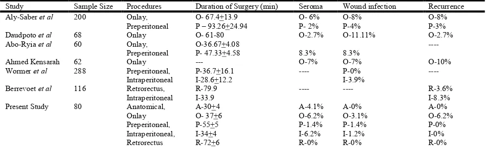
Table 15. Comparison of different parameters in various studies
Study Sample Size Procedures Duration of Surgery (min) Seroma Wound infection Recurrence Aly-Saber et al 200 Onlay,
Preperitoneal
O- 67.4+13.9 P – 93.26+24.94
O- 6% P- 2%
O-8% P-4%
O-8% P-3% Daudpoto et al 68 Onlay O- 61-80 O-2.7% O-11.11% O-2.7% Abo-Ryia et al 60 Onlay,
Preperitoneal
O-36.67+4.08
P- 47.33+4.58 8.3% 8.3%
----
Ahmed Kensarah 62 Onlay --- O-7% O-7% O-10%
Wormer et al 288 Preperitoneal, Intraperitoneal
P-36.7+16.1 I-28.6+12.2
---- P-0% I-3.9%
----
Berrevoet et al 116 Retrorectus, Intraperitoneal
R-79.9 I-33.9
---- ---- R-3.6%
I-8.3% Present Study 80 Anatomical,
Onlay Preperitoneal, Intraperitoneal, Retrorectus
A-30+4 O- 37+6 P-55+5 I-34+4 R-72+6
A-4.1% O-6.2% P-1.4% I-6.2% R-0%
A-0% O-3.1% P-1.4% I-1.2% R-0%
A-0% O-6.2% P-0% I-0% R-0% A-Anatomical repair, O-Onlay, P- Preperitoneal, I- Intraperitoneal, R- Retrorectus Mesh Repair
Youngest patient who presented with paraumbilical hernia in our study was 16 years old. It was found that paraumbilical hernia is infrequent after 70 years as only three patients were more than 70 years old. Our results are in comparison with studies by Prasad Shah et al. (2016), Kensarah et al. (2011)& Daudpoto et al. (2013). Fifty percent of patients were in 30-50 years age group in study by Prasad Shah and co. Paraumbilical hernia is more common in females. Fifty three patients were females and thirty seven patients were males. In our study ratio between female and male sex is 1.4:1. There is no significance difference in age distribution in males and females, as disease is more common between 3rd and 5th decade in both sex. Again the sex ratio found in our study is consistent with other studies by Abo-Ryia et al. (2015), Sangwan et al. (2013), Daudpoto et al. (2013), Kensarah et al. (2011)& Malik et al. (2008) among
which the study by Sangwan et al shows a female to male sex
ratio of 1.6:1 which closely matches our study. Whereas the study by Prasad Shah et al. (2016)and Sheikh et al. (2013) has a male predominance for paraumbilical hernia with a female to male ratio of 1:1.9 and 1:3.1 respectively which is in contrast with our study. All 90 patients presented with chief complaint of swelling around umbilicus. Fifty one patients had pain in the swelling or dragging type of pain abdomen. One patient presented with symptoms of intestinal obstruction and was excluded from the study. Most of the patients had swelling for 1 to 3 years before they presented to hospital. Maximum duration of symptoms was 10.5 years, minimum duration was 1 month. In our series swelling was present in infraumbilical position in majority of the patients (62%) and Supraumbilical swelling was found in 38%. On the other hand approximately 75% of patients had supraumbilical swelling as noted by Daudpoto et al. (2013). Even though it was stated in literature that most of the paraumbilical hernias are irreducible or partially reducible, in our study cough impulse was present and swelling was reducible in 93% of patients. Only 6 patients had absent cough impulse and irreducible swelling. Overlying skin changes were present in longstanding cases (5). Twenty six percentage of patients had poor abdominal muscle tone. In females, most common precipitating factor was multiparity. Out of 53 patients, 44 (83%) were multiparous. This can be attributed to stretching and weakening of anterior abdominal wall musculoaponeurotic layer. Next common precipitating factor was obesity seen in 19 patients (35.8%). Pathogenesis can be attributed to theory explained by Mayo; obesity causes downward traction on the abdominal wall bearing on a fixed point on umbilicus associated with an increase in vertical dimension of abdominal wall. Fat penetrates muscle bundles and layers, weakens aponeurosis and predisposes to hernia. Other less common precipitating factors were chronic cough and constipation. In males most common precipitating factor was smoking (24 patients) followed by obesity (16 patients).
Smoking is an important predisposing factor in development of inguinal hernia as it causes degeneration of collagen fibres, same theory applies to paraumbilical hernia. Other precipitating factors are chronic cough (COPD), constipation and heavy manual work. Some patients had more than one precipitating factors and some did not have any precipitating factor. Studies by Malik et al., 2008; Daudpoto et al., 2013; Kensarah, 2011; Shah et al., 2016 show similar results regarding the risk factors in either sex. Nineteen patients were hypertensive, 14 patients were diabetic and 3 patients were anemic. These associated diseases were treated adequately before surgery, hence there was no much effect on the outcome following surgery. In this series, 24 patients (27%) underwent Anatomical repair, 66
patients (73%) underwent polypropylene mesh repair. Out of 66 patients who underwent Mesh repair, 32 were Onlay procedure, 14 were preperitoneal, 16 intraperitoneal and 4 retrorectus (retromuscular) mesh repair. Although cases were selected intraoperatively for particular surgical procedure, size of defect, age of patient and tone of abdominal muscles has been considered. Mesh repair has been done for most of the large defects (>2cm). Anatomical repair was done for defects less than 2cm.Most common postoperative complications were seroma (6.6%) followed by wound infection (5.5%). No patient required removal of mesh because of infection, as infection was superficial and responded well to antibiotics. Incidence of immediate postoperative complication is high compared to study conducted by A. Arryo, P.Garcia (Toms et al., 1999) et al
during 2001. But there is no difference in postoperative complication between anatomical repair and Mesh repair similar to that study. In the study by Aly-Saber (Saber and Bayumi, 2015), 8% patients had seroma formation and 12% developed wound infection. This may be attributed to the larger sample size in this study compared to our study. Two (6.2%) out of thirty two developed seroma and one (3.1%) out of 32 developed wound infection following onlay repair in our study. In the study by Daudpoto (2013) wound infection (11.11%) was the commonest complication followed by seroma (2.7%) following onlay repair. On the other hand wound infection and seroma rates were equal (7%) in the study by Ahmed M. Kensarah (2011). In this series mean postoperative hospital stay following anatomical repair was 3.1 days with SD 0.6 days, mean postoperative hospital stay was 4.8+0.4 days following mesh repair. There was no statistical difference of postoperative hospital stay following anatomical repair and Mesh repair. Recurrence was seen in two patients who underwent onlay mesh repair in the study period and there was no recurrence noted in other patients. Mean follow up period following anatomical repair was 13.15 months with SD of 5.98 months. Mean follow up period following Mesh repair was 11.74±6.39 months.
Conclusion
There is no significance difference in recurrence following anatomical repair and mesh repair (p value = 0.207), but there is statistical trend towards the difference between two procedures regarding recurrence, this trend may be converted to significance difference, if sample size and follow up period is increased. To conclude, it can be said that sample size and follow up period in our study is small to show significance difference between two procedures and hence a larger study is required with bigger sample size and longer follow up period.
REFERENCES
Abo-Ryia M H, El-Khadrawy O H, Moussa G I, Saleh A M. 2015. Prospective randomized evaluation of open preperitoneal versus preaponeurotic primary elective mesh repair for paraumbilical hernias. Surgery Today, 45(4) : 429-433.
Brunicardi F C. 2015. Schwartz’s Principles of Surgery.10th ed. McGraw-Hill Education.
Daudpoto A Q, Mirani S, Memon R A, Abbas Q. 2013. A long term follow up: mesh versus mayo’s repair in paraumbilical hernia. JUMDC, 4:1.
Kensarah A M. 2011. A long Term Follow Up: Suture Versus Mesh Repair for Adult Umbilical Hernia in Saudi Patients. A Single Center Prospective Study. Surgical Science, 2:155-8.
Kensarah A M. 2011. A Long-Term Follow-Up: Suture Versus Mesh Repair for Adult Umbilical Hernia in Saudi Patients. A Single Center Prospective Study. Surgical Science, 2: 155-8.
Malik AM, Jawaid A, Talpur AH, Laghari AA, Khan A. 2008. Mesh versus non-mesh repair of ventral abdominal hernias.
J Ayub Med Coll Abbottabad., 20(3): 54-6.
Purushotham B. and Madhu S. 2015. Comparative study between laparoscopic and open repair of umbilical and para umbilical hernia. IntSurg J., 2(2):204-13.
Saber A. and Bayumi E K. 2015. Onlay versus Sublay Mesh Repair for Ventral Hernia. Journal of Surgery, 4(1-1): 1-4. Sangwan M, Sangwan V, Garg M, Mahendirutta P, Garg U.
2013. Abdominal wall hernia in a rural population in
India—Is spectrum changing? Open Journal of
Epidemiology, 3: 135-8.
Shah P, Shaikh S. and Panchabhai S. 2016. Prevalence of Anterior Abdominal Wall Hernia and its Associated Risk Factors. International Journal of Anatomy, Radiology and Surgery, 5(3): SO07-10.
Shaikh I, Willder J M, Kumar S. 2013. Same day discharge, surgical training and early complications after open and laparoscopic repair of primary paraumbilical hernia.
Hernia, 17: 505-9.
Toms A P, Dixon A K, Murphy J M P and Jamieson N V. 1999. Illustrated review of new imaging techniques in the diagnosis of abdominal wall hernias. Br J Surg., 86: 1243-50.
Williams N S, Bulstrode C J K, O’Connell P R. 2013. Bailey & Love’s Short Practice of Surgery. 26th ed. Boca Raton: CRC Press.
Wormer B A, Stefanidis A, Williams K B, Bradley J F, Augenstein V A, Henniford T. 2013. The impact of mesh position on open umbilical hernia repair outcomes: a comparison of preperitoneal and intraperitoneal placement in a prospective multicenter study. JACS, 217(3): S25.