RESEARCH ARTICLE
MINIMALLY INVASIVE TECHNIQUE OF MIDSHAFT CLAVICLE FRACTURE FIXATION BY
TITANIUM ELASTIC NAIL
*,1
Dr. Deepak Srivastav,
2Mithlesh Kumar and
3Dr. Arpita Mohan
1
Department of Orthopaedics, Hind Institute of Medical College, Barabanki, U.P., India
2Department of Orthopaedics, Azamgarh Government Medical College, India
3Department of Dentistry, Hind Institute of Medical Sciences, Barabanki, India
ARTICLE INFO ABSTRACT
Introduction: Fractures of the clavicle account for 2.6–4 % of all adult fractures, 35 % of all injuries to the shoulder girdle, and 69–82 % of these fractures occur in the middle-third and are usually treated non-operatively. There is an increasing trend toward their surgical fixation. The aim of our study was to evaluate the clinical, functional and radiological outcome following titanium elastic stable intramedullary nailing (ESIN) for midshaft non-comminuted clavicle fractures with >20 mm shortening/displacement.
Materials and Methods: 50 patient with mid shaft clavicular fracture, which met inclusion criteria, were fixed with titanium intramedullary elastic nail under image intensifier control between July 2013 and June 2017. There were 33 males and 17 females. The mean age was 33 years. Elastic nail introduce from 1cm lateral to sternoclavicular joint to fix the fracture. Outcomes assessed on the basis of Constant score.
Results: All patients achieved complete healing at a mean of 10.3 weeks.17 fracture reduced by closed means but 33 needs open reduction. Common size of elastic nail used was 2mm. no major complication was recorded all were minor and can be taken care off. Most common was pin tract infection recorded in 35 patients. 76% had excellent result and 24% had good result. 100% had nail removal.
Conclusion: In our hands, ESIN is safe and minimally invasive with good patient satisfaction, cosmetic appearance, and overall outcome.
Copyright©2017, Deepak Srivastavet al.This is an open access article distributed under the Creative Commons Attribution License, which permits unrestricted use, distribution, and reproduction in any medium, provided the original work is properly cited.
INTRODUCTION
Fractures of the clavicle account for 2.6–4 % of all adult fractures, 35 % of all injuries to the shoulder girdle, and 69–82 % of these fractures occur in the middle-third (Robinson, 1998; Khan et al., 2009). The midshaft clavicle fractures account for 3 to 5% of all injuries and 70 to 80% of all clavicle fractures (Duan et al., 2011; Schiffer et al., 2010). Displacement occurs in about 73 % of all midshaft clavicle fractures (Khan et al., 2009). The average age of patients sustaining a midshaft clavicular fracture is 33 years; 70 % of the patients are male (Pearson et al., 2010). In young adults, these fractures are usually related to sports or vehicle accidents, whereas in children and elderly, they are usually related to falls (Duan et
al., 2011; Schiffer et al., 2010). A fall or a direct blow to the
shoulder, giving an axial compressive force on the clavicle, is the most common trauma mechanism of injury for any clavicular fracture (Nowak et al., 1990; Stanley et al., 1988). Traditionally, the midshaft clavicle fractures have been treated
*Corresponding author: Dr. Deepak Srivastav
Department of Orthopaedics, Hind Institute of Medical College, Lucknow, India.
conservatively with a sling or figure-of-eight bandage (Neer, 1960; Rowe, 1968; Jeray, 2007). Functional outcome of midshaft clavicle fractures is not only related to its union, but also to its length (Lazarides, 2006). Clavicle acts as a "strut", that keeps the upper limb away from the torso for efficient shoulder and upper limb function, while also transmitting forces from upper limb to the trunk. Thus, displaced or comminuted fractures carry a risk of symptomatic malunion, non-union and poor functional outcome with cosmetic deformity (Canadian Orthopaedic Trauma Society, 2007; Hill
et al., 1997; Wild et al., 2006). The recent trend is shifting
towards internal fixation of these displaced midshaft clavicle fractures (DMCF) (Lazarides, 2006; Hill et al., 1997; Wild et
al., 2006; Smekal et al., 2009; McKee et al., 2003; Zlowodzki et al., 2005). Displaced midshaft fractures have traditionally
been treated non-operatively because of early reports suggesting that clavicular non-union were very rare and clavicular mal-union, being of radiographic interest only, was without clinical importance (Neer, 1960; Canadian Orthopaedic Trauma Society, 2007). However, recent studies
ISSN: 0975-833X
International Journal of Current Research Vol. 9, Issue, 10, pp.58827-58833, October, 2017
INTERNATIONAL JOURNAL OF CURRENT RESEARCH
Article History: Received 22ndJuly, 2017 Received in revised form 06thAugust, 2017
Accepted 28thSeptember, 2017 Published online 17thOctober, 2017
Citation: Dr. Deepak Srivastav, Mithlesh Kumar and Dr. Arpita Mohan, 2017.“Minimally invasive technique of midshaft clavicle fracture fixation by
titanium elastic nail”,International Journal of Current Research, 9, (10), 58827-58833. Key words:
Clavicle fracture,
Elastic stable intramedullary nailing, Midshaft fracture,
RESEARCH ARTICLE
MINIMALLY INVASIVE TECHNIQUE OF MIDSHAFT CLAVICLE FRACTURE FIXATION BY
TITANIUM ELASTIC NAIL
*,1
Dr. Deepak Srivastav,
2Mithlesh Kumar and
3Dr. Arpita Mohan
1
Department of Orthopaedics, Hind Institute of Medical College, Barabanki, U.P., India
2Department of Orthopaedics, Azamgarh Government Medical College, India
3Department of Dentistry, Hind Institute of Medical Sciences, Barabanki, India
ARTICLE INFO ABSTRACT
Introduction: Fractures of the clavicle account for 2.6–4 % of all adult fractures, 35 % of all injuries to the shoulder girdle, and 69–82 % of these fractures occur in the middle-third and are usually treated non-operatively. There is an increasing trend toward their surgical fixation. The aim of our study was to evaluate the clinical, functional and radiological outcome following titanium elastic stable intramedullary nailing (ESIN) for midshaft non-comminuted clavicle fractures with >20 mm shortening/displacement.
Materials and Methods: 50 patient with mid shaft clavicular fracture, which met inclusion criteria, were fixed with titanium intramedullary elastic nail under image intensifier control between July 2013 and June 2017. There were 33 males and 17 females. The mean age was 33 years. Elastic nail introduce from 1cm lateral to sternoclavicular joint to fix the fracture. Outcomes assessed on the basis of Constant score.
Results: All patients achieved complete healing at a mean of 10.3 weeks.17 fracture reduced by closed means but 33 needs open reduction. Common size of elastic nail used was 2mm. no major complication was recorded all were minor and can be taken care off. Most common was pin tract infection recorded in 35 patients. 76% had excellent result and 24% had good result. 100% had nail removal.
Conclusion: In our hands, ESIN is safe and minimally invasive with good patient satisfaction, cosmetic appearance, and overall outcome.
Copyright©2017, Deepak Srivastavet al.This is an open access article distributed under the Creative Commons Attribution License, which permits unrestricted use, distribution, and reproduction in any medium, provided the original work is properly cited.
INTRODUCTION
Fractures of the clavicle account for 2.6–4 % of all adult fractures, 35 % of all injuries to the shoulder girdle, and 69–82 % of these fractures occur in the middle-third (Robinson, 1998; Khan et al., 2009). The midshaft clavicle fractures account for 3 to 5% of all injuries and 70 to 80% of all clavicle fractures (Duan et al., 2011; Schiffer et al., 2010). Displacement occurs in about 73 % of all midshaft clavicle fractures (Khan et al., 2009). The average age of patients sustaining a midshaft clavicular fracture is 33 years; 70 % of the patients are male (Pearson et al., 2010). In young adults, these fractures are usually related to sports or vehicle accidents, whereas in children and elderly, they are usually related to falls (Duan et
al., 2011; Schiffer et al., 2010). A fall or a direct blow to the
shoulder, giving an axial compressive force on the clavicle, is the most common trauma mechanism of injury for any clavicular fracture (Nowak et al., 1990; Stanley et al., 1988). Traditionally, the midshaft clavicle fractures have been treated
*Corresponding author: Dr. Deepak Srivastav
Department of Orthopaedics, Hind Institute of Medical College, Lucknow, India.
conservatively with a sling or figure-of-eight bandage (Neer, 1960; Rowe, 1968; Jeray, 2007). Functional outcome of midshaft clavicle fractures is not only related to its union, but also to its length (Lazarides, 2006). Clavicle acts as a "strut", that keeps the upper limb away from the torso for efficient shoulder and upper limb function, while also transmitting forces from upper limb to the trunk. Thus, displaced or comminuted fractures carry a risk of symptomatic malunion, non-union and poor functional outcome with cosmetic deformity (Canadian Orthopaedic Trauma Society, 2007; Hill
et al., 1997; Wild et al., 2006). The recent trend is shifting
towards internal fixation of these displaced midshaft clavicle fractures (DMCF) (Lazarides, 2006; Hill et al., 1997; Wild et
al., 2006; Smekal et al., 2009; McKee et al., 2003; Zlowodzki et al., 2005). Displaced midshaft fractures have traditionally
been treated non-operatively because of early reports suggesting that clavicular non-union were very rare and clavicular mal-union, being of radiographic interest only, was without clinical importance (Neer, 1960; Canadian Orthopaedic Trauma Society, 2007). However, recent studies
ISSN: 0975-833X
International Journal of Current Research Vol. 9, Issue, 10, pp.58827-58833, October, 2017
INTERNATIONAL JOURNAL OF CURRENT RESEARCH
Article History: Received 22ndJuly, 2017 Received in revised form 06thAugust, 2017
Accepted 28thSeptember, 2017 Published online 17thOctober, 2017
Citation: Dr. Deepak Srivastav, Mithlesh Kumar and Dr. Arpita Mohan, 2017.“Minimally invasive technique of midshaft clavicle fracture fixation by
titanium elastic nail”,International Journal of Current Research, 9, (10), 58827-58833. Key words:
Clavicle fracture,
have found higher rates of delayed union, non-union, shoulder pain, and shoulder weakness and residual pain with non-operative treatment (Hill et al., 1997). The indications for surgery include the need for earlier functional mobilization in the patient with an isolated injury, in addition to open fractures, floating shoulders and patients with polytrauma (Denard et al., 2005). Hill et al. showed that displacement of more than 20 mm resulted in 15% non-union and 18% of the patients had thoracic outlet syndrome following union (Hill et
al., 1997). McKee et al. noted reduced patient satisfaction due
to asymmetry and cosmesis following malunion in patients with more than 20 mm shortening (McKee et al., 2006). Hence, more recently, there has been a trend toward surgical fixation. Surgery has been indicated for completely displaced fractures, potential skin perforation, shortening of clavicle by more than 20 mm, and floating injury (Frigg et al., 2009). For operative treatment, the available methods of fixation are fixation with Kirschner wires, pins (Rush pin, Knowles pin, Rockwood pin), plates with screws and external fixation (Lee
et al., 2007; Mudd et al., 2011; Mueller et al., 2008).
Intramedullary fixation for clavicular fractures was first described by Peroni in 1950 (Peroni, 1950). A systematic review showed relative risk reduction of 72% and 57% for non-union when using intramedullary fixation and plate fixation, respectively, when compared with non-operative treatment of midshaft clavicle fractures (Zlowodzki et al., 2005). Intramedullary devices behave as internal splints that maintain alignment without rigid fixation. The use of an intramedullary device carries advantages of a smaller incision, less soft tissue dissection, load sharing fixation with relative stability that encourages copious callus formation (Millett et
al., 2011). One advantage of the titanium ESIN is that it can
block itself in the bone and provide a three-point fixation within the S-shaped clavicle (Frigg et al., 2009; Mueller et al., 2008). However, some studies have shown a relatively high complication rate and technical difficulties with intramedullary nailing (Frigg et al., 2009). The aim of this study was to investigate the union rate and complication rate of our patients with displaced midshaft clavicle fractures treated with titanium ESIN.
MATERIALS AND METHODS
It is a prospective study, which was conducted in the department of orthopaedic, Hind Institute of Medical Sciences, Lucknow, Uttar Pradesh, India between July 2013 and June 2017; between 50 patients with midshaft clavicle fracture (33 male and 17 female) were treated by internal fixation with intramedullary titanium elastic nails.
Inclusion Criteria
Age >20 and <60 years of age
Shortening & Displacement >2cm in any view
Severe displacement causing tenting of the skin with the risk of puncture
open upto Gustillo grade II fractures Symptomatic non-union
Segmental fractures Floating shoulder
Exclusion criteria
Shortening & Displacement <2cm
Medial and lateral third fractures Comminuted fractures
Gustillo grade III fractures
Associated with neurovascular deficit Atrophic non-union
The patients were operated by different surgeons in the department. All surgeons used ESIN in patients meeting the inclusion criteria.
Operative technique
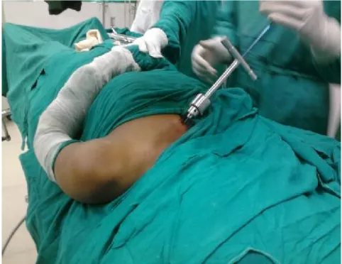
[image:2.595.313.554.586.772.2]After administration of general anaesthesia, the patient was placed in beach chair position with injured extremity prepared and draped from the midline to the upper arm with folded sheet under the affected shoulder. Care was taken to make sure that the sternoclavicular joint was accessible for the entry point. Elastic intramedullary nailing was done using the technique described first by Jubel et al., (2003).A vertical skin incision was made 1cm lateral to the sternoclavicular joint. The subcutaneous fat was incised along with platysma. The pectoral fascia was divided in line with the skin incision followed by careful elevation of the underlying musculature from the clavicle. The medullary cavity of the clavicle was opened using an all pointed laterally and angled at about 30° to the coronal plane in line with the clavicle. Care was taken not to perforate the dorsal cortex in order to avoid major complications. Single appropriate sized titanium ESIN was inserted (the diameter varied from 1.5 to 3 mm depending on the width of the bone). Attempt was made to close reduce the fracture. If the fracture could not be reduced by closed means, then a separate vertical incision was used at the fracture site to aid fracture reduction. Vertical incision was used as it was parallel to Langer's lines and minimized the risk of damage to supraclavicular nerves to avoid dysesthesia of skin and scar neuromas. At that time, the nail was used to create a path in the lateral end of the clavicle for subsequent easy access. The nail was then passed from the medial side and across the reduced fracture into the lateral end of clavicle. Care was taken to avoid perforation of the dorsolateral cortex of the lateral clavicle. After reaching the end point, the fracture was compressed and the nail was cut close to the entry point to minimize soft tissue irritation, at the same time leaving sufficient length behind for easy extraction later on. The fascia and skin were closed in layers.
All the patients were put in a shoulder sling postoperatively and followed same rehabilitation regime of early gentle mobilization when pain allows, with no overhead abduction for first two weeks. The shoulder sling was discarded at 2 weeks and active-assisted exercises were started, but the patients were advised not to lift any heavy object for 6 weeks. At that time, passive and strengthening exercises were started. All the patients were reviewed postoperatively in the clinic at 2, 6, 12 weeks and 6 months or until the fracture had healed clinically and radiologically. All the patients were assessed for clinical and radiological union and radiographs were taken at each clinic visit. We defined radiological union as visible bridging callus or absent fracture line. The clinical union was described as no bony tenderness on clinical examination. Time to achieve union was recorded.
RESULTS
A total of 50 patients with mean age 33yrs (range 20–60 yrs) met the inclusion criteria of diaphyseal midshaft, non-comminuted clavicle fractures with more than 20 mm shortening/displacement. Of these 50 patients, 13 patients had a fall on outstretched hand, eight had a fall from height and 29 had road traffic accident. Among 50 patients 35 (70%) had right clavicle fracture and 30% had left sided. Most of them (84%) were closed fracture but 16% (8 fracture) were open upto Gustillos grade I & II. Patients who presented to us early (1-7days) were reduced by closed means but who were presented late was required open reduction. In our study only 34% (17 fractures) were reduced closed and fix by ESIN but 66% (33 fractures) required open reduction. The average operative time was 45 min (range 20-75 minutes). 80% (40) patient were discharged within 3 days of operation on oral drugs , but one patient who develops infection post-operatively had stay for 7 days till the infection controlled. All the patients achieved clinical and radiological union at a mean of 10.3 weeks (Range, 6-20 weeks) i.e. 100% union achieved. Union achieved early in the fractures that were reduced by closed method in comparison to those required open reduction. In none of the fracture bone grafting done. The average follow up was 12 months (Range, 6-24 weeks). The average size of the titanium flexible nail used was 2 mm (range, 1.5 -3 mm). Full shoulder range of motion on an average gained at 7.4 weeks (6weeks - 12weeks). After clinical and radiological fracture healing routine removal of nail was done in all patients.
[image:3.595.337.532.53.197.2]Diag. 1. Distribution of cases according to time of union
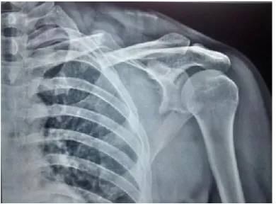
[image:3.595.339.529.232.449.2]Fig. 2. Pre-op xray 30 yr male with fracture clavicle and scapula left side
[image:3.595.338.528.488.642.2]Fig. 3. Post-op xray 30 yr male with fracture clavicle and scapula left side, clavicle fixed with TENs nail
[image:3.595.340.530.680.787.2]Fig. 4. 14-week post-op xray 30 yr male with fracture clavicle and scapula left side after-union TENs nail removed
Fig. 6. Shoulder ROM after 8 week post fixation
Complications
[image:4.595.331.534.53.363.2]Most common complication found was pin tract infection in 35 patients and entry site skin irritation (medial irritation) in 10 patients which was subsided after nail removal. Others were lateral protrusion of nail in one patient which was subsequently removed, superficial infection develops in one patient, who was controlled with antibiotics and nail removed early and subsequently settles. In one patient perforation of lateral cortex happens during nail insertion which was corrected under image intensifier. There was no instance of loss of reduction, joint stiffness, scar neuroma, perforation of posterior cortex. There were no cases of delayed union, non-union or malunion, or refracture.
Table 1. Distribution of cases according to post operative complication
S. No Complications No. of cases (n=50)
1 Entry site irritation 10
2 Superficial infection 1
3 Deep infection None
4 Pin tract infection 35
5 Refracture None
6 Non union None
7 Posterior cortex perforation None
8 Lateral migration of nail 1
9 Neuro vascular damage None
10 Iatrogenic perforation of lateral cortex 1
[image:4.595.337.530.54.182.2]Fig. 7. Lateral migration of nail
Fig. 8. Perforation of lateral cortex
Fig. 9. Pin tract infection
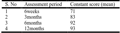
Functional outcome was assessed by the Constant score. In the Constant scoring system, the overall grading is excellent if the total score ranges from 90 to 100, good for 80–89, fair for 70– 79, and poor if the scores are 69 or less. At final evaluation, the overall results using the constant score were 38 excellent and 12 good results.
Table 2. Constant score
S. No Assessment period Constant score (mean)
1 6weeks 71
2 3months 83
3 6months 92
4 12months 93
DISCUSSION
Traditionally midshaft clavicle fractures had been treated non-operatively. Neer (1960) and Rowe (1968) in the 1960's recommended conservative treatment for clavicular fractures in view of very small incidence of non-union rates in their studies (0.1% and 0.8% respectively). No one has been able to reproduce these results so far. However, more recent studies have shown significantly higher non-union rates in conservatively treated patients (ranging between 10% and 15%). Current recommendation for treatment of displaced midshaft clavicle fractures is operative fixation (Lazarides, 2006; Canadian Orthopaedic Trauma Society , 2007; Hill et
al., 1997; Wild et al., 2006; Smekal et al., 2009; McKee et al.,
[image:4.595.338.529.207.361.2] [image:4.595.41.289.477.790.2] [image:4.595.335.528.488.541.2]clavicle fractures that were treated non-operatively (Hill et al., 1997; Robinson et al., 2004; Wild and Potter, 2006) in comparison to surgically treated patients (Jubel et al., 2003; Ali and Lucas, 1978; Jeray, 2007). In a multicenter prospective randomized trial, plate osteosynthesis had better functional outcome than non-operative treatment of displaced clavicle fractures with decreased rate of non-union and symptomatic malunion (Canadian Orthopaedic Trauma Society, 2007). Severe complications occur in 10% of the patients and include deep infection, non-union, implant failure, and fracture after implant removal. Lesser complications include superficial infection, keloid scar, dysesthesia in the region of scar, as well as implant loosening with loss of reduction (Smekal et al., 2011). Schuind et al. in 1988 reported results of 20 patients treated with Hoffmann external fixation with no non-union and return to full range of movements of the shoulder. However, there was no objective measurement of patient satisfaction (Schuind et al., 1988). Intramedullary stabilization is an established alternative fixation method. Intramedullary implants are optimal from biomechanical point of view as the tension side of clavicle changes with respect to rotation of arm and direction of loading (Mueller et al, 2008; Smekal et al., 2009). The potential benefits of this technique include smaller incision, minimal periosteal stripping, and load sharing device properties (Millett et al., 2011). Its relative stability allows copious callus formation during the healing process. The frequent complication includes skin irritation from the prominent medial end of the nail and this frequently leads to either trimming of the nail or its premature removal (Smekal et
al., 2011). Multifragmentary fracture can lead to telescoping of
the nail with shortening of the clavicle. The comminuted fractures were excluded because we believe that this fixation system cannot maintain length of the clavicle in these situations. Smekal et al. hence do not recommend use of ESIN in comminuted fractures with severe shortening (Smekal et al., 2011). Duan et al. in a meta-analysis of randomized controlled trials demonstrated similar functional outcome when comparing plating with intramedullary fixation (Duan et al., 2011). They, however, showed higher symptomatic hardware-related problems with plating (Duan et al., 2011). Zolowodzki
et al. in a systematic review of 2144 cases found non-union
rate of 1.6% with intramedullary fixation as compared with 2.5% with plate fixation (Zlowodzki et al., 2005).
The intramedullary fixation provides an alternative and less invasive way of surgically treating clavicle fractures. Murray published his technique of intramedullary krischner wire fixation in 1940 (Murray, 1940). Since then, numerous technical variations have been published (Lengua et al., 1987; Leppilahti and Jalovaara, 1999; Ngarmukos et al., 1998; Wllkins and Johnston, 1983). The use of titanium elastic nails in the treatment of midshaft clavicle fractures was first described by Jubel et al., (2003) Major complications described in the literature for other modes of intramedullary fixation of clavicle fractures (Kirschner wire, Rush pin etc.), like injury to neurovascular structures and implant migration into the chest cavity (Schwarz and Leixnering, 1984; Liu et al., 1993) were not observed in our study. No such complication has been described in the literature using TENs in clavicle fractures (Jubel et al., 2003). In a retrospective analysis between titanium elastic nails and reconstruction plates, Chen
et al. showed a significantly shorter time to union with the
TEN group with no significant difference in non-union or malunion rate between TEN and plating. They showed faster functional recovery with greater patient satisfaction with
cosmesis and overall outcome in the TEN group (Chen et al., 2012). Smekal et al. showed, in a randomized control trail between intramedullary nailing and non-operative treatment, better DASH and Constant scores and 100% union rate with intramedullary nailing (Smekal et al., 2009). Liu et al. found no significant difference between functional outcome and non-union rate following plate fixation and intramedullary fixation (titanium elastic nails) of displaced midshaft clavicle fractures (Liu et al., 2010). Frigg et al. in their study expressed concerns about the increased complication rate (Frigg et al., 2009). In one of our patients, the nail was left slightly proud and required trimming later on. The other patient with lateral protrusion (without any involvement of the acromioclavicular joint) had minimal discomfort from it and this nail was removed once the fracture had healed. Migration of intramedullary implant has been reported in a number of studies (Mueller et al., 2008; Liu et al., 2010; Smekal et al., 2011). Smekal et al. reported that 23% of their patients had medial nail protrusion and 89% of the patients required implant removal (Smekal et al., 2009). 47% of our patients had the implant removed, most of them for symptomatic medial irritation. Smekal et al. reported rate of closed vs open fracture reduction when using elastic flexible intramedullary nailing of 64:36 (Smekal et al., 2009). We do not consider open reduction of the fracture as unsatisfactory as despite its high rate, in our series, we achieved 100% union. In our series, the procedure was performed by various surgeons using a standardized surgical technique as detailed earlier. Despite this, we achieved good functional and cosmetic outcome in diaphyseal midshaft, non-comminuted clavicle fractures with more than 20 mm shortening/displacement with titanium ESIN with no major complications.
Conclusion
Antegrade flexible intramedullary nailing techniques have advantages like less soft tissue injury, shorter operating time and hospital stay, less blood loss, more cosmetic satisfaction and minor surgery needed to remove the implant. EIN is a safe, minimally invasive surgical technique with a lower complication rate, faster return to daily activities, excellent cosmetic and comparable functional result. In conclusion, the intramedullary fixation of midshaft clavicle fractures is a safe minimally invasive technique in indicated cases and in our hands it provides good functional outcome and cosmetic results. In selected case TENs is a safe & effective method for mid clavicular fracture with a low complication rate once potential technical pitfalls are appreciated
REFERENCES
Ali, K.M. and Lucas, H.K. 1978. Plating of fractures of the middle third of the clavicle. Injury, 9(4):263–267.
Canadian Orthopaedic Trauma Society, 2007. Nonoperative treatment compared with plate fixation of displaced midshaft clavicular fractures. A multicenter, randomized clinical trial. J Bone Joint Surg Am., 89:1-10.
Chen, Y.F., Wei, H.F., Zhang, C., Zeng, B.F., Zhang, C.Q. and Xue, J.F. 2012. Retrospective comparison of titanium elastic nail (TEN) and reconstruction plate repair of displaced midshaft clavicular fractures. J Shoulder Elbow
Surg., 21:495–501.
Denard, P.J., Koval, K.J., Cantu, R.V. and Weinstein, J.N. 2005. Management of midshaft clavicle fractures in adults.
Duan, X., Zhong, G., Cen, S., Huang, F. and Xiang, Z. 2011. Plating versus intramedullary pin or conservative treatment for midshaft fracture of clavicle: A meta-analysis of randomized controlled trials. J Shoulder Elbow Surg., 20:1008–15.
Ferran, N.A., Hodgson, P., Vannet, N., Williams, R. and Evans, R.O. 2010. Locked intramedullary fixation vs plating for displaced and shortened mid-shaft clavicle fractures: A randomized clinical trial. J Shoulder Elbow
Surg., 19:783–9.
Frigg, A., Rillmann, P., Perren, T., Gerber, M. and Ryf, C. 2009. Intramedullary nailing of clavicular midshaft fractures with the titanium elastic nail: Problems and complications. Am J Sports Med., 37:352–9.
Hill, J.M., McGuire, M.H. and Crosby, L.A. 1997. Closed treatment of displaced middle-third fractures of the clavicle gives poor results. J Bone Joint Surg Br., 79:537–539. doi: 10.1302/0301-620X.79B4.7529.
Jeray, K. Acute midshaft clavicular fracture. J Am Acad
Orthop Surg., 15:239–248.
Jeray, K.J. 2007. Acute midshaft clavicular fracture. J Am
Acad Orthop Surg., 15:239-48.
Jubel, A., Andemahr, J., Bergmann, H., Prokop, A. and Rehm, K.E. 2003. Elastic stable intramedullary nailing of midclavicular fractures in athletes. Br J Sports Med., 37:480–3. discussion 4.
Jubel, A., Andermahr, J., Schiffer, G., Tsironis, K. and Rehm, K.E. 2003. Elastic stable intramedullary nailing of midclavicular fractures with a titanium nail. Clin Orthop
Relat Res., 408:279–285. doi: 10.1097/00003086-200303000-00037.
Khan, L.A., Bradnock, T.J., Scott, C. and Robinson, C.M. Fractures of the clavicle. 2009. J Bone Joint Surg (Am), 91:447–460. doi: 10.2106/JBJS.H.00034.
Kleweno, C.P., Jawa, A., Wells, J.H., O’Brien, T.G. Higgins, L.D. and Harris, M.B. 2011. Midshaft clavicular fractures: Comparison of intramedullary pin and plate fixation. J
Shoulder Elbow Surg., 20:1114–7.
Lazarides, S. and Zafiropoulos, G. 2006. Conservative treatment of fractures at the middle third of the clavicle: The relevance of shortening and clinical outcome. J
Shoulder Elbow Surg., 15:191-4.
Lee, Y.S., Lin, C.C., Huang, C.R., Chen, C.N. and Liao, W.Y. 2007. Operative treatment of midclavicular fractures in 62 elderly patients: Knowles pin versus plate. Orthopedics., 30(11):959–964.
Lengua, F., Nuss, J.M., Lechner, R. et al., 1987. Treatment of fractures of the clabicle by closed pinning inside-out without back and forther. Rev. Chir. Orthop., 73: 377-380. Leppilahti, J. and Jalovaara, P. 1999. Migration of Kirschner
wires following fixation of the clavicle: A report of 2 cases.
Acta Orthop Scand., 70: 517-519.
Liu, H.H., Chang, C.H., Chia, W.T., Chen, C.H., Tarng, Y.W. and Wong, C.Y. 2010. Comparison of plates versus intramedullary nails for fixation of displaced midshaft clavicular fractures. J Trauma., 69:E82–7.
Liu, H.P., Chang, C.H., Lin, P.J., Chu, J.J., Hsieh, H.C., Chang, J.P., et al. 1993. Pulmonary artery perforation after Kirschner wire migration: case report and review of the literature. J Trauma., 34:154–156. doi: 10.1097/00005373-199301000-00031.
McKee, M.D., Pedersen, E.M., Jones, C., Stephen, D.J., Kreder, H.J. and Schemitsch, E.H. 2006. Deficits following nonoperative treatment of displaced midshaft clavicular fractures. J Bone Joint Surg Am., 88:35–40.
McKee, M.D., Wild, L.M. and Schemitsch, E.H. 2003. Midshaft malunions of the clavicle. J Bone Joint Surg Am., 85-A:790-7.
Millett, P.J., Hurst, J.M., Horan, M.P. and Hawkins, R.J. 2011. Complications of clavicle fractures treated with intramedullary fixation. J Shoulder Elbow Surg., 20:86–91. Mudd, C.D., Quigley, K.J. and Gross, L.B. 2011. Excessive complications of open intramedullary nailing of midshaft clavicle fractures with the Rockwood clavicle pin. Clin
Orthop Relat Res., 469(12):3364–3370. doi: 10.1007/ s11999-011-1867-1.
Mueller, M., Rangger, C., Striepens, N. and Burger, C. 2008. Minimally invasive intramedullary nailing of midshaft clavicular fractures using titanium elastic nails. J Trauma., 64(6):1528–1534. doi: 10.1097/TA.0b013e3180d0a8bf. Mullaji, A.B. and Jupiter, J.B. 1994. Low-contact dynamic
compression plating of the clavicle. Injury, 25:41–5. Murray, G. 1940. QA method of fixation for fracture of the
clavicle. J. Bone Joint Surg., 22:616-620
Neer, C.S. 1960. Nonunion of the clavicle. J Am Med Assoc., 172:1006–1101. doi: 10.1001/jama.1960.03020100014003. Ngarmukos, C., Parkpian, V. and Patradul, A. 1998. Fixation of fractures of the midshaft of the clavicle with Kirschner wires. Result in 108 patients. J. Bone Joint Surg., 80-B: 106-108.
Nowak, J., Mallmin, H. and Larsson, S. 2000. The aetiology and epidemiology of clavicular fractures. A prospective study during a two-year period in Uppsala, Sweden.
Injury., 31:353–358. doi: 10.1016/S0020-1383(99)00312-5. Pearson, A.M., Tosteson, A.N.A., Koval, K.J., McKee, M.D., Cantu, R.V., Bell, J.E. and Vicente, M. 2010. Is surgery for displaced, midshaft clavicle fractures in adults cost-effective? Results based on a multicenter randomized, controlled trial. J Orthop Trauma., 24:426–433. doi: 10.1097/BOT.0b013e3 181c3e505.
Peroni, L. 1950. Medullary osteosynthesis in the treatment of clavicle fractures. Arch Ortop., 63:398–405.
Robinson, C.M., Court-Brown, C.M., McQueen, M.M. and Wakefield, A.E. 2004. Estimating the risk of nonunion following nonoperative treatment of a clavicular fracture. J
Bone Joint Surg., 86-A:1359–1365.
Robinson, C.M. 1998. Fractures of the clavicle in the adult. Epidemiology and classification. J Bone Joint Surg Br., 80:476–484. doi: 10.1302/0301-620X.80B3.8079.
Rowe, C.R. 1968. An atlas of anatomy and treatment of midclavicular fractures. Clin Orthop Relat Res., 58:29-42. Schiffer, G., Faymonville, C., Skouras, E., Andermahr, J. and,
Jubel, A. 2010. Midclavicular fracture: not just a trivial injury: Current treatment options. Dtsch Arztebl Int., 107:711–7.
Schuind, F., Pay-Pay, E., Andrianne, Y., Donkerwolcke, M. Rasquin, C. and Burny, F. 1988. External fixation of the clavicle for fracture or non-union in adults. J Bone Joint
Surg Am., 70:692–5.
Schwarz, N. and Leixnering, M. 1984. Failures of clavicular intramedullary wire fixation and their causes. Aktuelle
Traumatol., 14:159–163.
Smekal, V., Irenberger, A., Attal, R.E., Oberladstaetter, J., Krappinger, D. and Kralinger, F. 2011. Elastic stable intramedullary nailing is best for mid-shaft clavicular fractures without comminution: Results in 60 patients.
Injury., 42:324–9.
displaced midshaft clavicular fractures-a randomized, controlled, clinical trial. J Orthop Trauma, 23:106–12. Smekal, V., Oberladstaetter, J., Struve, P. and Krappinger, D.
2009. Shaft fractures of the clavicle: Current concepts.
Arch Orthop Trauma Surg., 129:807-15.
Stanley, D., Trowbridge, E.A. and Norris, S.H. 1988. The mechanism of clavicular fracture. A clinical and biomechanical analysis. J Bone Joint Surg Br., 70:461– 464.
Wild, L.M. and Potter, J. 2006. Deficits following nonoperative treatment of displaced midshaft clavicular fractures. J Bone Joint Surg., 88-A:35-40.
Wild, L.M. and Potter, J. 2006. Deficits following nonoperative treatment of displaced midshaft clavicular fractures. J Bone Joint Surg., 88-A:35–40.
Wllkins, R.M. and Johnston, R.M. 1983. Ununited fractures of the clavicle. J Bone Surg., 65-A: 773-778.
Zlowodzki, M., Zelle, B.A., Cole, P.A, Jeray, K. and McKee, M.D, 2005. Evidence-Based Orthopaedic Trauma Working Group. Treatment of acute midshaft clavicle fractures: Systematic review of 2144 fractures: On behalf of the Evidence-Based Orthopaedic Trauma Working Group. J
Orthop Trauma., 19:504-7.