Food and Nutrition Sciences, 2011, 2, 837-843
doi:10.4236/fns.2011.28115 Published Online October 2011 (http://www.SciRP.org/journal/fns)
837
Non-Toxic Effect of Monascus purpureus Extract
on Lactic Acid Bacteria Suggested Their
Application in Fermented Foods
Mohan-Kumari Honganoor Puttananjaiah1, Mohan Appasaheb Dhale2, Vijayalakshmi Govindaswamy1
1Department of Food Microbiology, Central Food Technological Research Institute, Council of Scientific and Industrial Research,
Mysore, India; 2Biological Oceanography Division, National Institute of Oceanography, Council of Scientific and Industrial
Re-search, Dona Paula, India.
Email: {vij19_99, dhalemohan}@yahoo.com
Received May 9th, 2011; revised August 2nd, 2011; accepted August 9th, 2011.
ABSTRACT
The effect of Monascus purpureus extract (MPE) on probiotic lactic acid bacteria (LAB) was investigated to ascertain its application in fermented foods. Viable count of LAB was not affected after 24 hours of incubation in Man Rogosa Sharpe (MRS) broth containing MPE. The agar well-diffusion assay did not show any inhibition zone. The biotransfor-mation of isoflavone glycosides by LAB in culture medium supplemented with MPE increased antioxidant activities. These data suggest that, nutritive and biological functionality of fermented foods can be improved by the use of MPE.
Keywords: M. purpureus, Fermented Foods, Lactobacillus, Antioxidant
1. Introduction
Monascus sp. have been used commercially to produce valuable secondary metabolites viz., pigments [1,2], mo- nacolin K (lovastatin), hypotensive agent, γ-aminobutyric acid, 3-hydoxy-4-methoxy-benzoic acid [3,4]. Feeding of M. purpureus red mould rice demonstrated significant decrease TC, TG, and LDL-C levels in hyperlipidemic hamsters. Monacolin K (lovastatin) is a potent competi-tive inhibitor of 3-hydroxy-3-methylglutaryl coenzyme A (HMG-CoA) reductase enzyme [5]. Dimerumic acid, dihydromonacolin-MV and dehydromonacolin-MV2 iso- lated from Monascus sp. have been characterized for their antioxidant action [6-8]. Our earlier study on toxic-ity of RMR did not show any adverse effect on rats and significantly reduced the cholesterol and triacylglycerol levels in serum and liver [9]. Orange pigments (monas- corubrin and rubropunctatin) inhibited the growth of Ba-cillus subtilis, Escherichia coli, some filamentous fungi and yeasts [10]. Amino acid derivatives of Monascus pigments L-Phe, D-Phe, L-Tyr, and D-Tyr exhibited high activities against Gram + ve and Gram -ve bacteria. De-rivatives with L-Asp, D-Asp, L-Tyr and D-Tyr were ef-fective against the filamentous fungi Aspergillus niger, Penicillium citrinum and Candida albicans [11]. The use of Monascus cultures as food additives is not approved
either in the EU or in the USA, though it is currently permitted in Japan. It has been traditionally used for ma- nufacturing food colorants (e.g. red rice) and fermented foods and beverages in Southern and Far Eastern Asia [3]. Monascus pigments were used as colouring agents in preparation of sausage, hams, fish paste, surimi and tomato ketchup [12]. However, there are no reports on the M. purpureus isoflavones and their bioconversion. In this study we have identified the non-toxic effect of MPE on lactic acid bacteria (LAB) and biotransformation of isoflavone glycosides by LAB in culture medium sup-plemented with MPE. Consequently, providing food pro- ducts with aglycones would be considered as a novel trend for the food industry.
2. Materials and Methods
2.1. Microorganisms and Culture Conditions
respectively at 4˚C - 8˚C. Seed culture was prepared ac-cording to method of Su et al., by inoculating M. pur-pureus spores into a 500 ml flask containing 100 ml basal medium and incubated for 48 h at 30˚C. The seed culture inoculum was used for solid state fermentation [3]. The medium pH was adjusted to 6.0 prior to sterilization.
Sterile, debraned and cooked rice was inoculated with 5% (v/w) seed culture of M. purpureus in 500 ml conical flasks. The inoculum was incubated at 30˚C in a slanting position for 14 days with intermittent shaking of the flask. After incubation the fermented red rice was dried at 45˚C - 50˚C for 24 h and powdered. The powder was used to prepare MPE using methanol (1:4 w/v). The extraction was carried on rotary shaker at 150 rpm for 60 min at 70˚C. This was centrifuged at 20˚C with 18,000 rpm for 30 min and supernatant was collected. The supernatant was vacuum concentrated and lyophilized to completely remove methanol and water (Buchi, Flawil, Switzerland) and stored at 4˚C for further analysis. The MPE dis-solved in DMSO (5 mg/ml) was used to determine viable counts and inhibitory activity.
Five strains of LAB cultures maintained in MRS agar stabs were activated after two successive transfers in MRS broth at 37˚C for 12 h - 15 h. Again these activated cultures were inoculated into MRS broth. After incuba-tion at 37˚C for 16 h, approximately 7 - 8 log10 CFU/ml
were used for experimental purpose.
2.2. Determination of Viable Counts of LAB
Viable cell counts of LAB were determined by pour plate method [13]. Each 1 ml LAB suspension and MPE were added to 8 ml of sterile 0.85% saline and incubated at 37˚C for 24 hours. After 24 hours the cultures were seri-ally diluted with 9 ml saline and 1 ml of the appropriate dilution was used for selective enumeration by pour plate technique. After 24 h of incubation plates containing 25 to 250 colonies were counted and recorded as CFU. The control set of experiment for each LAB was maintained without MPE. The experiment was carried out in triplicate. 2.3. Agar Well-Diffusion Method
In vitro antibacterial activity was determined by the agar well-diffusion method [14]. The 16 hour LAB cultures were centrifuged at 8000 rpm for 10 min at 4˚C. The su- pernatant was discarded and bacterial cells were resus-pended in saline to make a suspension of approximately 7 - 8 log10 CFU/ml. Plating was carried out by
transfer-ring the bacterial suspension (120 μl) to a 20 ml sterile soft MRS agar medium and allowed to solidify. About 30, 40 and 50 μl of MPE (5 mg/ml) was placed in the wells and allowed to diffuse for 1 h at room temperature. Plates were incubated at 37˚C for 24 h to determine inhibition
zones. The assay was carried out in triplicate.
2.4. Analysis of Isoflavones
Isoflavones were analysed according to the method of Chiou and Cheng [15]. The stock solutions were prepa- red by dissolving daidzin, genistin, daidzein and gen-istein in 80% aqueous methanol (1 mg/10 ml, w/v). Each isoflavone standard solution was injected into the HPLC and the peak areas were determined. One ml of appropri-ately diluted MPE sample was filtered (0.45 μm) and 20 μl of sample was injected into the HPLC system (Shi-madzu, LC 10A, Japan). A reversed-phase water C18 Co-
lumn (Spherisorb ODS 2, 4.6 × 250 mm) was used. A gradient solvent system started with 20% solvent A (me- thanol) and 80% solvent B (water) and progressed to 80% of A and 20% of B within 16 min followed by hold- ing for an additional 2 min was followed with a flow rate of 1ml/min. The UV detection was carried out at 265 nm using Shimadzu diode array detector. The isoflavones in MPE were identified using standards injected under si- milar conditions. The concentrations of isoflavones, in 24 hours LAB culture broth supplemented with and with- out MPE were estimated as above.
2.5. DPPH Radical Scavenging Assay
The DPPH radical scavenging [16] activity was meas-ured according to the method of Moon and Terao [17]. The reaction mixture was containing 1ml DPPH (500 µM in ethanol), 0.9 ml Tris-HCl buffer (100 mM, pH 7.4) and 0.1 ml LAB culture supernatant. The reaction mix-ture was shaken vigorously and incubated at room tem-perature for 30 min. The absorbance of the resulting so-lution was measured at 517 nm. All the experiments were carried out by maintaining appropriate blanks and con-trols. Antioxidant activity was calculated using following formula.
sample 517nm
control 517nm
Antioxidant activity % 1 100
A
A
2.6. Inhibition of Ascorbate Autoxidation
Non-Toxic Effect of Monascus purpureus Extract on 839 Lactic Acid Bacteria Suggested Their Application in Fermented Foods
sample controlInhibition % A 1 100 A
2.7. Determination of Reducing Activity
The reducing power was determined according to the method of Oyaizu [19]. Briefly, the reaction mixture was containing 0.5 ml LAB culture supernatant, 0.5 ml so-dium phosphate buffer (200 mM, pH 7.0) and 0.5 ml potassium ferricyanide (1%). This mixture was incubated at 50˚C for 20 min and 0.5 ml trichloroacetic acid (10%) was added. The mixture was centrifuged at 780 g for 10 min. The upper layer (1.5 ml) was mixed with 0.2 ml of 0.1% ferric chloride and the absorbance was measured at 700 nm against appropriate blanks. A higher absorbance indicates a higher reducing power.
3. Statistical Analysis
Data was analyzed using Microsoft Excel Windows Vista (Microsoft Co., Redmond, WA, USA) and statistical analysis was performed with statistical analysis software SPSS-10 (SPSS Inc., Chicago, IL, USA). All determina-tions were carried out in triplicate. Statistical differences between means were determined by ANOVA and Dun-can’s test for multiple comparisons. P-values of <0.05 was considered significant.
4. Results
[image:3.595.308.538.92.264.2]The comparative growth of the five isolates of LAB and its combination with MPE at 37˚C for 24 h is shown in Table 1. The five LAB isolates grown in MRS broth, the highest viable count was observed in Lc (9.8 log10
CFU/ml) followed by Lp (9.5 log10 CFU/ml) and Lh (8.6
log10 CFU/ml). The isolates grown in MRS broth
sup-plemented with MPE did not show any significant dif-ference in their viable counts. While increase in the vi-able count of Lh (8.8 log10 CFU/ml) and Lp (9.7 log10
[image:3.595.56.288.610.723.2]CFU/ml) was observed. The MPE did not show any in-hibitory effects (agar well-diffusion assay) on LAB at different concentrations (Figure 1). These results revealed
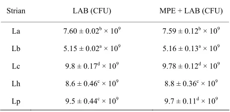
Table 1. Cell viability of LAB grown in combination with MPE at 37˚C.
Strian LAB (CFU) MPE + LAB (CFU)
La 7.60 ± 0.02b × 109 7.59 ± 0.12b × 109
Lb 5.15 ± 0.02a × 109 5.16 ± 0.13a × 109
Lc 9.8 ± 0.17d × 109 9.78 ± 0.12d × 109
Lh 8.6 ± 0.46c × 109 8.8 ± 0.36c × 109
Lp 9.5 ± 0.44c × 109 9.7 ± 0.11d × 109
Figure 1. Nontoxic effect of MPE against Lactobacillus
aci-dophilus, L. bulgaricus, L. casei, L. helviticus and L. planta-rum was identified by agar well-diffusion assay.
the nontoxic effect of MPE on LAB, even though it inhib-ited gram (+) ve and gram (–)ve bacteria. This indicates that supplementation of MPE will not affect the fermen-tation process.
The DPPH radical scavenging activity, inhibition of ascorbate autoxidation, reducing activity of MPE and LAB were shown in Table 2. The culture filtrates of Lc, Lp and Lh have shown higher DPPH scavenging activity (41.68%), inhibition of ascorbate (10.01%) and reducing activity (0.83 μM) respectively.
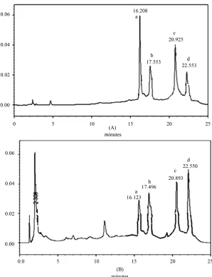
The concentration of isoflavones in MPE fermented with LAB were identified and estimated by HPLC (Figure 2).
The isoflavone isomers were eluted according to their polarity and hydrophobic interaction with the reversed phase HPLC column. Glucosidic isoflavones eluted first and then the aglycones. Within each glucosidic form of isoflavones, daidzin eluted first followed by genistin and in aglyconic form of isoflavones daidzein eluted first and then the genistein (Figure 3).
Changes occurred in the concentration of glucoside and aglycone isoflavone isomers in MPE fermented by La, Lb, Lc, Lp and Lh for 24 h at 37˚C was represented in Table 3. The MPE contained total amount glucosides (20.39 mg/100 ml) and aglycones (0.37 mg/100 ml). Af-ter 24 h of incubation, the concentration of total amo- unt aglycones in culture media of LAB was in the range of 27.98 mg/100 ml - 39.56 mg/100 ml and decrease in the concentration of glucosides isoflavones were observed. The highest concentrations of aglycones were measured in the culture medium of La followed by Lh and least concentration was estimated in Lp.
5. Discussion
Table 2. Antioxidant activities LAB cultured in MPE.
Antioxidant activity
Cultures
DPPH scavenging (%) Inhibition of ascorbate autoxidation (%) (equivalent cysteine, Reducing activity μM)
MPE 35.81 ± 0.11a 7.31 ± 0.24a 0.65 ± 0.03a
MPE + La 39.08 ± 0.13d 9.98 ± 0.15d 0.70 ± 0.04ab
MPE + Lb 36.19 ± 0.20a 8.65 ± 0.10c 0.73 ± 0.03ab
MPE + Lc 41.68 ± 0.15e 8.18 ± 0.17b 0.77 ± 0.02ab
MPE + Lh 36.90 ± 0.15b 9.80 ± 0.02d 0.83 ± 0.02b
MPE + Lp 38.35 ± 0.28c 10.01 ± 0.02d 0.75 ± 0.04ab
Mean values within each column with different superscripts are significantly different by Duncan’s test at P < 0.05.
[image:4.595.149.447.306.694.2]Non-Toxic Effect of Monascus purpureus Extract on 841 Lactic Acid Bacteria Suggested Their Application in Fermented Foods
[image:5.595.60.541.367.540.2]Figure 3. The isoflavone molecules daidzin (a), daidzein (b), genistin (c) and genistein (d) were identified in MPE by HPLC using standard isoflavones.
Table 3. Isoflavone contents were estimated in LAB culture media containing MPE before and after 24 hours of incubation.
Glucosides (mg/100 ml) Aglycones (mg/100 ml) Cultures
Daidzin Genistin Sub-total Daidzein Genistein Sub-total Total (mg/100 ml)
MPE 3.38 ± 0.04d 17.01 ± 0.07d 20.39 ± 0.11e 0.06 ± 0.01a 0.31 ± 0.03a 0.37 ± 0.02a 20.76 ± 0.12a
MPE + La 0.13 ± 0.03c 0.79 ± 0.01c 0.92 ± 0.011d 7.89 ± 0.03f 30.75 ± 0.04f 38.64 ± 0.02f 39.56 ± 0.02f
MPE + Lb 0.11 ± 0.03bc 0.59 ± 0.03b 0.70 ± 0.06c 5.97 ± 0.04c 21.98 ± 0.03c 27.95 ± 0.12c 28.65 ± 0.08c
MPE + Lc 0.05 ± 0.03a 0.30 ± 0.01a 0.35 ± 0.04a 4.72 ± 0.06b 17.16 ± 0.03b 21.88 ± 0.03b 22.23 ± 0.01b
MPE + Lh 0.07 ± 0.02ab 0.52 ± 0.02b 0.59 ± 0.02b 7.04 ±0.05e 26.86 ± 0.07e 33.90 ± 0.07e 34.49 ± 0.06e
MPE + Lp 0.11 ± 0.03bc 0.55 ± 0.06b 0.66 ± 0.03bc 6.49 ± 0.07d 24.17 ± 0.11d 30.66 ± 0.04d 31.32 ± 0.07d
Mean values within each column with different superscripts are significantly different by Duncan’s test at P < 0.05.
a limited number of cases such as sausage, hams, fish paste, surimi and tomato ketchup. However, there are no reports on the usage of Monascus pigment in fermented foods. For preparation of fermented foods, lactic acid bacteria including Lactobacilli are the most common ba- cterial species considered as potential probiotics and im- portant commensal members of the healthy human mi- crobiota. They are useful in promotion of human health and prevention or treatment of several diseases [20]. While, antibacterial property of Monascus sp and effec- tiveness of monascidin-A against Bacillus, Streptococcus and Pseudomonas has been reported [21]. So, prior to increase the nutritive and nutraceutical values of fermented
foods using MPE, it is necessary to evaluate the effect of MPE on LAB.
The agar well diffusion assay and viable count results re- vealed that, MPE do not have any bacteriostatic or bactericidal effect on LAB. Probiotic microorganisms are increasingly incorporated into food as dietary adjuncts for the purpose to benefit the human health [20] and there are several reports on applications of Monascus metabolites as a col- ouring [12], and antioxidant agent [6,7]. The aglycone forms of isoflavones are formed by the catalytic action of
of glucosidic isoflavones aglycone isoflavone (genistein and daidzein). Hence, the fermented foods prepared us- ing MPE will be more functional to reduce the oxidative stress related diseases as it contains active aglycone iso- flavones. The bioconversion suggested the application of MPE along with probiotic cultures can improves the bio- logical functionality of the fermented food products.
The MPE contains high concentration of glucoside isoflavones that are poorly bioavailable in intestine. The biological effects of M. purpureus isoflavones are not as a result of the glucosides but mainly from their aglycones such as daidzein and genistein. In intestine, the isoflavo- ne aglycones are absorbed in greater amounts than their glycosides and are hydrolysed by the intestinal bacteria [24,25]. The variation in intestinal bacteria may occur because of illnesses, diet or age. The bioconversion and bioavailability of isoflavones depends upon the relative ability of gut microflora. Thus the intestinal bacteria cannot always be relied upon for glucoside deconjugation to release aglycones. Our results confirmed the biocon- version of biologically inactive glucosides to biologically active aglycones and nontoxic effect of MPE on LAB. The fermented foods prepared by using M. purpureus extracts may have more health beneficial effects.
6. Acknowledgements
Mohan-Kumari H. P. acknowledges Council of Scientific and Industrial Research, New Delhi, India and University of Mysore for supporting research through the award of Senior Research Fellowship.
REFERENCES
[1] M. A. Dhale, H. P. Mohan-Kumari, S. Umesh-Kumar and G. Vijayalakshmi, “Production of Monascus Purpureus Pigments; Influenced by Amidase and Acid Protease Ac-tivity,” Journal of Food Biochemistry, Vol. 35, No. 4, 2011, pp. 1231-1241.
[2] H. C. Wong and P. E. Koehler, “Production and Isolation of an Antibiotic from Monascus Purpureus and Its Rela-tionship to Pigment Production,” Journal of Food Science, Vol. 46, 1981, No. 2, pp. 589-592.
doi:10.1111/j.1365-2621.1981.tb04917.x
[3] Y. Su, J. J. Wang, T. T. Lin and T. M. Pan, “Production of the Secondary Metabolites c-Aminobutyric Acid and Monacolin K by Monascus,” Journal of Industrial Mi-crobiology and Biotechnology, Vol. 30, 2003, pp. 40-46. [4] G. F. Wu and X. C. Wu, “Screening DPPH Radical Scav-
Engers from Monascus sp,” Acta Microbiologica Science, Vol. 40, No. 4, 2000, pp. 394-399.
[5] C. L. Lee, T. Y. Tsai, J. J. Wang and T. M. Pan, “In Vivo
Hypolipidemic Effects and Safety of Low Dosage Mo- nascus Powder in a Hamster Model of Hyperlipidemia,”
Applied Microbiology and Biotechnology, Vol. 70, No. 5,
2006, pp. 533-540. doi:10.1007/s00253-005-0137-0 [6] Y. Aniya, T. Yokomakura, M. Yonamine, K. Shimada, T.
Nagamine, M. Shimabukuro and H. Gibo, “Screening of Antioxidant Action of Various Molds and Protection of Monascus Anka against Experimentally Induced Liver Injuries of rats,” General Pharmacology, Vol. 32, No. 2, 1999, pp. 225-231. doi:10.1016/S0306-3623(98)00183-9 [7] M. A. Dhale, S. Divakar, S. Umesh-Kumar and G. Vija-
yalakshmi, “Isolation and Characterization of Dihydro- monacolin-MV from Monascus Purpureus for Antioxi-dant Properties,” Applied Microbiology and Biotechnol-ogy, Vol. 73, No. 5, 2007, pp. 1197-1202.
doi:10.1007/s00253-006-0578-0
[8] M. A. Dhale, S. Divakar, S. Umesh-Kumar and G. Vija- yalakshmi, “Characterization of Dehydromonacolin-MV2 from Monascus Purpureus Mutant,” Journal of Applied Microbiology, Vol. 130, No. 3, 2007, pp. 2168-2173. doi:10.1111/j.1365-2672.2007.03457.x
[9] H. P. Mohan-Kumari, K. A. Naidu, S. Vishwanatha, K. Narasimhamurthy and G. Vijayalakshmi, “Safety Evalua-tion of Monascus Purpureus Red Mould Rice in Albino Rats,” Food and Chemical Toxicology, Vol. 47, No. 8, 2009, pp. 1739-1746. doi:10.1016/j.fct.2009.04.038 [10] L. Martinkova, P. Juzlova, V. Kren, Z. Kucerouva, V. Ha-
vlicek, P. Olsovsky, O. Hovorka, B. Rihova, D. Vesly, D. Vesela, J. Ulrichova and V. Prikrylova, “Biological Acti- Vities of Oligoketide Pigments of Monascus Purpureus,”
Food Additive and Contaminants, Vol. 16, No. 1, 1999, pp. 15-24. doi:10.1080/026520399284280
[11] C. Kim, H. Jung, Y. O. Kim and C. S. Shin, “Antimicro-bial Activities of Amino Acid Derivatives of Monascus Pigments,” FEMS Microbiology Letters, Vol. 264, No. 1, 2006, pp. 117-124.
doi:10.1111/j.1574-6968.2006.00451.x
[12] H. Jung, C. Kim and C. S. Shin, “Enhanced Photostability of Monascus Pigments Derived with Various Amino Ac-ids via Fermentation,” Journal of Agriculture and Food Chemistry, Vol. 53, No. 18, 2005, pp. 7108-7114. doi:10.1021/jf0510283
[13] J. C. De Man, M. Rogosa and M. E. Sharpe, “A Medium for the Cultivation of Lactobacilli,” Journal of Applied Bacteriology, Vol. 23, No. 1, 1960, pp. 130-135. doi:10.1111/j.1365-2672.1960.tb00188.x
[14] P. K. Mukherjee, R. Balasubramanian, K. Saha, B. P. Saha and M. Pal, “Antibacterial Efficiency of Nelumbo nucifera (Nymphaeaceae) Rhizomes Extract,” Indian Drugs, Vol. 32, No. 6, 1995, pp. 274-276.
[15] R. Y. Y. Chiou and S. L. Cheng, “Isoflavone Transforma-tion during Soybean Koji PreparaTransforma-tion and Subsequent Miso Fermentation Supplemented with Ethanol and NaCl,” Journal of Agriculture and Food Chemistry, Vol. 49, No. 8, 2001, pp. 3656-3660. doi:10.1021/jf001524l [16] M. Blois, “Antioxidant Determinations by the Use of a
Stable Free Radical,” Nature, Vol. 181, 1958, pp. 1199- 1200. doi:10.1038/1811199a0
Non-Toxic Effect of Monascus purpureus Extract on Lactic Acid Bacteria Suggested Their Application in Fermented Foods
843
Acid and Dihydrocaffeeic Acid in Lard and Human Low-Density Lipoprotein,” Journal of Agriculture and Food Chemistry, Vol. 46, No. 12, 1998, pp. 5062-5065. doi:10.1021/jf9805799
[18] G. B. Kovachich and O. P. Mishra, “Stabilization of As- Corbic Acid and Norepinephrine in Vitro by the Subcel- lular Fractions of Rat Cerebral Cortex,” Neuroscience Letters, Vol. 52, No. 1-2, 1984, pp. 153-158.
doi:10.1016/0304-3940(84)90366-5
[19] M. Oyaizu, “Studies on Product of Browning Reaction Prepared from Glucose Amine,” Japanese Journal of Nutri- tion, Vol. 44, No. 6, 1986, pp. 307-315.
doi:10.5264/eiyogakuzashi.44.307
[20] F. Guarner and J. R. Malagelada, “Gut Flora in Health and Disease,” Lancet, Vol. 361, No. 9356, 2003, pp. 512- 519. doi:10.1016/S0140-6736(03)12489-0
[21] H. C. Wong and Y. S. Bau, “Pigmentation and Antibacte- rial Activity of Fast-Neutron and X-Ray Induced Strains of M. purpureus Went,” Plant Physiology, Vol. 60, No. 4, 1977, pp. 578-581. doi:10.1104/pp.60.4.578
[22] D. O. Otieno, J. F. Ashton and N. P. Shah, “Stability of
β-Glucosidase Activity Processing by Bifidobacterium and Lactobacillus spp in Fermented Soymilk during Pro- cessing and Storage,” Journal of Food Science, Vol. 70, No. 4, 2005, pp. 236-241.
doi:10.1111/j.1365-2621.2005.tb07194.x
[23] W. H. Hsu, B. H. Lee and T. M. Pan, “Red Mold Dio-scorea-Induced G2/M Arrest and Apoptosis in Human Oral Cancer Cells,” Journal Science and Food Agriculture, Vol. 90, No. 15, 2010, pp. 2709-2715.
doi:10.1002/jsfa.4144
[24] K. Setchell, N. Brown, N. L. Zimmer, W. Brashear, B. Wolfe, A. Kirschour and J. Heubi, “Evidence for Lack of Absorption of Soy Isoflavone Glycosides in Humans, Supporting the Crucial Role of Intestinal Metabolism for Bioavailability,” American Journal of Clinical Nutrition, Vol. 76, No. 2, 2002, pp. 447-453.