DETACHMENT OF POLYMICROBIAL
WITH SURFACE MODIFIERS CONTAINING FLUOROCARBON CHAIN
1
Ayako Teranaka,
2,3Kiyoshi Tomiyama,
1Hirotoshi Iwai,
6Nobushiro Hamada,
2,3
Toshio Teranaka,
1
Department of Operative Dentistry, Nihon University School of Dentistry at Matsudo,
2
Division of Cariology and Restorative Dentistry, Dentistry for Oral Interdisciplinary Medicine, Graduate School
of Dentistry, Kanagawa Dental University, Kanagawa, Japan
3Institute of Oral Regenerative Medicine, Kanagawa Dental University, Ja
4
Division of Clinical Biomaterials, Department of Oral Science, Graduate School of Dentistry, Kanagawa Dental
5
Division of Prosthodontics and Oral Implantology, Dentistry for Oral Interdisciplinary Medicine, Graduate
School of Dentistry, Kanagawa Dental University, Kanagawa, Japan
6
Division of Microbiology, Department of Oral Science, Graduate School of Dentistry, Kanagawa Dental
7
Departmentof Industrial Chemistry, Tokyo University of Science, Tokyo
ARTICLE INFO ABSTRACT
The purpose of this study was to evaluate the adherence and detachment of bacterial biofilms formed on glass surface modified with 10F2S
unmodified cover glasses were imme
and theywere cultured to develop PM biofilms. There were separated six experimental groups (n = 6), designated WC: cover glass washing withCPW, WI: modified with 10F2S
with CPW, WF: modified with FF01 cover glass washing with CPW, NC: cover glass without washing, NI: modified with 10F2S
glass without washing.The modified and unmodified groups were further divided b washing withCPW or without washing.
which were then cultured on blood agar medium to count CFU.PM biofilms were easily detached from 10F2S
difference of CFU among NC, NI and NF. But WI and WF were significantly lower than in WC. It was concluded that PM biofilms grown on the glass surface modified were easily detached compared with the unmodifiedglass groups
Copyright©2017, Ayako Teranaka et al. This is an open access article distributed under the Creative Commons Att use, distribution, and reproduction in any medium, provided the original work is properly cited.
INTRODUCTION
Owing to the low birthrate and aging issues, importance of providing an oral care method which preferably cleans soil in the mouth is increasing to prevent the occurrence of aspiration
*Corresponding author: Tomotaro Nihei,
3
Institute of Oral Regenerative Medicine, Kanagawa Dental University, Japan
4Division of Clinical Biomaterials, Department of Oral Science,
Graduate School of Dentistry, Kanagawa Dental University, Kanagawa, Japan.
ISSN: 0975-833X
Article History: Received 25th June, 2017
Received in revised form 26th July, 2017
Accepted 18th August, 2017
Published online 29th September, 2017
Citation: Ayako Teranaka, Kiyoshi Tomiyama, Kaori Miyake, Katsura Ohashi, Tota Shimizu, Hirotoshi Iwai, Nobushiro Hamada, Norio Yoshino Yoshiharu Mukai, Toshio Teranaka, Satoshi Hirayama and Tomotaro Nihei
surface modifiers containing fluorocarbon chain”, International Journal of Current Research
Key words:
Polymicrobial Biofilm, Surface Modification, Plaque Detachability.
RESEARCH ARTICLE
POLYMICROBIAL BIOFILMS ON GLASS SURFACE TREATED
WITH SURFACE MODIFIERS CONTAINING FLUOROCARBON CHAIN
Kiyoshi Tomiyama,
3,4Kaori Miyake,
3,4Katsura Ohashi,
Nobushiro Hamada,
7Norio Yoshino,
2,3Yoshiharu Mukai,
Toshio Teranaka,
1Satoshi Hirayama and
3,4,*Tomotaro Nihei
Department of Operative Dentistry, Nihon University School of Dentistry at Matsudo,
Division of Cariology and Restorative Dentistry, Dentistry for Oral Interdisciplinary Medicine, Graduate School
of Dentistry, Kanagawa Dental University, Kanagawa, Japan
Institute of Oral Regenerative Medicine, Kanagawa Dental University, Ja
Division of Clinical Biomaterials, Department of Oral Science, Graduate School of Dentistry, Kanagawa Dental
University, Kanagawa, Japan
Division of Prosthodontics and Oral Implantology, Dentistry for Oral Interdisciplinary Medicine, Graduate
of Dentistry, Kanagawa Dental University, Kanagawa, Japan
Division of Microbiology, Department of Oral Science, Graduate School of Dentistry, Kanagawa Dental
University, Kanagawa,Japan
Departmentof Industrial Chemistry, Tokyo University of Science, Tokyo
ABSTRACT
The purpose of this study was to evaluate the adherence and detachment of bacterial biofilms formed on glass surface modified with 10F2S-3I or FF01 using a PMbiofilm model. Modified and unmodified cover glasses were immersed in buffered McBain medium containing stimulated saliva, and theywere cultured to develop PM biofilms. There were separated six experimental groups (n = 6), designated WC: cover glass washing withCPW, WI: modified with 10F2S
CPW, WF: modified with FF01 cover glass washing with CPW, NC: cover glass without washing, NI: modified with 10F2S-3I cover glass without washing, NF: modified with FF01 cover glass without washing.The modified and unmodified groups were further divided b
washing withCPW or without washing. Serial dilutions were conducted of the bacterial cultures, which were then cultured on blood agar medium to count CFU.PM biofilms were easily detached from 10F2S-3I and FF01 modified surfaces by washing with CPW. There were no significant difference of CFU among NC, NI and NF. But WI and WF were significantly lower than in WC. It was concluded that PM biofilms grown on the glass surface modified were easily detached compared with the unmodifiedglass groups.
is an open access article distributed under the Creative Commons Attribution License, which use, distribution, and reproduction in any medium, provided the original work is properly cited.
Owing to the low birthrate and aging issues, importance of providing an oral care method which preferably cleans soil in prevent the occurrence of aspiration
Regenerative Medicine, Kanagawa Dental
Division of Clinical Biomaterials, Department of Oral Science, Graduate School of Dentistry, Kanagawa Dental University,
pneumonia, endocarditisa and other diseases that can severely diminish quality of life (Abe et al
2002). However, the self-care ability of the elderly and aged people who need nursing care is low. Furthermore, the provision of proper oral hygiene by no
such as their family members and caregivers, is not an easy task. As a result, poor oral hygiene among such peopleare frequently seen (Petersen et al
accumulation on dentures increases the risk of aspira pneumonia (Yoneyama et al., 2002
70% of pneumonia in elderly people is considered to be
International Journal of Current Research Vol. 9, Issue, 09, pp.57249-57253, September, 2017
Ayako Teranaka, Kiyoshi Tomiyama, Kaori Miyake, Katsura Ohashi, Tota Shimizu, Hirotoshi Iwai, Nobushiro Hamada, Norio Yoshino
Yoshiharu Mukai, Toshio Teranaka, Satoshi Hirayama and Tomotaro Nihei, 2017. “Detachment of polymicrobial
International Journal of Current Research, 9, (09), 57249-57253.
GLASS SURFACE TREATED
WITH SURFACE MODIFIERS CONTAINING FLUOROCARBON CHAIN
Katsura Ohashi,
5Tota Shimizu,
Yoshiharu Mukai,
Tomotaro Nihei
Department of Operative Dentistry, Nihon University School of Dentistry at Matsudo, Chiba, Japan
Division of Cariology and Restorative Dentistry, Dentistry for Oral Interdisciplinary Medicine, Graduate School
of Dentistry, Kanagawa Dental University, Kanagawa, Japan
Institute of Oral Regenerative Medicine, Kanagawa Dental University, Japan
Division of Clinical Biomaterials, Department of Oral Science, Graduate School of Dentistry, Kanagawa Dental
Division of Prosthodontics and Oral Implantology, Dentistry for Oral Interdisciplinary Medicine, Graduate
of Dentistry, Kanagawa Dental University, Kanagawa, Japan
Division of Microbiology, Department of Oral Science, Graduate School of Dentistry, Kanagawa Dental
Departmentof Industrial Chemistry, Tokyo University of Science, Tokyo, Japan
The purpose of this study was to evaluate the adherence and detachment of bacterial biofilms formed 3I or FF01 using a PMbiofilm model. Modified and rsed in buffered McBain medium containing stimulated saliva, and theywere cultured to develop PM biofilms. There were separated six experimental groups (n = 6), designated WC: cover glass washing withCPW, WI: modified with 10F2S-3I cover glass washing CPW, WF: modified with FF01 cover glass washing with CPW, NC: cover glass without 3I cover glass without washing, NF: modified with FF01 cover glass without washing.The modified and unmodified groups were further divided by either gently Serial dilutions were conducted of the bacterial cultures, which were then cultured on blood agar medium to count CFU.PM biofilms were easily detached ith CPW. There were no significant difference of CFU among NC, NI and NF. But WI and WF were significantly lower than in WC. It was concluded that PM biofilms grown on the glass surface modified were easily detached compared
ribution License, which permits unrestricted
a and other diseases that can severely
et al., 2006; Paju, 2007; Mojon,
care ability of the elderly and aged people who need nursing care is low. Furthermore, the provision of proper oral hygiene by non-dental professionals, such as their family members and caregivers, is not an easy task. As a result, poor oral hygiene among such peopleare
et al., 2010). In addition, plaque
accumulation on dentures increases the risk of aspiration ., 2002), one study reported that 70% of pneumonia in elderly people is considered to be
INTERNATIONAL JOURNAL OF CURRENT RESEARCH
Ayako Teranaka, Kiyoshi Tomiyama, Kaori Miyake, Katsura Ohashi, Tota Shimizu, Hirotoshi Iwai, Nobushiro Hamada, Norio Yoshino,
aspiration pneumonia (Teramoto et al., 2008). Aspiration pneumonia is one of the leading causes of death in elderly, andit can be prevented by proper oral care (Scannapieco, 2014). Therefore, preventing plaque accumulation could reduce the risk of aspiration pneumonia and endocarditis. Plaque accumulation begins with attachment of bacteria on a thin layer of salivary pellicle on the tooth surface. Pellicle forms immediately, even after proper tooth cleaning, and the following plaque formation is inevitable. It is known that attachment of pellicle and plaque is affected by surface free energy (SFE) on the solid surfaces (Glantz, 1971; Van Dijk et al., 1987; Quiynen et al., 1989). Busscher et al. (1984) reported that when SFE on tooth surfaces is lower than 50mN/m, plaque adherence should decrease. Thus, modification of the tooth surface to lower SFE is considered an effective means to decrease plaqueformation.
We have conducted to develop a surface modifying agent to reduce bacterial adherence, plaque formation, and demineralization of tooth surface to assist in the prevention of caries and periodontal disease (Ogiwara et al., 1995; Kurosaka
et al., 2000; Omoto et al., 2005; Nihei et al., 2013). Ogiwara
et al. (1995) and Kurosaka et al. (2000) reported that resin
composite and tooth surfaces modified by a silane coupling
agent with high water and oil repellency, 1H,1H,2 H, 2H-henicosafluorododecyltriisocyanatosilane (10F2S-3I),
developed by Yoshino et al. (1996; 1997), discouraged adherence and promoted detachment of Streptococcus mutans. While antibacterial studiesusing a few species of bacteria have made a dramatic advance, there are few studies on oral biofilms including many species of bacteria. In this study, polymicrobial (PM) biofilm modeldeveloped by Exterkate et al. (2010) was used. The purpose of this study was to evaluate the adherence and detachment of biofilms formed on glass surfacesmodified with 10F2S-3I and newly developed heptadecafluorodecylhydroxyethylmethacrylate (FF01) using PM biofilm model (Exterkate et al., 2010; Macbain, 2009; Exterkate et al., 2014; Tomiyama et al., 2015; Hasegawa et al., 2015).
MATERIALS AND METHODS
Surface modifying agent
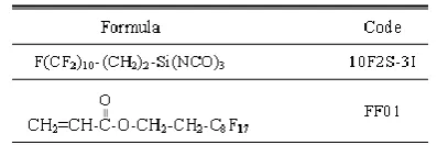
[image:2.595.63.262.646.712.2]10F2S-3I (Tokyo University of Science,Tokyo, Japan)prepared at a concentration of 3mmol/Lwith hydrofluoroether (Novec HFE 7200, 3M, Tokyo, Japan) and newly developed FF01 (Daikin Industries, Ltd., Osaka, Japan)were used as a surface modifying agent. (Table 1)
Table 1. Formula amd code of modifiers user in this study
Preparation of the saliva sample
Saliva was collected under ice application from a healthy adult donor, with natural dentition without active caries or periodontal disease. The inclusion criteria were as follows: not having administered antibiotics or antibacterial agents for 3 months, not having brushed their teeth for the last 24 hours,
and not having had anything to eat or drink since 2 hours before saliva collection. Subjects chewed paraffin wax (Parafilm M Barrier Films, Pechiney Plastic Packaging, Chicago, IL, USA), and the stimulated saliva was collected into a plastic container which was cooled down with ice. The saliva was kept at 80°C after being diluted into a 70 vol% solution with glycerol following filtration by glass wool (NRK GRW-10, Nippon Rikagaku Kikai CO.LTO., Tokyo, Japan). The study protocol was approved by the Institutional Review Board of Kanagawa Dental University (Approval number: 206).This study was ensured compliance with the Helsinki Declaration for the consideration of the human rights of research subjects and informed consent.
Glass coverslips modifications
Fifty-fourglass coverslips (diameter 12mm, thickness 0.15mm, Menzel, Braunschweig, Germany) were soaked in 1mol/L sodium hydroxide and 1mol/L hydrochloric acid for one day each to remove contaminants. Each eighteenof the glass coverslipswere immersed in 3mmol/L10F2S-3I (group I) and FF01 (group F) for 1 hour to allow surface modification. After modification, the glass coverslips were rinsed with hydrofluoroether and dried at room temperature. The remaining 18 glass coverslips were unmodified (group C) as control. All glass coverslips were mounted on the lids of 24-well culture plates and autoclaved at 121ºC for 15 minutes.
Measurement of contact angle and SFE
Contact angle measurements employing the drop technique were carried out at 25ºC using distilled water and diiodomethane (Wako, Osaka, Japan) by automatic contact angle meter (DCA-VZ Type, Kyowa Interface Science Co, Saitama, Japan). Contact angle data on a given surface yields the surface free energy by SFE calculation software using the Owents /Wendt theory.
PM biofilms formation
Retained saliva was diluted 50-fold with buffered McBain semidefined medium (0.2wt% sucrose, 50 mmol PIPES, 2.5 g/l mucin, 2.0 g/l Bacto peptone, 2.0 g/l Trypticase peptone, 1.0 g/l yeast extract, 0.35 g/l NaCl, 0.2 g/l KCl, 0.2 g/l CaCl2,
0.001 g/l hemin, and 0.0002 g/l vitamin K1) (18-22). After
medium was prepared, biofilms were produced by adding 1.5 ml of the inoculation medium with saliva to each well of standard polystyrene 24-well plates (multiwell plates, Greiner Bio One, Muunich, Germany), and the glass coverslipsfixed ontothe dedicated lids of the culture plates were immersed and anaerobically cultured for 10 hours at 37ºC (10% CO2, 10% H2
[image:2.595.324.543.673.747.2]and 80% N2).
Table 2. Abbreviations of combined treatments for CFU and SEM
into the 24-well plates with antibiotic agents or DW for 5 minutes at 22°C.
Table 3. Contact angle and SFE for each group
To wash off various antibiotics, plates were soaked in wells into which 2 ml of cysteine peptone water (CPW) was added; then, the plates were shaken up and down 10 times within 10 seconds. This process was repeated three times. The remaining halves of group I, F and C were without washing. The combinations for CFU and SEM studies were shown in Table 2.
Calculation of viable cell counts formed on glass coverslips of each group
Six each from WC, WI, WF, NC, NI and NFglass coverslipswith biofilms were transferred into tubes containing CPW at 2 ml, and subjected to ultrasonic wave for 90 seconds (Transsonic T780, Elma electric GmbH, Stuttgart, Germany), followed by vortexing for 30 seconds (Tube mixer, VTX-3500, LMS, Tokyo, Japan) to disintegrate and disperse bacterial cells. Conducting serial dilutions using CPW, 50µL of each sample was cultured in tryptic soy blood agar medium at 37ºC under anaerobic conditions (10% CO2, 10% H2 and 80% N2)
for 4 days, after which CFUs were counted.
SEM observation
The glass coverslips with PM biofilms of each group (n=3) were soaked in 0.01mol/Lphosphate buffered saline (PBS) for 30 seconds followed by a 30 seconds wash with 0.1mol/L cacodylate buffer. The glasses were then stored in a mixture of 0.1 mol/L cacodylate buffer and 1% glutaraldehyde for 1 hour for fixation. Each glass was then rinsed twice (30 seconds each) with 0.1 mol/L cacodylate buffer, and then, immersed in serial concentrations of ethanol solutions (50, 70, 80, 90, and 100%) for 15 minutes each at room temperature. After they were immersed in isoamyl acetate, conducted drying at the critical point. The glasses were fixed on a brass stage with a carbon tape and sputter coated with platinum (200Ǻ in thickness). The glass surfaces were observed using a scanning electron microscope (SEM, JSM-820, JEOL, Tokyo, Japan) at an accelerating voltage of 5.0kV.
Statistical analysis
The average and standard deviation of the experimental data were calculated, and subjected to one-way ANOVA and Tukey multiple comparison assay. The level of significance was taken as p <0.05. All statistical analyses were performed by IBM SPSS Ver. 20.0 software (IBM Japan, Ltd., Tokyo, Japan).
RESULTS
Contact angle and SFE
Contact angle and SFE for each group are displayed in Table3. Contact angle of distilled water were14.7°, 114.4° and 108.7° in group C, I and F respectively. Contact angle of
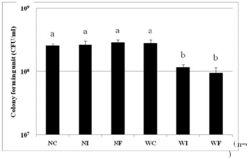
[image:3.595.307.554.181.339.2]dioodomethane were 50.5°, 98.7° and 90.9° in group C, I and F respectively. Contact angle of distilled water for group I and F were significantly higher than group C (p<0.05). Contact angle of diiodomethane for group I and F were significantly higher than group C (p<0.05). SFE of each group were shown 70.5mN/m at group C, 9.5mN/mat group I, and 12.8mN/mat group F. Surface modified group I and F were 50mN/m or less. It was significantly lower compared to group C (p<0.05. Live bacterial count of PM biofilms on each groupare displayed in Figure 1.
Fig.1. CFU counts of PM biofilm for each group there was no significant difference in the CFU counts between NC, NI, NF
and WC (p>0.05). However, the CFU count of WI and WF
was significantly lower that that of WC (p>0.05)
Fig.2. Scanning electorn micrographs of the PM biofilm of each group WI and WF demonstrated abacteria compared to the other
groups. Bars; 10µm
The CFU counts of NI, NF and WC were similar (2.56×10⁸, 2.62×10⁸, 2.88×10⁸ and 2.82×10⁸ CFU/disk, respectively). However, the CFU counts of WI (1.17×10⁸ CFU/disk) and WF (9.48×10⁷ CFU/disk) were significantly lessthan those of other groups (p<0.05).
SEM observation
Images of PM biofilms attached to the glass coverslipsare presented in Figure 2. The glass surface ofWI and WF were showeddecreased bacteria than the other groups.
DISCUSSION
[image:3.595.315.551.421.562.2]of floating microorganisms as initial colonizer in the oral fluid to the pellicle. Formation of initial colonies on the surface and subsequentaccumulation, growth microorganism leads to the development of a mushroom-shaped mature biofilm. The slimy main body of a biofilm consists of extracellular polysaccharide (EPS). Oral bacteria synthesize EPS to develop a population (Xiao et al., 2012). The PM biofilm used in the present study is a novel, high-throughput and active attachment model (Hung
et al., 2012). It is an effective method of forming biofilms
consisting of multiple bacterial types similar to those typically found in the oral cavity under the same conditions; this is done by storing stimulated human saliva at 80°C following sterilized glycerin preparation and by defrosting and using it for each test (Exterkate et al., 2010).
In addition, the method can easily be implemented, and therefore, the present biofilms consisting of multiple bacterial types are the most suitable biofilm model for the investigation of antibiotic agents, dental materials, and surface modifiers. The adherence test of PM biofilms on glass surfaces modified by 10F2S-3I and FF01 revealed no significant differences in the CFUs between NC, NI and NF, both groups were not washed with CPW, indicating 10F2S-3I and FF01 haveno antibacterial activity. After extremelygentle washing with CPW, WCwas showed no significant difference in the CFU counts with NC, NI and NF, whereas the CFU counts for WI and WF were significantly lower. It was found that after modification with 10F2S-3I and FF01, PM biofilmsadhered to the glass surface became more prone to detachment by extremely gentle washing with CPW. Although insoluble glucan is produced from sucrose at the interface, it is considered that bacteria are not able to establish a strong adherence to the modified surfacebecause of the high water and oil repellency of 10F2S-3I (Yoshino et al., 1996; Yoshino and Teranaka, 1997). These findings suggested that the surface modification effect of 10F2S-3I and FF01 are maintained even after completion of culturing. Moreover, effective detachment of the biofilm would be obtained due to the continuous modification effect after water storage for long periods of time.
Yoshino et al. (1996, 1997) demonstrated that surface modification with 10F2S-3I showed excellent oxidation and acid resistance against hotnitric acid. Ogiwara et al. (1995) and Kurosaka et al. (2000) reported that glass and resin composite surfaces modified with the 10F2S-3I inhibitedthe attachment and promoted detachment of S. mutans. Furthermore, Omoto et al. (2005) and Nihei et al. (2013) found that 10F2S-3I applied to glass, hydroxyapatite and enamel maintained high water and oil repellency in water at 37°C for 90 days, indicating that there is no notable change of surface free energy values after the completion for 24hours culturing. Also Omoto et al. (2005) reported that high SFE was maintained even if it was kept underwater for a long period. Consequently,it is considered that 10F2S-3I obtained high detachment of biofilms. Newly developed heptadecafluorodecylhy droxyethylmethacrylate (FF01) has characterized antifouling, antifouling edurance, water and oil repellency, excellent slidingproperty, and low effect on optical property. Chemical plaque controlling agents such as mouth rinse have been considered as a secondary preventive measure in addition to mechanical control (Baehni, 2003; Michael, 2006; Marsh, 2010), and several studies have reported that the bactericidal effect of mouthwash inhibit the increasing of biofilm thickness (Stewart, 2001; Watson et al., 2005; Sliepen et al., 2010; Yamaguchi et al., 2013; Wakamatsu et al., 2014). Even if bacteria in a biofilm are
disinfected, the remaining matrix structure consisting of EPS can act as a scaffold for formation of new biofilms (Takenaka
et al., 2012). Because 10F2S-3I and FF01were facilitated the
detachment of bacteria with gentle washing, and adhered bacteria are removed by regular oral hygiene, we suggest that application of 10F2S-3I and FF01arean effective measure for preventing caries and periodontal disease. And, 10F2S-3I and FF01 may have an application as an oral care measure for used by evacuees in the event of a large-scale disaster. However, for applying FF01 in a clinical setting, it is necessary to evaluate the cytotoxicity and biocompatibility of FF01. In molecular structure of FF01, it is considered possible that to add to polymerization initiators, forming a bond with intermolecular andforming ultrathin film polymer or add to crosslinking monomer. It was considered possible development in water repellent monomer. In the future, it is necessary to examine the physical properties and durability of these modifiers.
Conclusion
It was concluded that after modification with 10F2S-3I and FF01, PM biofilms adhered to the glass surface became more prone to detachment by extremely gentle washing with CPW. From these results, it was suggested that it was the modifier which kept intraoral environment well.
Acknowledgement
The authors would like to acknowledge support of the part of this study by the strategic research infrastructure construction support project for private colleges by the Ministry of Education, Culture, Sports, Science, and Technology (2012 to 2014, and 2016 to 2017), and the scientific research subsidy infrastructure research (B) and (C) (task No. 25462973, and No. 16K20520). There is no conflict of interest regarding the materials and method used in this study.
REFERENCES
Abe S, Ishihara K, Adachi M, Okuda K. 2006. Oral hygiene evaluation for effective oral care in preventing pneumonia in dentate elderly. Arch Gerontol Geriat., 43, 53-64. Baehni PC, Takeuchi Y 2003. Anti-plaque agents in the
prevention of biofilm-associated oral diseases; Oral Dis., 9, 23-29.
Busscher HJ, Weerkamp AH, van der Mei HC, van Pelt AWJ, de Jong HP, Arends J 1984. Measurement of the surface free energy of bacterial cell surfaces and its relevance for adhesion. Appl Environ Microbiol., 48, 980-983.
Exterkate RAM, Crielaard W, ten Cate JM 2010. Different response to amine fluoride by Streptococcus mutans and polymicrobial biofilms in a novel high-throughput active attachment model. Caries Res., 44, 372-379.
Exterkate RAM, Zaura E, Buijs MJ, Koopman J, Crielaard W, ten Cate JM 2014. The effects of propidium monoazide treatment on the measured composition of polymicrobial biofilms after treatment with chlorhexidine. Caries Res.,
48, 291-298.
Glantz P-O 1971. The adhesiveness of teeth. J Colloid
Interface Sci., 37, 281-290.
Hung X, Extekate RAM, ten Cate JM 2012. Factors associated with alkali production from arginine in dental biofilms. J
Dent Res., 91, 1130-1134.
Kurosaka N, Nihei T, Mitsuhashi S, Kumada H, Umemoto T, Kondo Y, Yoshino N, Teranaka T. 2000. Inhibitory effect of surface modification with polyfluorotriisocyanatosilane on plaque accumulation to resin composite in vitro. Jpn J
Conserv Dent., 43(5), 1083-1089.
Macbain AJ 2009. Chapter 4 in vitro biofilm models. Adv Appl
Microbiol., 69, 99-132.
Marsh PD 2010. Controlling the oral biofilm with antimicrobials. J Dent., 38, 11-15.
Michael LB 2006. The rationale for daily use of an antimicrobial mouthrinse. J Am Dent Assoc 137, 165-215. Mojon P. 2002. Oral health and respiratory infection. J Can
Dent Assoc., 68, 340-345.
Nihei T, Omoto N, Ohashi K, Kondo Y, Yoshino N, Teranaka T 2013. Effect of enamel surface modification by novel aqueous phosphate-type fluoride surfactants. Dent Mater J.,
32, 83-87.
Ogiwara S, Teranaka T, Kumada H, Yoshino N 1995. Effect of a fluorocarbon chain containing silane coupling agent on accumulation and detachment of Streptococcus mutans on and from resin composite in vitro. Jpn J Conserv Dent., 38, 1071-1081.
Omoto N, Nihei T, Kurata S, Kondo Y, Umemoto K, Yoshino N, Teranaka T. 2005. Modification effect of novel phosphate type hybrid surfactant – Modification effect of surfactants for hydroxyapatite plate. Jpn J Conserv Dent., 48(1), 128-136.
Paju S, Scannapieco FA. 2007. Oral biofilms periodontitis and pulmonary infections. Oral Dis., 13, 508-512.
Petersen PE, Kandelman D, Arpin S, Ogawa H 2010. Global oral health of older people - Call for public health action.
Community Dent Health, 27, 257-268.
Quiynen M, Marechal M, Busscher HJ, Weerkamp AH, Arends J, Darius PL, van Steenberghe D 1989. The influence of surface free-energy on planimetric plaque growth in man. J Dent Res., 68, 796-799.
Scannapieco FA, Shay K 2014 Oral health disparitiesinolder adults: oralbacteria, inflammation, and aspirationpneumonia. Dent Clin North Am., 58, 771-82. Sliepen I, Essche MV, Quirynen M, Teughels W. 2010. Effect
of mouthrinses on Aggregatibacter actinomycetem comitans biofilms in a hydrodynamic model. Clin Oral
Investig., 14, 241-250.
Stewart PS, Costerton JW 2001. Antibiotic resistance of bacteria in biofilms. The Lancet, 358, 135-138.
Takenaka S, Ohshima H, Ohsumi T, Okiji T. 2012. Current and future strategies for the control of mature oral biofilms-Shift from a bacteria-targeting to a matrix-targeting approach. J Oral Biosci., 54, 173-179.
Teramoto S, Fukuchi Y, Sasaki H, Sato K, Sekizawa K, Matsuse T 2008. High incidence of aspiration pneumonia in community and hospital-acquired pneumonia in hospitalized patients: a multicenter, prospective study in Japan. J Am Geriatr Soc., 56, 577-579.
Tomiyama K, Mukai Y, Kumada H, Watanabe K, Hamada N, Teranaka T 2015. Formation of subsurface dentin lesions using a polymicrobial biofilm model. Am J Dent., 28, 13-17.
van Dijk J, Herkströter F, Busscher HJ, Weerkamp AH, Arends J 1987 Surface-free energy and bacterial adhesion An in vivo study in beagle dogs. J Clin Periodontol., 14, 300-304.
Wakamatsu R, Takenaka S, Ohsumi T, Terao Y, Ohshima H, Okiji T. 2014. Penetration kinetics of four mouthrinses into Streptococcus mutans biofilms analyzed by direct time-lapse visualization. Clin Oral Investig., 18, 625-634. Watson PS, Pontefract HA, Devine DA, Shore RC, Nattress
BR, Kirkham J, Robinson C 2005. Penetration of fluoride into natural plaque biofilms. J Dent Res., 84, 451-455. Xiao J, Klein MI, Falsetta ML, Lu B, Delahunty CM,Yates JR
3rd, Heydorn A, Koo H 2012. The exopolys accharidematrix modulates the inter action between 3 Darchitecture and virulence of amixed-speciesoral bio film. PLoS Pathog.8: e1002623. doi: 10.1371.
Yamaguchi M, Noiri Y, Kuboniwa M, Yamamoto R, Asahi Y, Maezono H, Hayashi M, Ebisu S. 2013. Porphyromonas gingivalis biofilms persist after chlorhexidine treatment.
Eur J Oral Sci., 121, 162-168.
Yoneyama T, Mukaiyama H, Ihara S, Mizuno Y, Hashimoto K 2002. Oral care reduce pneumonia in older patients in nursing homes. J Am Geriatr Soc., 50, 430-433.
Yoshino N, Kondo Y, Yamauchi T 1996. Syntheses and reactions of metal organics. XXI. syntheses of (1H,1H,2H, 2H-polyfluoroalkyl) trimethoxy silanes and surface modification of glass. J Fluorine Chem., 79, 87-91. Yoshino N, Teranaka T 1997. Synthesis of silane coupling
agents containing fluorocarbon chain and applications to dentistry: Plaque-controlling surface modifiers. J Biomater
Sci Polymer Ed., 8, 623-653.