http://dx.doi.org/10.4236/jbbs.2013.32027 Published Online May 2013 (http://www.scirp.org/journal/jbbs)
Correlative Analysis of Data and Functions of
Neuronal Synapse
T. R. Gopalakrishnan Nair1, A. Baby Jerald2* 1
Vice President—RIIC,Dayananda Sagar Institutions,ARAMCO Endowed Chair—Technology, PMU, Alkhobar, KSA 2
Research Associate—RIIC, Neuroscience Research Group Dayananda Sagar Institutions,
ARAMCO Endowed Chair—Technology, PMU, Alkhobar, KSA
Email: [email protected], *[email protected]
Received February 15, 2013; revised April 1, 2013; accepted May 9,2013
Copyright © 2013 T. R. Gopalakrishnan Nair, A. Baby Jerald. This is an open access article distributed under the Creative Commons Attribution License, which permits unrestricted use, distribution, and reproduction in any medium, provided the original work is properly cited.
ABSTRACT
Until recently, the synaptic transmission and excitatory amino acid transporters activation of neurons are very well dis- cussed in the previous studies and are considered to be the two distinct features of Synapse. It is also found that a large number of interactions take place in the domain of ionic exchanges and protein interactions in synapses. It is evolu- tionary to have destined to release of Neurotransmitters to conduct an impulse to the other consecutive neurons, which forms the most important characteristic of synapse. From the popular perspective, it has been identified that detailed the- oretical closer correlation of data produced through various studies about synapse can unravel many mysteries related to functions of synapse. Hence, this research paper tries to concentrate on a selected group of prominent characteristics and properties of synapse and also highlights some noteworthy discoveries, emphasizing the influential capabilities of them in the thought process and improving the knowledge of the field. It also highlights the expressive properties and forms of synapse brought out through the evidences available in sparse to dense data in a correlational way.
Keywords: Synapse; Synapse Formation; Synapse Gap; Synaptic Transmission; Synaptic Plasticity; Synapse Proteins; Functions
1. Introduction
It is commonly known that the mossy fiber system (MFBs) is a complex structure morphologically [1] and all the polarized nerve cells communicate via synapse, thus forms the developmental, functional, structural and trophic units of the nervous system [2]. Many known and unknown interactions occur within the central nervous system through synaptic contacts and transports. The study of synapse morphology and characteristics has re- cently taken under the domain of systems biology and computational biology as it has got the highest increased research interest. In this paper, the following subsections cover the knowledge on morphology and functions, along with certain other features like signal transmission and synaptic proteins. It has been a long term goal for many researchers to investigate neural and brain archi- tecture and function to find appropriate reasons for many mental disorders, as there is a lack of deep information regarding the above mentioned topics. Hence, the pur-
pose of the paper is to provide a crisp and combined in- formation of neuron synapse which is being pulled out from the available resources till today. This even handy information will help to indicate other properties such as synapse connectivity, synapse formation, synapse gap, transmission of signals via synapse and the role of some proteins in the synapse region and various disfunctions. Nevertheless even with these information, several other studies are required to take advantage of more diversified information on neuron synapse.
2. Introduction on Synapse
emerge out to show that there are lots more strategies involved in the functioning of synapse. The formation of memories and the acquisition of new behaviors are thought to occur through the activity dependant regulation of sy- naptic connections in the brain [4]. Moreover while con- sidering synapse as a unit, the first thing that strikes is the presence of the two major regions that are distinc- tively present with presynaptic and postsynaptic proper- ties. It also involves the characteristic nature of synapse’s ability to pass both chemical and electrical signal. The mechanism of the protein interaction and the release of neurotransmitter are considered as a fable and the infor- mation related studies are vast and diverse.
3. Relative Findings
Memory is one of the critical interest factors as far as brain properties are concerned. It can be dealt with start- ing from the superstars of clustered regions like Hippo- campus, to the threads of single neuron and finally the proteins and its folding with ionization characteristics. Through the earlier studies and findings, enormous infor- mation has been brought out about the synapse origin and its functions including the perspective aspects of the for- mation of memory and the activity dependant regulation of synapse connections. However, certain readings say that the formation of memories and the acquisition of new behaviors are thought to occur through the activity dependant regulation of synaptic connections in the brain [4]. It is also found that the nearest neighbor distance fa- vors the crosstalk of synapse and observed the distance between the presynapse and postsynapse in adults as 0.48 µm in MFBs [1]. In hippocampus MFBs, the plasticity of synapse is controlled by Munc 13 - 2 and hence it plays an important role in normal release probabilities [5]. The substantial evidence shows that the long term synaptic plasticity depends on the synthesis of proteins and also the inherent maintenance and modification of synapse enables long long-term memory [6]. As proteins play mar- kable role in neurons, the unfolding of proteins can cause some disorders and may lead to certain mental disorders. The study of FMR1 (Fragile X Mental retardation 1) prone protein indicates that it accelerates the formation of pro- teins in the synapse and its low carbon content leads to disorder and therefore results in mental disorder [7].
In the recent past, the author [8] proposed that the pre- synaptic muting is induced by calcium independant inhi- bitory G-protein signaling. On the basis of neural circuits, the observation of adaptive presynaptic silencing indi- cates that it functions over a range of physiological con- ditions [9]. Similarly the author [10] noted some evi- dence on presynaptic silencing based on cAMP signaling. These findings hint at the possibility that prolonged cAMP signaling create presynaptic silencing. Though, the study reveals the cause for presynaptic silencing, does the exis-
tence of presynaptic silencing induce any functional me- chanisms? But it is clear that there is inadequate infor- mation on induction and expression mechanisms of pre- synaptic silencing [11-13]. Furthermore, intense theore- tical and in vitro studies can unravel the difficulties in understanding the neural circuits. For this reason some of the qualitative researches have been analyzed to provide a novel outlook in a fair and evenhanded way.
4. Synapse Formation
The neuron doctrine started in the 19th century and later hit many new discoveries along with the introduction of neuronal networks. It led to neuron mapping, whereas the previous findings provided the platform to identify the molecular changes in the brain and bring up the revo- lutions in the corresponding field. It was Santiago Ramon Y. Cajal (1852-1934) [14] who suggested that the neuron was the anatomical and functional unit of the nervous system, and it is largely because of his work that the Neuron Doctrine eventually got accepted. Cajal was an outstanding neuroanatomist who is regarded as the father of modern neuroscience. He made many contributions to our understanding of the organization of the nervous system. The structural information and the parts of neu- ron are well known now but still there are several ques- tions like how the transformation of signals from one cell to another cell take place and the communication be- tween them get established. There are evidences of the fact that the postsynaptic density contains high concen- trations of cell adhesion molecules, neurotransmitter re- ceptors, ion channels, and signal transduction proteins and the occurrence of the synaptic gap [15-17]. For now, there were no clear closures on synaptic gap and also on the communication of nerve cells. Arguments still conti- nue on the same topic but as of now we know that there is a gap between the nerve cells and that contains the sec- rets of information transduction, which was apparently stressed by Cajal and other scientists. Viewed as a whole the human nervous system is primarily concerned with the processing of sensory input or with the execution of motor output which is a physical action.
The author [18] said that the over expression of the two Abp1 [Filamentous actin (F-actin) binding protein] F-actin-binding domains increases the length of thin, fi- lopodia-like and mushroom-type spines but dramatically reduces mushroom spine density, attributable to lack of the Abp1 Src homology 3 (SH3) domain. In contrast, overexpression of full length Abp 1 increases mushroom spine and synapse density. In fact this has been analyzed after doing staining techniques and also with high mi- croscopic and quantitative analyzes. He himself demons- trates experimentally that Abp1 mediated effects on spine head and synapse formation depend on ProSaps (proline rich-Synapse associated proteins). For the experiment, he used RNAi-mediated Knockdown of ProSAP2 [22]. On the whole when we think about the formation of synapse, it has been apparently known that Abp1 executes the spine head and synapse formation.
The Author [18] concludes with effective information. The protein modification is another complex topic in which unknown interaction occurs but it plays a major role in the formation and function of synapse. Though the experimental techniques are cost effective, the Au- thor has justified his work with the valid results. Here, a hope of positive indication is derived and it helps to hold on a rope to further inventions that make evolutionary effect. At this instance it is important to hunt the truth behind synapse and to analyze what exactly is happening in synapse. In particular, there are several cellular and molecular processes control the formation, function and remodeling of chemical synapse (see review of [23])
Recent advancements show the formation of synapse enabled by lithium. It induced the formation of synapse via inositol depletion and subsequent down regulation of the phophoinositide signaling cascade in the hippocam- pus region [24]. In case of previous Research, it shows that Glial cells participate in the synapse formation and it can detect neuronal activity and transform synaptic func- tion, as well as endorse synapse formation, repair, and stabilization [25-30].
5. Synapse Connectivity
Most of the recent studies focus on faster and more prevalent synaptic connections as the information is also sparse. The excitatory neurons on cerebral cortex when compared with MFBs are found to connect with the den- dritic spines and the reason; more axo-dendritic connec- tions were observed and named as potential synapses [31-33]. Determining which of the strategies signify the connectivity pattern is found to be difficult as more fac- tors are irresolute. Generally excitatory connectivity de- pend upon the neuron morphology and the recent study tried to get the different connectivity patterns by convert- ing potential synapses into actual within the stipulated area [34]. To the interest, the author [34] also says that
the cells decide the synaptic connections with the neigh- boring synapse and the cells that are compatible synap- tically will endeavor converting potential synapse into actual and facilitate multiple connections.
The second factor influences the synaptic connectivity is the ability to cooperate and the connection strength between the positively correlated neurons after spine ma- turation [35-38]. Indeed, it takes place when the synapse is electrically activated. Henceforth, the potential synap- ses abide some molecular activity and some activity de- pendant rules [35]. Thus, the synaptic connectivity appears to be induced by the external outgrowth which is again directed by the pre and postsynaptic activity of the neu- ron within the predetermined area of the brain [39,40]. Recalling the earlier discussion, the Intercortical and in- tracortical connections depend on synapse potentiation and specifically intracortical connections are found to have ten release sites. Subsequently the connection mechanism be- tween different types of neurons and connection specific connections are still being indefinite [41].
Searching for biologically more relevant conformation on synaptic connectivity is still in progress and the very recent research compares the amplitudes of action poten- tial dependant spontaneous synaptic currents with the amplitude of the synaptic currents that are free of action potentials to analyze the average connectivity of potential synapse against a postsynaptic neuron [42]. It is also been derived that GABAergic interneurons and the pyramidal cells shows equivalent development of GABAergic con- nectivity but differs with glutamatergic connectivity in the recent research by the author [42]. This portion of re- search seemingly poses great challenge for the resear- chers and has surprisingly large effect on neuron investi- gations based on mental complications.
6. Short History on Synapse Gap
mossy fiber system and the complexity in predicting the structure of synapse gap. Before we conclude with any previous inventions, we should walk a mile in their shoes to get a catch with the history.
The Author [46] is the one to describe the synaptic gap as the second system of axon and it is also been observed that this issue is one of the most captivating for decades [44,47]. It is illustrated that [45,46] tried to work on all components of the cell to show the neural processing and the author’s [48] work focused on partial staining of the nerve cell which was not done formerly and he could ob- serve the discontinuity of free ending axons. To the sur- prise, these free terminating axons indicates the presence of synaptic gap using silver staining methods but he didn’t speak about synapse gap instead he said the axons fuses to form a reticulum. The author [48] is one of the very old leading scientists to throw a vision on neuron doctrine and in particular in view of the renewed inter- national interest with exciting research methodologies. Later Cajal (1954) [14] proposed a neuron theory based on Golgi methods and he is the one to describe on sy- naptic gap between the axon terminal and the dendrites. The author [49] found the intercellular grey matter which has integrated with many cellular functions [50]. The current research shows the adhesion of various molecules in the presynapse and the postsynapse which are likely to be involved in synapse formation. The NGL-3 (Netrin G Ligand-3) that found in the postsynapse adheres with the presynaptic LAR (leukocyte antigen related) family pro- teins and thus regulates the formation of synapse bidirec- tionally [51].
By promoting electrical and biochemical coupling be- tween the neurons, the synaptic junction accelerates the development of neural circuits and favors potentiation of synapse [52-56].
7. Synapse Stability
Once the signal passes, the transmission occurs via sy- napse to reach the other cell. The receptors are the recog- nition site in the post synapse and these are among the first molecules to accumulate at sites of nascent synapses [57]. This process is similar to molecular docking but here the neurotransmitters are the one to bind to the re- ceptors. It is believed that many forms of activity depen- dant regulation of synapses, established in vitro and in
vivo require activation of NMDA-type glutamate recep- tors (NMDARs) [58-60]. These receptors play a main role in LTD (Long Term Depression) and LTP (Long Term Potentiation). It is found that in immature synapses the induction of NMDARs-dependant results in long term potentiation [61,62] and in mature synapses it is seen as the removal of AMPARs after the induction of NMDAR- dependant long term depression [63,64]. Henceforth it has been very clear that NMDAR, by regulating LTD and
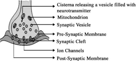
LTP induction, control the maturation and stabilization of synapses over longer time scales [65]. There is volu- minous information given by the Author and a peer group of more than hundred eminent scientists are work- ing on synapse functions. Figure 1 gives the general view of synapse and its components and the release of neu- rotransmitters from the vesicle diffuses through the cell membrane which is attached to the receptors in signal propagation.
8. Transmission of Signals
Sudden changes in the ionic concentrations induce ion channels to intake sodium ions. This brings the change in the ionic gradients around the cell membrane and the im- pulsion transferred downwards from the dendrites to sy- napses and these impulses are referred as signals. The previous studies [66] shows that the presynaptic nerve terminals and the postsynaptic target cells signals to each other as they establish precisely aligned synaptic specia- lizations. It’s a question how this signal transmission occurs within a network of neurons and how does it reaches the target neuron. It can be answered on pre- dicting the behavior of these signals when they propagate through complex neuron network. Earlier studies reveal that there are some specific signaling pathways through which Signals has been transferred and it is been dis- tinguished after the introduction of the signaling through the second messengers and the intracellular pathways. It has taken more than three decades to find the measure- ments of changes in cytosolic cAMP and free Ca2+ con- centrations [67].
[image:4.595.309.536.522.624.2]Cellular signaling is a huge complex network and the signaling pathways carry and process information be- tween the networks and exhibit bodily actions. There can be many inputs of information such as summation and
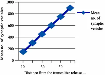
temporal integration during the propagation of signals. It is apparent that the release of neurotransmitters in to the cleft propagates the signal transmission. The reuptake of neurotransmitter, in this case its glutamate in MFBs is carried out with the help of glutamate transporters [68, 69]). The signal transmission follows specific pathways to reach the target. Recently the development of compu- tational simulations can resolve the complexity in under- standing the signal propagation as they consider all mo- tifs as nodes and there have been emerged many simu- lation tools to analyze signal propagation. Generally Mo- ssy Fiber synapses considered as strong synapses. It is observed in generating high postsynaptic currents and potentials in CA3 pyramidal neurons and interneurons [70,71]. Synaptic transmission is observed to depend mainly on the active zones, where the release of trans- mitters takes place and it is observed mainly within the puncta adherentia, putative adhesion complexes. It is also understood that the distance between the active zones provoke the fast transmission of signals and the efficacy of the synapses is directly proportional to the synaptic vesicles. In adult it is approximately 900 vesicles located within the 60 nm from the active zone which was ob- served from the quantitative analyzes by the author [1]. It is clearly represented in Figure 2, it shows the mean num- ber of synaptic vesicles within the active zone. For more information related to efficacy of synapse (see review of [72]).
[image:5.595.71.273.555.699.2]Amongst the various reasons, the major cause for sig- nal transmission is the release of neurotransmitters. It mainly depends on the flow of ca2+ ions in to the pre- synaptic region through ca2+ ion channels. The distance between the ca2+ channels and the sensor triggers the breakdown of vesicles, ultimately transfer the signals. Other important factors like buffering kinetics of ca2+ and the number of docked and primed vesicles are also plays an important role in the release of neurotransmitters [73].
Figure 2. The correlation of synaptic vesicles within the ac- tive zone (Transmitter release site) in adults. Resketched from the data obtained by the author [1].
There are cases where the retrograde signaling takes place in synapses [74].
9. Synapse Proteins
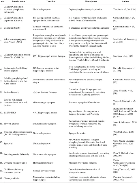
The multiple functions of the synapse depend on mo- lecular interaction that guided through synaptic proteins. The analysis of multivariate proteins involved in synaptic connections is an ongoing endeavor in proteomics. The protein database facilitates the collection of information about synaptic proteins. It is comprised of protein struc- ture, function, interaction, pathways and expression (SynDB, http://syndb.cbi.pku.edu.cn) [75]. There exist some unco- vered proteins which were detected after the year 2006 till today. Probably that might have attained much impor- tance in recent research in neuroscience. The protein mo- dulations and the derived functions of different synapses are listed in the table (Table 1). Almost all protein inte- ractions results in specific function like the interaction of dentate gyrus and CA3 morphologically different regions influences the information processing in hippocampas region which involve series of protein interactions [76- 78].
On the other hand degradation of proteins acts as a modulator of synaptic physiology [79-81]. The degra- dation process streams down through selective pathway named Ubiquitin Proteasome System (UPS). The UPS controls the half life and the activity state of protein by linking the proteins with ubiquitin through enzytmatic activities [82]. Other than degradation of proteins, the UPS inhibition found to raise the release of neurotrans- mitters in mammals. In fact it is proved that it takes part in the regulation of synaptic transmission [83].
10. Improved Outlook on Synapse
Table 1. Proteins found in different synapses and their functions.
S. no Protein name Location Description Author
1.
Calcium/Calmodulin activated phosphatase calcineurin
Neuronal synapses Dephosphorylate endocytic proteins Tao Sun et al., 2010 [84]
2. Calcium/Calmodulin dependant Kinase II
It’s a component of electrical synapse in the mauthner cell
It is requires for the induction of changes in both forms of transmission
Carmen E Flores et al., [85]
3. Connexin (Cx35) Synaptic terminals on the teloest
mauthner cell It undergoes activity dependant potentiation
Carmen E Flores et al., [85]
4. Adenomatous polyposis Coli Protein (APC)
It organizes a complex multiprotein that directs nicotinic acetylcholine receptor (nAChR) localization at postsynaptic sites in avian ciliary ganglion neurons in vivo
It coordinates presynaptic and postsynaptic maturation and promotes synaptic efficacy. It also promotes retrograde signals via postsynaptic neuroligin that interacts with presynaptic neurexin extracellularly
Madelaine M. Rosenberg
et al., [86]
5. Calcium/Calmodulin protein
kinase IIα (CaMk IIα) CA1 hippocampal neuron Synapse
Central role in regulating neuronal excitability and now it is observed to directly phosphorylate the inhibitory GABAA receptor (GABAAR) α1, β2 and γ2 subunits
Marsdena et al., [87]
6. Postsynaptic Scaffolding protein gephrin
GABAergic synapses in the hippocampal neurons
It’s a synaptogenic molecule regulating GABAergic synaptic plasticity and contributes the therapeutic action of lithium
Shiva K. Tyagarajan et al., [88]
7.
Soluble guanylyl cyclase/ Protein kinase G and rho A/rho kinase
Motoneurons at adult and neonatal stages.
Neurodegenerative process/Synapse elimination
Carmen R. Sunico et al., [89]
8. Protein kinase C Aplysia Sensory Neurons
Formation of specific synapses and maturation of the synapse by activating the additional signaling pathways
Jiang-Yuan Hu et al., [90]
9.
Leucine rich repeat transmembrane neuronal proteins
Glutamatergic synapses Promotes synaptic differentiation Tabrez J. Siddiqui et al., [91]
10. BDNF/TrKB CA1 hippocampal neuron Key mediators of axon guidance, Synapse formation and Plasticity
Huang and Reichardt 2003; Luikart and Parada 2006; Lu et al., 2008 [92-94]
11. Phocein proteins Neuromuscular synapses
Regulation of axonal transport, neurite elongation, synapse formation, and microtubule organization
Joost Schulte et al., 2010. [95]
12. Synaptic adhesion like olecule
(SALM family proteins) Neuronal synapses Synapse formation
Won Mah et al., 2010. [96].
13.
Synapsin Neuronal synapses
MAPK/ERk dependant synapsin phosphorylation initiates the functional synaptic connections and their short term plasticity
Carlo Natale Giuseppe Giachello et al., 2010 [97]
14.
Docking protein 7 (Dok-7) Neuromuscular synapses Involves in synapse formation by recruiting adaptor proteins named Crk and Crk-L
Peter T. Hallock et al., 2010 [98]
15. Cysteine string protein α Hippocampal synapse Maintain presynaptic function
Pablo
García-Junco-Clemente
et al., 2010 [99]
16. Cc2d1a-evolutionary
conserved protein Central nervous system
Controls functional maturation of synapses in mouse
Meng Zhao et al., 2011. [100]
17. Cholecystokinin Mammalian brains including hippocampus
Facilitates presynaptic glutamate release (glutamatergic transmission)
target cell specific and also it emphasis that it rely on phenotypic properties of the interneuron especially in receiving the input signals from the neighboring cell [106, 107]. As controversy proceeds on synapse transmission, it is evident that astrocytes regulate synaptic transmission by interacting with neurons. In recent times, an extensive research tried to experiment the role of gap junction pro- teins connexin 30 and connexin 40 in synaptic physio- logy. They extend the work by blocking the activities of connexin 30 and connexin 40 genes, thus evolved that there is a delay in synaptic transmission in CA1 pyrami- dal neurons and affirmed that connexins than any other proteins play equally important role in synaptic plasticity. It also stressed that the role of astrocytes in chemical sy- napses which is truly based on the modulation of astrog- lial clearance rate and ultimately controls the neuron ex- citation, neurotransmitter release, postsynaptic receptors, potassium ion removal from the extracellular matrix and the silencing synapses [108]. As with other important fac- tors that simulate the synapse potentiation, the ERK1/2 signaling plays a vital role in long term potentiation in hippocampal neurons [109].
Some factors do appear to elicit synaptic transmission. The high degree of receptor activation was found to be mediated by endocannabinoids especially in retrograde signaling system in the brain [110]. There also observed hyperactive interneurons associated with the increase of potassium ions from 2.5 to 10 mM in the extracellular matrix [111]. Finally, the laboratory experiment demon- strates that the increase of potassium ions in the extracel- lular matrix activates GABA receptors in the postsynapse of CA3 pyramidal neurons [112].
11. Common View on Synapse Today
Other than classical synapses, some group of neurons communicates in an unusual way. It is observed that neurogliaform cells inhibit the transmission of signals to the neighboring cortical neuron with GABA neurotrans- mitters. In this case, it is a revelation as there are no sy- napses involved in this transmission and it is purely by chemical transmission [113]. Upcoming research targets neuronal polarity because of its role in brain disorders. As a result, involvement of gene in the synaptic defects has been identified. Therefore, in existing neurons, these genetic evidence studies reveal that the level of phospho- ionositides (PIP2) control the neuronal polarity which in- turn controlled by the enzyme myo-inositol monopho- sphatase (IMPase) and thus believed to promote pola- rized synaptic components [114]. In glutamatergic synap- ses, excitatory amino acid transporters (EAATs) regulate AMPA receptor (AMPAR) accumulation in the postsy- napse and the synaptic efficacy depend on the AMPAR accumulation. If EAAT fail to function, glutamate disse- minates in to the synaptic cleft and get bind to the
NMDA receptor (NMDAR) [115]. The mechanism by the EAAT affects the AMPAR localization in synapse is questionable. For this reason the resources are becoming fewer and the difficulty and the complexities in under- standing the activation of AMPARs produce a delay in the report of data in the particular field.
12. Discussion
The Synapse and its function are intricately entangled with the neuron function and signal propagation. Resear- chers are yet to work for many years by being far apart in silos achieved less to form a hypothesis about the func- tions of brain and the model of neuron connections to re- alize the signal transmission between them. Now with technology advancements, it is easy to learn several phe- nomena within the broad restraint of neuroscience. This review correlates several well defined proteins and func- tions of synapse. It is not projecting the fact that synapse and related fields have been studied in an extraordinary way to recreate memory and learning. In fact, the poten- tial of this work lies in identifying certain hitherto, explor- ed evidences and project it for further evaluations and cross correlations for better perception of this field.
The science of neurons is still a new and developing area which calls for a special educational domain as Neu- ron Science rather than the conventional psychology em- bedded Neuroscience. It must deal with very fast fields like Electronics, Electrochemical, Chemical and Protein Dynamics for information encoding and decoding. The initiative behind this work focusing on the synapse for- mation and the signal transmission is to create certain perspectives. It gives a correlative presentation of pro- perties to the certain extent based on the latest achieve- ments in the field. Hence it is not covering certain earlier studies, but it positioned itself to cover recent expositions of synapse. It mainly highlights synapse formation with the actin binding protein as it increases the synaptic den- sity, the long term potentiation and the long term depres- sion which maintains the synaptic stability, the specia- lized pathways in neuronal network, and the receptor bin- ding sites. The basic mechanism is mediating between the presynaptic and the postsynaptic region and gives well perceptive on synaptic functions. On the whole, the author [116] rationalizes his work by presenting a review on neuron doctrine which is an eminent effort to trespass the past history on neuroscience and perhaps helped this writing more meaningful.
13. Conclusion
ning of early studies. The synapse formation involves many protein interactions and the modification of actin cytoskeleton elements. The synapse density is found to rely much on the actin binding protein which can provide further knowledge on the formation of synapse. The lack of standardization in assay procedures particularly on synaptic properties is highlighted here as a priority con- cern. Testing the hypothesis with experimental techni- ques is fundamentally complicated. For that reason, this review paper confers on previous eminent works on neu- ron synapse. It is possible to reach much deeper under- standing of neuronal capabilities and clustering with sig- nificant improvements by applying new technologies and incorporating challenging computer simulations.
14. Acknowledgements
This work was supported by the research and industry in- cubation centre, Dayananda Sagar Institutions. We thank Dr. Suma V. for her support in research methodology. We also thank Mrs Reena Phillips for her assistance in creat- ing the synapse diagram.
REFERENCES
[1] A. Rollenhagen, K. Satzler, E. P. Rodriguez, P. Jonas, M. Frotscher and J. H. R. Lübke, “Structural Determinants of Transmission at Large Hippocampal Mossy Fiber Synap- ses,” The Journal of Neuroscience, Vol. 27, No. 39, 2007, pp. 10434-10444.
doi:10.1523/JNEUROSCI.1946-07.2007
[2] R. W. Guillery, “Observations of Synaptic Structures: Ori- gins of the Neuron Doctrine and Its Current Status,” Phi-
losophical Transactions on Royal Society Lond B Biolo- gical Science, Vol. 360, No. 1458, 2005, pp. 1281-307. [3] W. X. Zhang, Y. Zhang, H. Zheng, C. Zhang, W. Xiong, J.
G. Olyarchuk, M. Walker, W. F. Xu, M. Zhao, S. Q. Zhao, Z. Zhou and L. P. Wei, “A Synapse Protein DataBase Bas- ed on Synapse Ontology 2006,” Nucleic Acids Research, Vol. 35, No. S1, 2006, pp. D737-D741.
doi:10.1093/nar/gkl876
[4] S. J. Martin, P. D. Grimwood and R. G. M. Morris, “Sy- naptic Plasticity and Memory: An Evaluation of the Hy- pothesis,” Annual Review of Neuroscience, Vol. 23, 2000, pp. 649-711. doi:10.1146/annurev.neuro.23.1.649 [5] J. Breustedt, A. Gundlfinger, F. Varoqueaux, K. Reim, N.
Brose and D. Schmitz, “Munc13-2 Differentially Affects Hip- pocampal Synaptic Transmission and Plasticity,” Cere-
bral Cortex, Vol. 20, 2011, pp. 1109-1120.
[6] S. I. Cohen-Matsliah, H. Motanis, K. Rosenblum and E. Bar- kai, “A Novel Role for Protein Synthesis in Long-Term Neuronal Plasticity: Maintaining Reduced Postburst After- hyperpolarization,” The Journal of Neuroscience, Vol. 30, No. 12, 2010, pp. 4338-4342.
doi:10.1523/JNEUROSCI.5005-09.2010
[7] A. B. Jerald, T. R. G. Nair and E. Rajasekaran, “Evalua- tion of the Structural Disorder of the Protein FMR1 with
Carbon Composition,” 2nd Annual Intrnational Conference
on Advances in Biotechnology, 2012.
[8] D. C. Crawford, C. Y. Chang, K. L. Hyrc and S. Mennerick, “Calcium-Independant Inhibitory G-Protein Signaling In- duces Persistent Presynaptic Muting of Hippocampal Sy- napses,” Journal of Neuroscience, Vol. 31, No. 3, 2011, pp. 979-991. doi:10.1523/JNEUROSCI.4960-10.2011 [9] K. L. Moulder, J. P. Meeks, A. A. Shute, C. K. Hamilton,
G. de Erausquin and S. Mennerick, “Plastic Elimination of Functional Glutamate Release Sites by Depolarization,”
Neuron, Vol. 42, No. 3, 2004, pp. 423-435. doi:10.1016/S0896-6273(04)00184-9
[10] K. L. Moulder, X. Jiang, C. Chang, A. A. Taylor, A. M. Benz, A. C. Conti, L. J. Muglia and S. Mennerick, “A Spe- cific Role for Ca2-Dependant Adenylyl Cyclases in Re- covery from Adaptive Presynaptic Silencing,” Journal of
Neuroscience, Vol. 28, No. 20, 2008, pp. 5159-5168. doi:10.1523/JNEUROSCI.5317-07.2008
[11] R. C. Malenka and R. A. Nicoll, “Silent Synapses Speak Up,” Neuron, Vol. 19, No. 3, 1997, pp. 473-476. doi:10.1016/S0896-6273(00)80362-1
[12] L. L. Voronin and E. Cherubini, “Deaf, Mute and Whispe- ring’ Silent Synapses: Their Role in Synaptic Plasticity,”
Journal of Physiology, Vol. 557, 2003, pp. 3-12. doi:10.1113/jphysiol.2003.058966
[13] D. Atasoy and E. T. Kavalali, “Presynaptic Unsilencing: Searching for a Mechanism,” Neuron, Vol. 50, No. 3, 2006, pp. 345-346. doi:10.1016/j.neuron.2006.04.018
[14] R. R. Linas, “The Contribution of Santiago Raman Y Ca- jal to the functional Neuroscience,” Nature, Vol. 4, 2003. [15] M. Sheng and C. Sala, “PDZ Domains and the Organi-
zation of Supramolecular Complexes,” Annuals of Review
Neuroscience, Vol. 24, 2001, pp. 1-29. doi:10.1146/annurev.neuro.24.1.1
[16] H. J. Kreienkamp, “Organisation of G-Protein-Coupled Re- ceptor Signalling Complexes by Scaffolding Proteins,”
Current Opinion in Pharmacology, Vol. 2, No. 5, 2002, pp. 581-586. doi:10.1016/S1471-4892(02)00203-5 [17] T. M. Boeckers, “The Postsynaptic Density,” Cell Tissue
Research, Vol. 326, No. 2, 2006, pp. 409-422. doi:10.1007/s00441-006-0274-5
[18] A. Haeckel, R. Ahuja, E. D. Gundelfinger, B. Qualmann and M. M. Kessels, “The Actin-Binding Protein Abp1 Con- trols Dendritic Spine Morphology and Is Important for Spine Head and Synapse Formation,” The Journal of Neu-
roscience, Vol. 28, No. 40, 2008, pp. 10031-10044. [19] C. Dillon and Y. Goda, “The Actin Cytoskeleton: Integra-
ting form and Function at Synapse,” Annuals of Review
Neuroscience, Vol. 28, 2005, pp. 25-55. doi:10.1146/annurev.neuro.28.061604.135757
[20] V. Schubert, J. S. Da Silva and C. G. Dotti, “Localized Re- cruitment and Activation of RhoA Underlies Dendritic Spine Morphology in a Glutamate Receptor-Dependant Manner,” Journal of Cell Biology, Vol. 172, No. 3, 2006, pp. 453-467. doi:10.1083/jcb.200506136
doi:10.1016/j.conb.2005.12.001
[22] G. Roussignol, F. Ango, S. Romorini, J. C. Tu, C. Sala, P. F. Worley, J. Bockaert and L. Fagni, “Shank Expression Is Sufficient to Induce Functional Dendritic Spine Synapses in Aspiny Neurons,” Journal of Neuroscience, Vol. 25, No. 14, 2005, pp. 3560-3570.
doi:10.1523/JNEUROSCI.4354-04.2005
[23] K. Shen and C. W. Cowan, “Guidance Molecules in Synapse Formation and Plasticity,” Cold Spring Harbor Laboratory Press, 2010. doi:10.1101/cshperspect.a001842 [24] H. J. Kim and S. A. Thayer, “Lithium Increases Synapse
Formation between Hippocampal Neurons by Depleting Phosphoinositides,” Molecular Pharmacology, Vol. 75, No. 5, 2009, pp. 1021-1030. doi:10.1124/mol.108.052357 [25] P. G. Haydon, “GLIA: Listening and Talking to the Syna-
pse,” National Review of Neuroscience, Vol. 2, 2001, pp. 185-193. doi:10.1038/35058528
[26] R. D. Fields and B. Stevens-Graham, “New Insights into Neuron-Glia Communication,” Science, Vol. 298, No. 5593, 2002, pp. 556-562.
doi:10.1126/science.298.5593.556
[27] D. S. Auld and R. Robitaille, “Perisynaptic Schwann Cells at the Neuromuscular Junction: Nerve- and Activity-De- pendant Contributions to Synaptic Efficacy, Plasticity, and Reinnervation,” The Neuroscientist, Vol. 9, No. 2, 2003, pp. 144-157. doi:10.1177/1073858403252229
[28] G. I. Hatton and V. Parpura, “Glial Neuronal Signaling. Kluwer Academic, Boston, 2004.
doi:10.1007/978-1-4020-7937-5
[29] N. J. Allen and B. A. Barres, “Signaling between Glia and Neurons: Focus on Synaptic Plasticity,” Current Opinion
in Neurobiology, Vol. 15, No. 5, 2005, pp. 542-548. doi:10.1016/j.conb.2005.08.006
[30] C.-P. Ko, Y. S. Sugiura and Z. Feng, “The Biology of Pe- risynaptic (Terminal) Schwann Cells,” In: P. J. Armati, Ed.,
The Biology of Schwann Cells: Development, Differentia-
tion and Immunomodulation, 2007, pp. 72-99. doi:10.1017/CBO9780511541605.006
[31] Z. F. Kisvárday, et al., “Synaptic Targets of HRP-Filled Layer III Pyramidal Cells in the Cat Striate Cortex,” Ex-
perimental Brain Research, Vol. 64, No. 3, 1986, pp. 541-552. doi:10.1007/BF00340492
[32] A. Stepanyants and D. B. Chklovskii, “Neurogeometry and Potential Synaptic Connectivity,” Trends Neuroscience, Vol. 28, No. 7, 2005, pp. 387-394.
doi:10.1016/j.tins.2005.05.006
[33] A. Stepanyants, P. R. Hof and D. B. Chklovskii, “Geome- try and Structural Plasticity of Synaptic Connectivity,” Neu-
ron, Vol. 34, No. 2, 2002, pp. 275-288. doi:10.1016/S0896-6273(02)00652-9
[34] Tarec Fares and Armen Stepanyants, “Cooperative Syna- pse Formation in the Neocortex,” PNAS, Vol. 106, No. 38, 2002, pp. 16463-16468.
www.pnas.org_cgi_doi_10.1073_pnas.0813265106. [35] N. Kalisman, G. Silberberg and H. Markram, “The Neo-
cortical Microcircuit as a Tabularasa,” Proceedings of
National Academy of Science of the USA, Vol. 102, No. 3, 2005, pp. 880-885. doi:10.1073/pnas.0407088102
[36] D. Feldmeyer, V. Egger, J. Lubke and B. Sakmann, “Re- liable Synaptic Connections between Pairs of Excitatory Layer 4 Neurones within a Single ‘Barrel’ of Developing Rat Somatosensory Cortex,” The Journal of Physiology, Vol. 521, No. 1, 1999, pp. 169-190.
doi:10.1111/j.1469-7793.1999.00169.x
[37] D. Feldmeyer, J. Lubke, R. A. Silver and B. Sakmann, “Synaptic Connections between Layer 4 Spiny Neu-rone-Layer 2/3 Pyramidal Cell Pairs in Juvenile Rat Bar-rel Cortex: Physiology and Anatomy of Interlaminar Sig-nalling within a Cortical Column,” The Journal of Physi-ology, Vol. 538, No. 3, 2002, pp. 803-822.
doi:10.1113/jphysiol.2001.012959
[38] H. Markram, J. Lubke, M. Frotscher, A. Roth and B. Sakmann, “Physiology and Anatomy of Synaptic Con- nections between Thick Tufted Pyramidal Neurones in the Developing Rat Neocortex,” The Journal of Physiol-
ogy, Vol. 500, No. 2, 1997, pp. 409-440.
[39] U. V. Nagerl, G. Kostinger, J. C. Anderson, K. A. Martin and T. Bonhoeffer, “Protracted Synaptogenesis after Ac- tivity-Dependant Spinogenesis in Hippocampal Neurons,”
The Journal of Neuroscience, Vol. 27, No. 30, 2007, pp. 8149-8156. doi:10.1523/JNEUROSCI.0511-07.2007 [40] K. Matsumoto-Miyai, et al., “Coincident Pre- and Post-
synaptic Activation Induces Dendritic Filopodia via Neu- rotrypsin-Dependant Agrin Cleavage,” Cell, Vol. 136, No. 6, 2009, pp. 1161-1171. doi:10.1016/j.cell.2009.02.034 [41] T. Branco and K. Staras, “The Probability of Neuro-
transmitter Release: Variability and Feedback Control at Single Synapses,” Nature Reviews of Neuroscience, Vol. 10, No. 5, 2009, pp. 373-383.
http://www.ncbi.nlm.nih.gov/pubmed/19377502/
[42] I. Riebe and E. Hanse, “Development of Synaptic Con- nectivity onto Interneurons in Stratum Radiatum in the CA1 Region of the Rat Hippocampus,” BMC Neurosci-
ence, Vol. 13, No. 1, 2012, p. 14. doi:10.1186/1471-2202-13-14
[43] E. De Robertis, “Submicroscopic Morphology of the Sy- napse,” International Review of Cytology, Vol. 8, 1959, pp. 61-96. doi:10.1016/S0074-7696(08)62728-X
[44] R. W. Guillery, “Early Electron Microscopic Observa- tions of Synaptic Structures in the Cerebral Cortex: A View of the Contributions Made by George Gray (1924- 1999),” Trends in Neuroscience, Vol. 23, No. 12, 2000, pp. 594-598. doi:10.1016/S0166-2236(00)01635-0 [45] J. Gerlach, “The Spinal Cord,” In: S. Stricker, Ed., Man-
ual of Histology, Williams and Wood & Co., New York, 1872, pp. 327-366.
[46] E. Deiters and R. W. Guillery, “Otto Deiters: 1834-1863,”
Experimental Neurology, Vol. 9, No. 1, 1963, pp. iii-vi. [47] E. G. Gary and R. W. Guillery, “Synaptic Morphology in
the Normal and Degenerating Nervous System,” Interna-
tional Review of Cytology, Vol. 19, 1996, pp. 111-182. doi:10.1016/S0074-7696(08)60566-5
1908, pp. 1-31.
[49] F. Nissl, D. Neuronenlehre and I. Anhänger, “Ein Beitrag zur Lo¨sung des Problems der Beziehungen zwischen Nervenzelle,” Faser und Grau, Gustav Fisher, Jena, 1903.
[50] L. Shapiro and D. R. Coleman, “The Diversity of Cad- herins and the Implications for a Synaptic Adhesive Code in the CNS,” Neuron, Vol. 23, No. 3, 1999, pp. 427-430. doi:10.1016/S0896-6273(00)80796-5
[51] S.-K. Kwon, J. Woo, S.-Y. Kim, H. Kim and E. Kim, “Trans-Synaptic Adhesions between Netrin-G Ligand-3 (NGL-3) and Receptor Tyrosine Phosphatases LAR, Pro- tein-Tyrosine Phosphatase_ (PTP_), and PTP_ via Spe- cific Domains Regulate Excitatory Synapse Formation,”
The Journal of Biological chemistry, Vol. 285, No. 18, 2010, pp. 13966-13978.
[52] M. V. Bennett and R. S. Zukin, “Electrical Coupling and Neuronal Synchronization in the Mammalian Brain,”
Neuron, Vol. 41, No. 4, 2004, pp. 495-511. doi:10.1016/S0896-6273(04)00043-1
[53] B. W. Connors and M. A. Long, “Electrical Synapses in the Mammalian Brain,” Annual Review of Neuroscience, Vol. 27, No. 1, 2004, pp. 393-418.
doi:10.1146/annurev.neuro.26.041002.131128
[54] S. Hestrin and M. Galarreta, “Electrical Synapses Define Networks of Neocortical GABAergic Neurons,” Trends
in Neuroscience, Vol. 28, No. 6, 2005, pp. 304-309. doi:10.1016/j.tins.2005.04.001
[55] R. Bruzzone and R. Dermietzel, “Structure and Function of Gap Junctions in the Developing Brain,” Cell and Tis-
sue Research, Vol. 326, No. 2, 2006, pp. 239-248. doi:10.1007/s00441-006-0287-0
[56] B. Sutor and T. Hagerty, “Involvement of Gap Junctions in the Development of the Neocortex,” Biochimia et Bio-
phys Acta, Vol. 1719, No. 1-2, 2005, pp. 59-68. doi:10.1016/j.bbamem.2005.09.005
[57] P. Washbourne, J. E. Bennet and A. K. McAllister, “Ra- pid Recruitment of NMDA Receptor Transport Packets to Nascent Synapses,” Nature Neuroscience, Vol. 5, No. 8, 2002, pp. 751-759.
[58] R. C. Malenka and R. A. Nicoll, “NMDA-Receptor-De- pendant Synaptic Plasticity: Multiple Forms and Mecha- nisms,” Trends in Neuroscience, Vol. 16, No. 12, 1993, pp. 521-527. doi:10.1016/0166-2236(93)90197-T
[59] K. Fox, B. L. Schlaggar, S. Glazewski and D. D. O’Leary, “Glutamate Receptor Blockade at Cortical Synapses Dis- rupts Development of Thalamocortical and Columnar Organization in Somatosensory Cortex,” Proceedings of
National Academy of Science of USA, Vol. 93, No. 11, 1996, pp. 5584-5589. doi:10.1073/pnas.93.11.5584 [60] L. Huang and S. L. Pallas, “NMDA Antagonists in the
Superior Colliculus Prevent Developmental Plasticity but Not Visual Transmission or Map Compression,” Journal
of Neurophysiology, Vol. 86, No. 3, 2001, pp. 1179-1194. [61] D. Liao, N. A. Hessler and R. Malinow, “Activation of
Postsynaptically Silent Synapses during Pairing-Induced LTP in CA1 Region of Hippocampal Slice,” Nature, Vol. 375, No. 6530, 1995, pp. 400-404. doi:10.1038/375400a0 [62] R. Malinow, Z. F. Mainen and Y. Hayashi, “LTP Mecha-
nisms: From Silence to Four-Lane Traffic,” Current Opi-
nion in Neurobiology, Vol. 10, No. 3, 2000, pp. 352-357. doi:10.1016/S0959-4388(00)00099-4
[63] M. Y. Xiao, H. Wigstrom and B. Gustafsson, “Long- Term Depression in the Hippocampal CA1 Region Is As- sociated with Equal Changes in AMPA and NMDA Re- ceptor-Mediated Synaptic Potentials,” European Journal of Neuroscience, Vol. 6, No. 6, 1994, pp. 1055-1057. doi:10.1111/j.1460-9568.1994.tb00600.x
[64] R. C. Carroll, D. V. Lissin, M. V. Zastrow, R. A. Nicoll and R. C. Malenka, “Rapid Redistribution of Glutamate Receptors Contributes to Long-Term Depression in Hip- pocampal Cultures,” Nature Neuroscience, Vol. 2, No. 5, 1999, pp. 454-460. doi:10.1038/8123
[65] V. A. Alvarez, A. D. Ridenour and L. B. Sabatini, “Dis- tinct Structural and Ionotropic Roles of NMDA Receptors in Controlling Spine and Synapse Stability,” The Journal of Neuroscience, Vol. 27, No. 28, 2007, pp. 7365-7376. doi:10.1523/JNEUROSCI.0956-07.2007
[66] J. R. Sanes and J. W. Lichtman, “Induction, Assembly, Maturation and Maintenance of a Postsynaptic Appara- tus,” Nature Reviews Neuroscience, Vol. 2, No. 11, 2001, pp. 791-805. doi:10.1038/35097557
[67] I. I. Moraru and L. M. Loew, “Intracellular Signaling: Spatial and Temporal Control,” Pysiology, Vol. 20, No. 3, 2005, pp. 169-179, doi:10.1152/physiol.00052.2004 [68] N. C. Danbolt, “Glutamate Uptake,” Progress in Neuro-
biology, Vol. 65, No. 1, 2001, pp. 1-105. doi:10.1016/S0301-0082(00)00067-8
[69] S. H. Oliet, R. Piet, D. A. Poulain and D. T. Theodosis, “Glial Modulation of Synaptic Transmission: Insights from the Supraoptic Nucleus of the Hypothalamus,” Glia, Vol. 47, No. 3, 2004, pp. 258-267.
doi:10.1002/glia.20032
[70] D. A. Henze, L. Wittner and G. Buzsáki, “Single Granule Cells Reliably Discharge Targets in the Hippocampal CA3 Network in Vivo,” Nature Neuroscience, Vol. 5, No. 8, 2002, pp. 790-795.
[71] J. J. Lawrence, Z. M. Grinspan and C. J. McBain, “Quan- tal Transmission at Mossy Fibre Targets in the CA3 Re- gion of the Rat Hippocampus,” The Journal of Physiology, Vol. 554, No. 1, 2003, pp. 175-193.
doi:10.1113/jphysiol.2003.049551
[72] J. Bischofberger, D. Engel, M. Frotscher and P. Jonas, “Timing and Efficacy of Transmitter Release at Mossy Fiber Synapses in the Hippocampal Network,” Pflügers Archiv, Vol. 453, No. 2, 2006, pp. 361-372.
doi:10.1007/s00424-006-0093-2
[73] H. L. Atwood and S. Karunanithi, “Diversification of Synaptic Strength: Presynaptic Elements,” Nature Re- views Neuroscience, Vol. 3, No. 7, 2002, pp. 497-516. [74] H. W. Tao and M.-M. Poo, “Retrograde Signaling at Cen-
tral Synapses,” Proceedings of National Academy of Sci- ence of USA, Vol. 98, No. 20, 2001, pp. 11009-11015. doi:10.1073/pnas.191351698
[75] W.-X. Zhang, et al., “SynDB: A Synapse Protein Data- Base Based on Synapse Ontology,” Nucleic Acids Re-
doi:10.1093/nar/gkl876
[76] K. Nakazawa, T. J. McHugh, M. A. Wilson and S. To- negawa, “NMDA Receptors, Place Cells and Hippocam- pal Spatial Memory,” Nature Reviews Neuroscience, Vol. 5, No. 5, 2004, pp. 361-372.
[77] J. K. Leutgeb, S. Leutgeb, M. B. Moser and E. I. Moser, “Pattern Separation in the Dentate Gyrus and CA3 of the Hippocampus,” Science, Vol. 315, No. 5814, 2007, pp. 961-966. doi:10.1126/science.1135801
[78] T. Nakashiba, J. Z. Young, T. J. McHugh, D. L. Buhl and S. Tonegawa, “Transgenic Inhibition of Synaptic Trans- mission Reveals Role of CA3 Output in Hippocampal Learning,” Science, Vol. 319, No. 5867, 2008, pp. 1260- 1264. doi:10.1126/science.1151120
[79] B. Bingol and E. M. Schuman, “A Proteasome-Sensitive Connection between PSD-95 and GluR1 Endocytosis,”
Neuropharmacology, Vol. 47, No. 5, 2004, pp. 755-763. doi:10.1016/j.neuropharm.2004.07.028
[80] J. J. Yi and M. D. Ehlers, “Emerging Roles for Ubiquitin and Protein Degradation in Neuronal Function,” Phar- macological Reviews, Vol. 59, No. 1, 2007, pp. 14-39. doi:10.1124/pr.59.1.4
[81] K. F. Haas and K. Broadie, “Roles of Ubiquitination at the Synapse,” Biochimica et Biophysica Acta, Vol. 1779, No. 8, 2008, pp. 495-506.
doi:10.1016/j.bbagrm.2007.12.010
[82] P. Kaiser and E. A. Fon, “Expanding Horizons at Big Sky. Symposium on Ubiquitin and Signaling,” EMBO Reports, Vol. 8, No. 9, 2007, pp. 817-822.
doi:10.1038/sj.embor.7401017
[83] G. V. Rinettil and F. E. Schweizer, “Ubiquitination Acu- tely Regulates Presynaptic Neurotransmitter Release in Mammalian Neurons,” The Journal of Neuroscience, Vol. 30, No. 9, 2010, pp. 3157-3166.
[84] T. Sun, X.-S. Wu, J.-H. Xu, B. D. McNeil, Z. P. Pang, W.-J. Yang, L. Bai, S. Qadri, J. D. Molkentin, D. T. Yue and L.-G. Wu, “The Role of Calcium/Calmodulin-Acti- vated Calcineurin in Rapid and Slow Endocytosis at Cen- tral Synapses,” The Journal of Neuroscience, Vol. 30, No. 35, 2010, pp. 11838-11847.
doi:10.1523/JNEUROSCI.1481-10.2010
[85] C. E. Flores, R. Cachope, S. Nannapaneni, S. Ene, A. C. Nairn and A. E. Peredal, “Variability of Distribution of Ca2+/Calmodulin-Dependant Kinase II at Mixed Syn- apses on the Mauthner Cell: Colocalization and Associa- tion with Connexin 35,” The Journal of Neuroscience, Vol. 30, No. 28, 2010, pp. 9488-9499.
[86] M. M. Rosenberg, F. Yang, J. L. Mohn, E. K. Storer and M. H. Jacob, “The Postsynaptic Adenomatous Polyposis Coli (APC) Multiprotein Complex Is Required for Local- izing Neuroligin and Neurexin to Neuronal Nicotinic Synapses in Vivo,” The Journal of Neuroscience, Vol. 30, No. 33, 2010, pp. 11073-11085.
[87] K. C. Marsdena, A. Shemesha, K. U. Bayerb and R. C. Carrolla, “Selective Translocation of Ca2+/Calmodulin Protein Kinase IIα (CaMKIIα) to Inhibitory Synapses,”
Proceedings of National Academy of Science of USA, Vol. 107, No. 47, 2010, pp. 20559-20564.
[88] S. K. Tyagarajana, H. Ghosha, G. E. Yévenesa, I. Nik-onenkob, C. Ebelinga, C. Schwerdela, C. Sidlera, H. U. Zeilhofera, B. Gerritsc, D. Mullerb and J.-M. Fritschya, “Regulation of GABAergic Synapse Formation and Plas- ticity by GSK3β-Dependant Phosphorylation of Gephy- rin,” Vol. 108, No. 1, 2011, pp. 379-384.
[89] C. R. Sunico, D. Gonzalez-Forero, G. Domínguez, J. M. García-Verdugo and B. Moreno-Lopezl, “Nitric Oxide Induces Pathological Synapse Loss by a Protein Kinase G-, Rho Kinase-Dependant Mechanism Preceded by My- osin Light Chain Phosphorylation,” The Journal of Neu- roscience, Vol. 30, No. 3, 2010, pp. 973-984.
[90] J.-Y. Hu, Y. Chen and S. Schacher, “Multifunctional Role of Protein Kinase C in Regulating the Formation and Maturation of Specific Synapses,” The Journal of Neuro-
science, Vol. 27, No. 43, 2007, pp. 11712-11724
[91] T. J. Siddiqui, R. Pancaroglu, Y. Kang, A. Rooyakkers and A. M. Craig, “LRRTMs and Neuroligins Bind Neur- exins with a Differential Code to Cooperate in Glutamate Synapse Development,” The Journal of Neuroscience, Vol. 30, No. 22, 2010, pp. 7495-7506
[92] E. J. Huang and L. F. Reichardt, “Trk Receptors: Roles in Neuronal Signal Transduction,” Annual Reviews of Bio-
chemistry, Vol. 72, 2003, pp. 609-642. doi:10.1146/annurev.biochem.72.121801.161629
[93] B. W. Luikart and L. F. Parada, “Receptor Tyrosine Ki- nase Bmediated Excitatory Synaptogenesis,” Progress in Brain Research, Vol. 157, 2006, pp. 15-24.
doi:10.1016/S0079-6123(06)57002-5
[94] Y. Lu, K. Christian and B. Lu, “BDNF: A Key Regulator for Protein Synthesis-Dependant LTP and Long-Term Memory?” Neurobiology of Learning and Memory, Vol. 89, No. 3, 2008, pp. 312-323.
doi:10.1016/j.nlm.2007.08.018
[95] J. Schulte, K. J. Sepp, R. A. Jorquera, C. Wu, Yun Song, P.-Y. Hong and J. T. Littleton, “DMob4/Phocein Regu- lates Synapse Formation, Axonal Transport, and Micro- tubule Organization,” The Journal of Neuroscience, Vol. 30, No. 15, 2010, pp. 5189-5203.
[96] W. Mah, J. Ko, J. Nam, K. Han, W. S. Chung and E. Kim, “Selected SALM (Synaptic Adhesion-Like Molecule) Family Proteins Regulate Synapse Formation,” The Jour- nal of Neuroscience, Vol. 30, No. 16, 2010, pp. 5559- 5568.
[97] C. N. G. Giachello, F. Fiumara, C. Giacomini, A. Corradi, C. Milanese, M. Ghirardi, F. Benfenati and P. G. Mon- tarolo, “MAPK/Erk-Dependant Phosphorylation of Syn- apsin Mediates Formation of Functional Synapses and Short-Term Homosynaptic Plasticity,” Journal of Cell
Science, Vol. 123, 2010, pp. 881-893. doi:10.1242/jcs.056846
[98] P. T. Hallock, C.-F. Xu, T.-J. Park, T. A. Neubert, T. Curran and S. J. Burden, “Dok-7 Regulates Neuromuscu- lar Synapse Formation by Recruiting Crk and Crk-L,”
Genes Development, Vol. 24, No. 21, 2010, pp. 2451- 2461. doi:10.1101/gad.1977710
Prevents Activity-Dependent Degeneration in GABAer-gic Synapses,” The Journal of Neuroscience, Vol. 30, No. 21, 2010, pp. 7377-7391.
[100]M. Zhao, J. Raingo, Z.-J. “James” Chen and E. T. Kava- lali, “Cc2d1a, a C2 Domain Containing Protein Linked to Nonsyndromic Mental Retardation, Controls Functional Maturation of Central Synapses,” Journal of Neurophysi-
ology, Vol. 105, No. 4, 2011, pp. 1506-1515. doi:10.1152/jn.00950.2010
[101]P.-Y. Deng, Z.-Y. Xiao, A. Jha, D. Ramonet, T. Matsui, M. Leitges, H.-S. Shin, J. E. Porter, J. D. Geiger and S. Lei, “Cholecystokinin Facilitates Glutamate Release by Increasing the Number of Readily Releasable Vesicles and Releasing Probability,” The Journal of Neuroscience, Vol. 30, No. 15, 2010, pp. 5136-5148.
[102]C. G. Evans, B. C. Ludwar, T. Kang and E. C. Cropper, “Effect of Presynaptic Membrane Potential on Electrical vs. Chemical Synaptic Transmission,” Journal of Neuro-
physiology, Vol. 106, No. 2, 2011, pp. 680-689.
[103]K. L. Todd, W. B. Kristan Jr. and A. Kathleen “French Gap Junction Expression Is Required for Normal Chemi- cal Synapse Formation,” The Journal of Neuroscience, Vol. 30, No. 45, 2010, pp. 15277-15285.
[104]C. G. Evans, B. C. Ludwar, T. Kang and E. C. Cropper, “Effect of Presynaptic Membrane Potential on Electrical vs. Chemical Synaptic Transmission,” Journal of Neuro-
physiology, Vol. 106, No. 2, 2011, pp. 680-689. doi:10.1152/jn.00340.2011
[105]D. M. Kullmann and K. P. Lamsa, “Long-Term Synaptic Plasticity in Hippocampal Interneurons,” Nature Reviews
Neuroscience, Vol. 8, No. 9, 2007, pp. 687-699.
[106]R. Scott, T. Lalic, D. M. Kullmann, M. Capogna and D. A. Rusakov, “Target-Cell Specificity of Kainate Autore- ceptor and Ca2+-store-Dependent Short-Term Plasticity at Hippocampal Mossy Fiber Synapses,” The Journal of
Neuroscience, Vol. 28, No. 49, 2008, pp. 13139-13149. doi:10.1523/JNEUROSCI.2932-08.2008
[107]K. A. Pelkey, L. Topolnik, J. C. Lacaille and C. J. Mc- Bain, “Compartmentalized Ca2+ Channel Regulation at Divergent Mossy-Fiber Release Sites Underlies Target Cell-Dependent Plasticity,” Neuron, Vol. 52, No. 3, 2006. pp. 497-510. doi:10.1016/j.neuron.2006.08.032
[108]U. Pannascha, L. Vargová, J. Reingruberd, P. Ezana, D.
Holcmand, C. Giaumea, E. Syková and N. Rouacha, “As- troglial Networks Scale Synaptic Activity and Plasticity,”
Proceedings of National Academy of Science of USA, Vol. 108, No. 20, 2011, pp. 8467-8472.
www.pnas.org/cgi/doi/10.1073/pnas.1016650108
[109]F. Canal, O. Palygin, Y. Pankratov, S. A. L. Correa and J. Muller, “Compartmentalization of the MAPK Scaffold Protein KSR1 Modulates Synaptic Plasticity in Hippoca- mpal Neurons,” The FASEB Journal Research Commu- nication, Vol. 25, No. 7, 2011, pp. 2362-2372.
doi:10.1096/fj.10-173153
[110]R. Cachope, “Functional Diversity on Synaptic Plasticity Mediated by Endocannabinoids,” Philosophical Transac-
tions of the Royal Society B, Vol. 367, No. 1607, 2012, pp. 3242-3253. doi:10.1098/rstb.2011.0386
[111]D. S. Shin, W. Yu, A. Fawcett and P. L. Carlen, “Charac- terizing the Persistent CA3 Interneuronal Spiking Activ- ity in Elevated Extracellular Potassium in the Young Rat Hippocampus,” Brain Research, Vol. 1331, 2010, pp. 39- 50. doi:10.1016/j.brainres.2010.03.023
[112]D. S. Shin, W. Yu, A. Sutton, M. Calos and P. L. Carlen, “Elevated Potassium Elicits Recurrent Surges of Large GABAA-Receptor Mediated Post-Synaptic Currents in Hippocampal CA3 Pyramidal Neurons,” Journal of Neu- rophysiology, Vol. 105, No. 3, 2011, pp. 1185-1198. doi:10.1152/jn.00770.2010
[113]S. Olah, M. Fule, G. Komlosi, et al., “Regulation of Cor- tical Microcircuits by Unitary GABA-Mediated Volume Transmission,” Nature, Vol. 461, No. 7268, 2009, pp. 1278-1281. doi:10.1038/nature08503
[114]T. Kimata, et al., “Synaptic Polarity Depends on Phos- phatidylinositol Signaling Regulated by Myo-Inositol Monophosphatase in Caenorhabditis Elegans,” Genetics, Vol. 191, No. 2, 2012, pp. 509-521.
[115]L. A. Jarzylo and H.-Y. Man, “Parasynaptic NMDA Re- ceptor Signaling Couples Neuronal Glutamate Trans- porter Function to AMPA Receptor Synaptic Distribution and Stability,” The Journal of Neuroscience, Vol. 32, No. 7, 2012, pp. 2552-2563.
doi:10.1523/JNEUROSCI.3237-11.2012