For Review Only
Development of proteolytically stable N-methylated peptide inhibitors of aggregation of the amylin peptide implicated in
type 2 diabetes
Journal: Interface Focus
Manuscript ID RSFS-2016-0127.R2 Article Type: Research
Date Submitted by the Author: n/a
Complete List of Authors: Obasse, Idira; Lancaster University, Division of Biomedical and Life Sciences
Taylor, Mark; Lancaster University, Division of Biomedical and Life Sciences Fullwood, Nigel; Lancaster University, Division of Biomedical and Life Sciences
Allsop, David; Lancaster University, Division of Biomedical and Life Sciences
Subject: Biochemistry < CROSS-DISCIPLINARY SCIENCES
Keywords: diabetes, amylin, islet amyloid polypeptide, N-methylated peptide, retro-inverso peptide, amyloid
For Review Only
Development of proteolytically stable N-methylated peptide inhibitors of aggregation of the
amylin peptide implicated in type 2 diabetes
Idira Obasse, Mark Taylor, Nigel J Fullwood and David Allsop*
University of Lancaster, Division of Biomedical and Life Sciences, Faculty of Health and Medicine,
Lancaster LA1 4YQ, UK.
*Corresponding author at: University of Lancaster, Division of Biomedical and Life Sciences, Lancaster
LA1 4YQ, UK. E-mail address: [email protected] (D. Allsop) 3
For Review Only
AbstractIslet amyloid polypeptide, also known as amylin, is the main component of the amyloid deposits
present in approximately 90% of people with type 2 diabetes mellitus (T2DM). In this disease, amylin
aggregates into multimeric β-pleated sheet structures which cause damage to pancreatic islet β-cells.
Inhibitors of early-stage amylin aggregation could therefore provide a disease-modifying treatment
for T2DM. In this study, overlapping peptides were designed to target the ‘binding’ region
(RLANFLVHSS, residues 11-20) of human amylin, and their effects on amyloid fibril formation were
determined by Thioflavin-T assay. The first generation peptides showed less than 50% inhibition of
aggregation, but a second generation peptide (H2N-RGANFLVHGR-CONH2) showed strong inhibitory
effects on amylin aggregation, and this was confirmed by negative stain electron microscopy.
Cytotoxicity studies revealed that this peptide protected human pancreatic 1.4E7 (ECACC 10070102)
insulin-secreting cells from the toxic effects of human amylin. Unlike the retro-inverso version of this
peptide, which stimulated aggregation, two N-methylated peptides (H2N-RGAmNFmLVmHGR-CONH2
and H2N-RGANmFLmVHmR-CONH2) gave very clear dose-dependent inhibition of fibril formation.
These two peptides were also stable against a range of different proteolytic enzymes, and in human
plasma. These N-methylated peptides could provide a novel treatment for slowing progression of
T2DM.
Keywords: diabetes, amylin, islet amyloid polypeptide, IAPP, N-methylated peptide, retro-inverso
peptide, amyloid. 3
For Review Only
IntroductionAmyloid is a generic term for pathological protein deposits that accumulate in many different organs
and tissues when protein molecules in a predominantly β-pleated sheet conformation self-associate,
mainly by hydrogen bonds, to form long and unbranching 8-10 nm diameter fibrils [1, 2]. More than
30 different proteins are known to form these fibrils in a wide variety of diseases in humans, including
various forms of systemic and inherited amyloidosis [3-5], Alzheimer’s disease (AD) [6] and other
neurodegenerative diseases [7], and type 2 diabetes mellitus (T2DM) [8-10]. One of the most
prevalent of these diseases (along with AD) is T2DM, where the amyloid deposits are found in the
islets of Langerhans of the pancreas, and are composed of islet amyloid polypeptide (IAPP), also known
as amylin [10]. Amylin is a 37 amino acid peptide belonging to the calcitonin family, members of which
have a disulphide bridge between Cys residues 2 and 7, as well as an amidated carboxyl terminus [10,
11]. Amyloid deposits have been reported in around 90% of cases of T2DM [12, 13] and amylin
aggregation has been strongly linked with the development of islet β-cell failure in this disease [13,
14]. Early studies demonstrated the toxicity of human amylin to cultured islet cells, through induction
of membrane damage, Ca2+ ion influx, and apoptosis, and suggested that this toxicity resides in the
amyloid fibrils themselves [15-18]. However, as is the case with other amyloids, more recent studies
have indicated that smaller ‘soluble oligomers’ could be the most toxic form of this molecule [19-21].
Currently, approximately 425 million people globally have diabetes and this figure is expected to rise
to 642 million by the year 2040 [22]. Moreover, total deaths from diabetes have been predicted to
increase by 50% in the next 10 years [23]. Diabetes leads to a number of secondary complications
including blindness, heart disease, kidney failure and stroke. A healthy diet, weight control, and
exercise, are all necessary for management of T2DM [24, 25]. In addition to these lifestyle changes, a
number of drug treatment options are available, including insulin therapy. However, these drugs do
not provide a cure for diabetes, or prevent secondary complications. There is, therefore, a great need
for more research to develop new and potentially more effective treatment options for diabetes. 3
For Review Only
Compounds that inhibit the self-assembly of amylin are a potential therapeutic target for limiting
damage to pancreatic islet cells in T2DM, and this would be expected to slow progression of this
disease (i.e. have a disease-modifying effect). The objective of this study was to develop novel
peptide-based inhibitors of amylin aggregation that impede the spontaneous assembly of amylin into
oligomers and fibrils in vitro. In general, it has been challenging to find suitable drug-like therapeutic
agents that inhibit the aggregation of amyloid proteins. However, small organic molecules, peptides,
peptidomimetics and nanoparticles have all been developed for this purpose. In the case of AD, where
this type of therapy is most advanced, a number of inhibitors of aggregation of the Aβ peptide found
in senile plaques, including small molecules and peptides, have been developed over the years, but
none of these compounds have been successful yet in human clinical trials [26, 27]. This is partly due
to the fact that inhibition of amyloid aggregation involves blocking the interactions between protein
monomers, and protein-protein interactions are recognized as difficult therapeutic targets [28, 29].
Generally, regions for protein-protein interactions are 1500–3000 Å in size [30, 31], while the region
for protein-small molecule interactions is only 300–1000 Å [32, 33]. Therefore, small molecules are
not able to build adequate steric interruptions to inhibit protein aggregation [34]. These challenges
make it difficult to develop potent and selective small molecule inhibitors of amyloid aggregation.
An alternative strategy for inhibition of amyloid aggregation is the use of peptide-based inhibitors.
Peptide-based inhibitors directed against specific amyloid sub-regions represent the first generation
of amyloid-based therapeutics, which can then be developed further into more drug-like molecules,
and this could be a promising avenue for development of a new disease-modifying therapy for T2DM.
In the case of amylin, previous studies along these lines have focused almost exclusively on the
primary ‘amyloidogenic’ region of the peptide (amino acid residues 22-28, with sequence NFGAILS),
which is the main region involved in protein misfolding into the toxic β-sheet conformational structure
[35, 36]. These peptide inhibitors are designed to act as ‘β-sheet breakers’ and are typically 3
For Review Only
compounds that consist of this amyloidogenic motif in combination with a β-sheet breaker element.
The latter can be comprised, for example, of methylated amino acids, or prolines [37, 38]. However,
these ‘β-breaker’ peptides do not completely inhibit fibril formation and their inhibitory effects are
often seen only at high concentrations, when the peptides are present in molar excess compared to
amylin [39-41]. In contrast, the peptide inhibitors described in this report are designed to interact with
amylin at the 11-20 ‘binding’ region (RLANFLVHSS), peptide derivatives from which show maximum
binding to full-length human amylin [42]. Many peptides face the challenge of insolubility in aqueous
solution and/or susceptibility to proteolytic degradation. To improve the solubility of the peptides
described here, and to limit their self-aggregation, arginine-glycine residues (RG-GR) were placed at
both N- and C-termini (Figure 1). This approach differs from the ‘β-sheet blockers’ presented in other
studies [43-45] and this rationale follows previous successful research where a peptide inhibitor (OR2)
with the sequence H2N-RGKLVFFGR-CONH2 was found to inhibit Aβ oligomer and fibril formation [46].
A proteolytically stable retro-inverso version of this peptide (RI-OR2), with sequence reversal and
substitution of L-amino acids with D-amino acids, was then developed [47]. The next step was to
attach a ‘TAT’ transit sequence (Trans-Activator of Transcription from HIV) to RI-OR2 to allow it to
penetrate into cells, and cross the blood-brain barrier [48]. In a final iteration, RI-OR2-TAT was
attached to nanoliposomes to produce a highly potent multivalent inhibitor [49, 50]. Here, the first
steps of a similar strategy are described for inhibition of aggregation of the amylin peptide in T2DM. 3
For Review Only
Materials and MethodsPeptides
Full length human amylin (1-37 amide) was purchased from American Peptide Company, California,
USA. Structures of the new peptides designed for this study are presented in Table 1. Seven peptide
inhibitors (IO1-IO7) derived from the 11-20 binding region of amylin (RLANFLVHSS), together with IO8
(the combined amino acid sequences of IO4 and IO5), and NFGAILS (H2N-RGNFGAILSGR-CONH2, from
the primary amyloidogenic region), were purchased from ChinaPeptide Company, Shanghai, China.
RI-IO8 (retro-inverso IO8), and two N-methylated peptides (N1-IO8, N2-IO8), were synthesised by
Cambridge Peptides, Birmingham, UK. The effects of two previously published inhibitors were also
assessed. The first of these is the hexapeptide H2N-NF(N-Me)GA(N-Me)IL-COOH (abbreviated here to
NMeG24 NMeI26) which is a modification of the amylin 22-27 fragment (NFGAIL), with an
N-methylation of the amide bonds at G24 and I26 [51], and was purchased from Anaspec EGT Group,
California, USA. The second of these peptides, with amino acid sequence H2N-ANFLVH-COOH [52], was
synthesised by ChinaPeptide Company. All peptides were analysed for purity (see Table 1) and had
their mass confirmed by HPLC-MS (Supplementary Figure 1).
Determination of peptide aggregation by thioflavin-T assay
Amylin was ‘deseeded’ in trifluoroacetic acid (TFA), followed by 1,1,1,3,3,3-hexafluoro-2-propanol
(HFIP), to remove any preformed aggregates prior to these experiments. ThT assays were carried out
in 384-well clear-bottomed microtiter plates (NUNC) by incubating the amylin peptide (25 μM) in the
presence of ThT (15 μM) in 10 mM phosphate-buffered saline (PBS), pH 7.4, at 30oC. The inhibitors,
when present, were at varying molar ratios relative to amylin, with the total volume of solution in
each well set at 60 μl. The plates were shaken every 10 mins, and the fluorescence was then read (λex
= 442 nm, and λem = 483 nm) in a BioTek Synergy 2 plate reader. Triplicate readings were taken for
each condition, with each experiment being repeated three times. 3
For Review Only
Cell toxicity experimentsHuman pancreatic 1.4E7 (ECACC 10070102) insulin-secreting cells were obtained from Public Health
England Culture Collection. 1.4E7 is a hybrid cell line formed by the electrofusion of a primary culture
of human pancreatic islets with PANC-1, a human pancreatic ductal carcinoma cell line (ECACC
87092802). These cells were routinely grown in RPMI-1640 medium with L-Glutamine (Gibco Life
Technologies), supplemented with 10% fetal calf serum and 1% penicillin/streptomycin. Monolayers
of cells were grown in 75 cm3 flasks with incubation at 37°C, 5% CO2. Cell viability was assessed using
the Promega CellTiter 96 aqueous one solution cell proliferation (MTS) assay. A confluent layer of cells
was detached using trypsin, washed, and then suspended and replated, at 250,000 cells/ml, in culture
medium. After 24 hrs, the medium was replaced with fresh medium containing amylin (10 or 20 μM),
with the required concentration of peptide inhibitor, with replicates of 6 wells. After incubation for 24
hrs, 20 μl of CellTiter 96 aqueous one solution reagent was added to each well and the plate was
incubated for a further 3 hrs. Absorbance at 490 nm was determined using a Wallac Victor2 1420
multilabel counter (PerkinElmer). Each experiment was repeated at least twice.
Determination of peptide stability
Reverse-phase high performance liquid chromatography (RP-HPLC) was used to determine the
stability of the peptide inhibitors in plasma, and in the presence of the stated proteolytic enzymes. A
C18 column (Phenomenex, 250 x 4 mm) was used for these experiments, with elution by a gradient of
acetonitrile, containing 0.01% trifluoro acetic acid (TFA). A sample of human plasma was obtained,
with ethical approval (Oldham Ethics Committee), from Prof. David Mann (University of Manchester).
Each peptide (5 µl of 100 µM peptide) was added to 95 µl of 50% plasma. To assess the stability of
peptides in the presence of proteolytic enzymes, 2 µl of enzyme (1 mg/ml) was added to 98 µL of
peptide (100 µM) in PBS. After incubation, the samples were injected onto the RP-HPLC column and
eluted with a linear gradient of 0-60% acetonitrile, with continuous monitoring of absorbance (λ =
220nm). 3
For Review Only
Transmission Electron MicroscopySolutions of amylin (25 μM), and amylin in the presence of inhibitors at varying concentrations, were incubated in PBS at room temperature for 48 hrs, with continuous orbital shaking, and a 5 μl sample was applied to a carbon-coated formvar grid. After 3 mins, the liquid was adsorbed by filter paper, then 5 μl of 2% aqueous phosphotungstic acid (PTA) (adjusted to pH 7.3 using 1N NaOH) was applied,
and left for 1 min. Excess liquid was removed, and the grid was allowed to dry overnight before observation under a Jeol JEM-1010 electron microscope. Five fields were photographed at random for each sample, after first examining the grids for uniformity.
Statistical analysis
Data for ThT and cell toxicity assays are expressed as mean ± standard error of mean (SEM), for one
representative experiment. Statistical analysis was performed using a two-tailed Student's t test.
Anova and confidence interval (CI) analysis (P < 0.05 + 95% CI) was used to compare mean values. 3
For Review Only
ResultsThe aggregation of human amylin at 25 µM in the presence of varying concentrations of peptides
IO1-IO7 was monitored by ThT assay. Figure 1 shows typical examples of ThT aggregation curves,
demonstrating the effects of one of these peptides (IO4) on fibril formation. Figure 2 presents data
for percentage aggregation of amylin when incubated in the presence of different concentrations of
each peptide, as determined by ThT fluorescence after 48 hrs incubation (corresponding to the level
of the final plateau phase of fibril formation). Surprisingly, IO1 (H2N-RGRLANFLVHSSGR-CONH2), which
spans the entire binding region of amylin, gave no significant inhibition. Lower concentrations (0.6 and
2 µM) of all of the peptides IO1-IO7 appeared to stimulate fibril formation, and no peptide inhibited
aggregation to <50% of the non-treated control. The most convincing inhibition of amylin aggregation
was obtained with IO4 and IO5, and particularly with I05 (H2N-RGNFLVHGR-CONH2) which inhibited at
all concentrations ≥12.5 μM, and so another inhibitor, IO8 (H2N-RGANFLVHGR-CONH2), was designed
by combining the sequences of these two peptides. In order to protect IO8 from proteolysis, a
retro-inverso version (RI-IO8, Ac-rGhvlfnaGr-CONH2) was also made, by reversing the peptide sequence and
replacing the L-amino acids with D-amino acids. IO8 displayed pronounced inhibitory effects on amylin
aggregation at all concentrations ≥1 μM (i.e. down to 1:25 molar ratio of inhibitor to amylin), with
100 μM IO8 decreasing ThT fluorescence to levels comparable with a buffer only control (Figure 3A).
In contrast, RI-IO8 showed no inhibitory effects on amylin aggregation, but appeared to enhance fibril
formation at all concentrations ≤50 μM (Figure 3A). In addition to retro-inversion, another method to
improve the physiochemical properties of IO8 is through N-methylation, and so the next step was to
carry out ThT assays with two different N-methylated peptides, N1-IO8 and N2-IO8. Both of these
peptides displayed a clear and almost identical dose-dependent inhibition of amylin aggregation
(Figure 3B). Results for I08 and related peptides from three independent experiments, each carried
out in triplicate, are presented in Supplementary Figure 2. Inhibitor IO8 was then compared with
peptide ‘NFGAILS’ (H2N-RGNFGAILSGR-CONH2) which was derived from the amyloidogenic region of
human amylin. NFGAILS enhanced amylin aggregation at all concentrations ≥25 μM. The effects of 3
For Review Only
NMeG24 NMeI26 [51] and ANFLVH [52], which are inhibitors reported in the literature to reduce
amylin fibril formation, were also assessed. ANFLVH did not dissolve in aqueous solution, and
NMeG24 NMeI26 showed no inhibitory effects (Figure 3D).
Figure 4 focusses on the early stages of amylin (25 µM) aggregation in the presence of varying
concentrations (0.1 – 100 µM) of N1-IO8 (Figure 4A) and N2-IO8 (Figure 4B). It can be seen that
increasing concentrations of these inhibitors were found to progressively reduce the steepness of the
curve during the fibril growth phase, indicating a reduction in rate of fibril growth. There was also a
progressive decrease in the level of the final plateau phase, indicating a reduction in amount of fibrils
formed (it has been demonstrated previously that ThT fluorescence correlates linearly with amyloid
fibril concentration [54]). The ThT curves, both in the absence and presence of inhibitors, showed
virtually no ‘lag’ phase, and so any effects of the inhibitors on the initial nucleation steps are not clearly
defined.
TEM was used to monitor the effects of IO8, RI-IO8, N1-IO8 and N2-IO8 peptides on amylin
aggregation, with samples being negatively stained by 2% phosphotungstic acid (PTA). Amylin (at
25 μM) was incubated with 100 μM, 50 μM, 25 μM, 5 μM and 0 µM (non-inhibited control) of each of
these peptides. Figure 5A shows the typical dense meshwork of amyloid fibrils that was observed after
48 hrs incubation of amylin alone. With addition of 100 μM, 50 μM or 25 µM of IO8 (i.e. 4:1, 2:1 and
1:1 molar ratios of IO8 to amylin), no fibrils were observed (illustrated for 25µM IO8 in Figure 5B). At
5 μM IO8 (1:5 molar ratio of IO8 to amylin) some fibrils were observed, but at a lower density than
those seen in the amylin control. On addition of 100 μM, 50 μM, 25 µM or 5 µM RI-IO8 to 25 µM
amylin, very dense fibrillar aggregates of amylin were observed (illustrated for 25 µM RI-IO8 in Figure
5C). In the presence of 100 μM, 50 μM or 25 μM of either N1-IO8 or N2-IO8, no fibrils were seen after
48 hrs incubation (illustrated for 25 µM N1-IO8 and N2-IO8 in Figures 5D, 5E). At 5 μM of N1-IO8, a
few fibrils were observed, but no fibrils were seen with 5 µM of N2-IO8. None of the peptides tested 3
For Review Only
showed any tendency to form oligomers or fibrils when incubated alone (Figure 5F, 5G, 5H, 5I). These
TEM results support the ThT data and confirm that IO8, N1-IO8 and N2-IO8 are effective inhibitors of
amylin aggregation, whereas RI-IO8 has no inhibitory effect, and may even stimulate fibril formation.
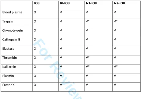
The stability of the most promising inhibitory peptides (IO8, N1-IO8 and N2-IO8) towards individual
proteolytic enzymes (see Supplemetary Figure 3), and in human plasma, was assessed by RP-HPLC.
The data are summarised in Table 2, with examples of RP-HPLC traces of peptides in plasma presented
in Figure 6. IO8 was highly susceptible to the effects of trypsin and chymotrypsin, which rapidly
degraded this peptide, but it was also degraded by cathepsin G, elastase, thrombin, kallikrein, plasmin,
and factor X. Not surprisingly, therefore, IO8 was very unstable in human plasma (Figure 6A). In
contrast, both N1-IO8 and N2-IO8 were stable for up to 24 hrs incubation in plasma (Figure 6B and
6C), and, unlike IO8, remained intact after 3 hrs incubation with each of the individual proteolytic
enzymes, although some degradation was noted after 24 hrs incubation (see Table 2).
The toxic effect of human amylin (20 µM and 10 µM) on human pancreatic 1.4E7 cells was determined
by MTS assay, in the presence and absence of 1:1 and 1:4 molar ratios (inhibitor:amylin) of the IO8,
N1-IO8 and N2-IO8 peptides (for results see Figure 7). Amylin at 20 μM was cytotoxic to PANC-1 cells
and consistently reduced cell viability to ~40% of untreated control cells, whereas at 10 μM amylin the
results were more variable and cell viability was reduced to 60-90%. All of these inhibitor peptides, at
both molar ratios, rescued the cells from the toxic effect of 20 µM amylin and 10 µM amylin. None of
the inhibitors alone, at concentrations of up 20 μM, had any effect on cell viability. Cell toxicity results
for I08 and related peptides from three independent experiments, each carried out in triplicate, are
presented in Supplementary Figure 4. 3
For Review Only
DiscussionT2DM is the most widespread endocrine disorder [53], and is characterised by a reduction in β-cell
mass, insulin resistance, and the presence of amyloid deposits in the pancreatic islets, the main
component being amylin [55]. The 22-28 (NFGAILS) segment of amylin is regarded as the most highly
amyloidogenic region of this peptide, and will itself assemble into amyloid fibrils [56, 39]. However,
residues 8-20 [57], 14-20 [58], and 30-37 [59] have also been reported to form β-sheet fibrils. Although
several amyloidogenic regions of human amylin have been proposed, this study was concerned with
developing peptide inhibitors from the ‘binding’ region of human amylin, corresponding to amino acid
residues 11-20 (RLANFLVHSS), and on studying their impact on the fibrillogenesis of full-length human
amylin. This region is thought to be involved in the initial interactions between two misfolded amylin
molecules, after which they begin to aggregate [42]. Thus, preventing this interaction should impede
aggregation. This strategy, to target the binding region, has been successfully applied to development
of inhibitors of Aβ aggregation as a potential disease-modifying treatment for AD [47-50].
Seven potential inhibitor peptides were derived from this binding region, and investigated for their
ability to influence amylin fibril formation, based on the ThT fluorescence assay [60]. Peptides IO2,
IO3, IO4, IO5 and IO7 (see Table 1) showed some inhibitory effects, but IO4 and IO5 gave the most
promising results, and were considered for further investigation. These two amino acid sequences
were combined to give IO8 (amino acid sequence: H2N-RGANFLVHGR-CONH2). Retro-inverso peptides
often retain bioactivity and are stable to proteolysis [61, 47], and so the retro-inverso version of IO8
(RI-IO8: Ac-rGhvlfnaGr-CONH2) was derived by sequence reversal and D-amino acid substitution. The
IO8 peptide showed a strong inhibitory effect on amylin aggregation, and, unlike peptides IO1-IO7,
did not stimulate amylin aggregation at low concentrations. However, RI-IO8 had no inhibitory effect
on amylin aggregation, except at a 4:1 molar ratio RI-IO8 to amylin, where the peptide reduced amylin
aggregation to only 77% of a non-inhibited control (Figure 3). At lower concentrations, RI-IO8 actually
stimulated amylin aggregation. This finding was unexpected and suggests that RI-IO8 does not interact 3
For Review Only
in the same way as IO8 to full-length human amylin. Congo red binding experiments have also
confirmed the inhibitory effect of IO8, and the stimulatory effect of RI-IO8, on amylin aggregation
(data not shown). This finding was further supported by TEM studies, where IO8 abolished and RI-IO8
increased amylin fibril formation (Figure 5). This result with RI-IO8 is contrary to a previous study,
where the retro-inverso peptide RI-OR2, developed against β-amyloid (Aβ) aggregation, was shown to
inhibit amyloid fibril formation, and also rescue cells from the toxic effects of Aβ, as well as being
highly resistant to proteolysis [47].
Since RI-IO8 did not inhibit amylin aggregation, N-methylation was considered as an alternative means
to improve its stability and pharmacokinetic properties. It is not surprising that IO8 was rapidly
degraded in plasma, and in the presence of proteolytic enzymes, because L-peptides are quickly
metabolized in this way [62]. IO8 would be cleaved after amino acid 5 (Phe) by high specificity
chymotrypsin, and after amino acids 5, 6 and 8 (Phe, Leu, His) by low specificity chymotrypsin, while
trypsin will cleave after position 1 (Arg). N-methylation has been shown to improve the
pharmacokinetic properties of peptides, by protecting them from proteolysis [63]. Also,
N-methylation of alternate amino acid residues gives one face of the peptide molecule that is not
available for H-bonding, which impedes amyloid fibril formation [64]. N-methylated derivatives of
Aβ(25–35) have been reported to impede the aggregation of fibrils and prevent Aβ cytotoxicity, and
N-methylated analogues of amylin do not form fibrils [18, 65]. Here, IO8 was stabilised against
proteolytic degradation through N-methylation of alternate amino acid residues, to give N1-IO8
(H2N-R-G-Am-N-Fm-L-Vm-H-G-R-CONH2) and N2-IO8 (H2N-R-G-A-Nm-F-Lm-V-Hm-G-R-CONH2). ThT and
TEM data showed that both NI-IO8 and N2-IO8 are excellent and highly convincing inhibitors of amylin
aggregation (Figures 3-5) and are relatively stable against proteolytic degradation (Figure 6).
New inhibitors of amylin aggregation are desired, as many of the reported inhibitors only work when
present in molar excess over amylin [65-67]. For example, a study on peptide fragments corresponding 3
For Review Only
to human amylin residues 20-25 (SNNFGA) and 24-29 (GAILSS) showed an inhibitory effect on β-sheet
transition and amyloid aggregation at 10:1 and 20:1 molar ratios of peptide to amylin, and the GAILSS
peptide had no significant effect on amylin-induced cytotoxicity [41]. The inadequacy of some
previously published inhibitors is also highlighted by the comparison of the effects of IO8, ANFLVH
[52] and NMeG24 NMeI26 [51] on amylin aggregation in this report. ANFLVH has the same amino acid
sequence as IO8, but without the flanking cationic arginine residues together with their glycine
spacers. Although ANFLVH was reported to inhibit amylin fibril formation in vitro and to protect
against amylin cytotoxicity, the latter effect was only observed at 10-fold and 20-fold molar excess
concentrations of the peptide [52]. In contrast, the IO8 peptide inhibits amylin aggregation at much
lower concentrations than this, and, given the inability to dissolve ANFLVH in aqueous solution, is
clearly much more soluble. Moreover, IO8, N1-IO8 and N2-IO8 were seen to rescue human pancreatic
1.4E7 cells from the toxic effects of amylin at a 1:4 molar ratio of these peptides to amylin. In contrast
to a previous report [51], no evidence of inhibition of aggregation was observed upon addition of
NMeG24 NMeI26, at reasonable concentrations, to amylin. In fact, NMeG24 NMeI26 was seen to
promote fibril formation (Figure 3). This discrepancy could be due to the fact that NMeG24 NMeI26
was reported to inhibit IAPP aggregation when added before nucleation [68] but the aggregation
system reported here lacks any lag-phase (see Figure 4), and so nucleation may be too rapid for this
inhibitor to be effective. Two human amylin-derived peptides, with sequences NFGAIL and
SNNFGAILSS, were unable to inhibit fibrillation of human amylin [69]. Another study has indicated that
NFGAIL causes an immediate shift of amylin to the β-sheet conformation, suggesting that this peptide
promotes fibril formation [41]. These results, together with the lack of effect of the
H2N-RGNFGAILSGR-CONH2 peptide in this study (Figure 3), justify the decision to focus on the
RLANFLVHSS (residues 11-20) amylin binding region.
As noted above, modified full-length amylin with N-methylation at positions 24 and 26 has been
shown to impede amylin aggregation and its associated cytotoxicity [65]. In addition, a human amylin 3
For Review Only
derived peptide marketed as pramlintide, with proline substitutions at positions 25, 28 and 29, has
undergone clinical trials [70-73] where it was administered, alongside insulin, for management of
T2DM. This combination of drugs was able to maintain near-normal glycaemic levels, but pramlintide
peptide does not appear to have been assessed as an inhibitor of human amylin aggregation. The short
peptides described in this report would be much easier and less expensive to synthesize than these
full-length human amylin analogues, and would, potentially, be less immunogenic. N1-IO8 and N2-IO8
in particular appear to be potent and stable aggregation inhibitors that are suitable for further
development and testing in human amylin transgenic rodent models as potential disease-modifying
agents for T2DM. However, the effects of these inhibitors on oligomer formation are not clear from
the data presented here, and will need to be examined in further studies. Also, it is emerging that a
significant component of amylin toxicity is mediated by inflammation [74], and so the ability of these
inhibitors to attenuate amylin-mediated macrophage activation and associated β-cell dysfunction will
also need to be determined. 3
For Review Only
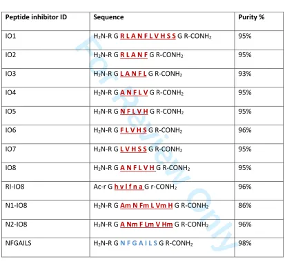
Table 1Design of peptide inhibitors employed for this study.
H
2N–K C N T A T C A T Q
R L A N F L V H S S
N
N F G A I L S
S T N V G S N T Y-CONH
2Peptide inhibitor ID Sequence Purity %
IO1 H2N-R G R L A N F L V H S S G R-CONH2 95%
IO2 H2N-R G R L A N F G R-CONH2 95%
IO3 H2N-R G L A N F L G R-CONH2 93%
IO4 H2N-R G A N F L V G R-CONH2 95%
IO5 H2N-R G N F L V H G R-CONH2 95%
IO6 H2N-R G F L V H S G R-CONH2 96%
IO7 H2N-R G L V H S S G R-CONH2 95%
IO8 H2N-R G A N F L V H G R-CONH2 95%
RI-IO8 Ac-r G h v l f n a G r-CONH2 96%
N1-IO8 H2N-R G Am N Fm L Vm H G R-CONH2 86%
N2-IO8 H2N-R G A Nm F Lm V Hm G R-CONH2 96%
NFGAILS H2N-R G N F G A I L S G R-CONH2 98%
The amino acid sequence of human amylin shows the binding site for amylin self-association [42] and
the main amyloidogenic region [35]. All of the short peptide inhibitors are designed to interact with
the binding region of full-length amylin, except for the last peptide. The arginine-glycine flanking
residues (RG-GR) impede peptide self-aggregation. In the retro-inverted peptide (RI-IO8), D-amino
acids are in lower case. N-methylated peptide residues are indicated by lower case ‘m’.
Binding region
Amyloidogenic region Disulphide
bridge
For Review Only
Table 2Stability of peptide-inhibitors to proteolysis
√ = stable, X = degraded, after 3 hrs incubation with the enzyme. *Some degradation seen
after 24 hrs incubation. HPLC traces for IO8, N1-IO8 and N2-IO8 are given in Supplementary
Figure 3.
IO8
RI-IO8
N1-IO8
N2-IO8
Blood plasma
X
√
√
√
Trypsin
X
√
√*
√*
Chymotrypsin
X
√
√
√
Cathepsin G
X
√
√
√
Elastase
X
√
√
√
Thrombin
X
√
√*
√
Kallikrein
X
√
√*
√*
Plasmin
X
√
√
√
Factor X
X
√
√
√
For Review Only
Figure LegendsFigure 1.
Example of ThT fluorescence curves for human amylin in the presence of different concentrations of
an inhibitor (IO4). Data are means for a single experiment carried out in triplicate, with readings taken
every 10 mins. Amylin alone (at 25 µM) displays a characteristic increase in fluorescence
corresponding to the ‘sigmoidal’ and ‘plateau’ phases of amyloid fibril formation, while the addition
of the inhibitor, at varying concentrations, has a dose-dependent effect on fibril formation. The buffer
control (‘Control’) contained neither amylin nor inhibitor. RFU = relative fluorescence units.
Figure 2.
ThT data showing effects of IO1, IO2, IO3, IO4, IO5, IO6 and IO7 peptides on amylin aggregation, after
48 hrs incubation. All peptides were assayed at 0.6 µM, 2.5 µM, 5 µM, 12.5 µM, 25 µM, 50 µM and
100 µM concentrations in the presence of 25 μM amylin. Results are means +/- SEM, n = 3, for a single
experiment. Student’s t-test was used to establish significance at *P<0.05, **P<0.01, or ***P<0.001,
compared to 100% control (amylin alone).
Figure 3.
ThT data showing the effects of IO8 and related peptides, as well NFGAILS and NMeG24 NMeI26, on
amylin aggregation, after 48 hrs incubation. (A) IO8 and RI-IO8. (B) N1-IO8 and N2-IO8. (C) NFGAILS
(H2N-RGNFGAILSGR-CONH2). (D) NMeG24 NMeI26. All peptides were assayed at 0.6 µM, 2.5 µM,
5 µM, 12.5 µM, 25 µM, 50 µM and 100 µM in the presence of 25 μM amylin. Results show means
+/- SEM, n = 3, for a single experiment. See Supplementary Figure 2 for the data from 3 independent
experiments. Note the clear dose-dependent effects of the two N-methylated peptides (N1-IO8 and
N2-IO8). 3
For Review Only
Figure 4.ThT fluorescence curves for the first 10 hrs of incubation of amylin in the presence of different
concentrations of (A) N1-IO8 and (B) N2-IO8. Data are means for a single experiment carried out in
triplicate, with readings taken every 10 mins. Amylin alone (at 25 μM) displays a characteristic increase
in fluorescence corresponding to the ‘sigmoidal’ and ‘plateau’ phases of amyloid fibril formation (top
curve in both cases). In both (A) and (B), the stepwise decrease in the final level of ThT fluorescence
in the 10 curves underneath is due to addition of the inhibitors at concentrations of 0.1, 0.3, 0.6, 1,
2.5, 5, 12.5, 25, 50 and 100 µM. RFU = relative fluorescence units.
Figure 5.
Negative stain EM images of amylin incubated in the presence and absence of inhibitors. (A) Sample
of amylin (25 µM) incubated for 48 hrs in PBS at room temperature and stained with phosphotungstic
acid (2% w/v). (B) Amylin (25 µM) + IO8 (25 μM). (C) Amylin (25 µM) + RI-IO8 (25 μM); (D) Amylin (25
µM) + N1-IO8 (25 μM); (E) Amylin (25 µM) + N2-IO8 (25 μM); (F) IO8 alone (100 µM); (G) RI-IO8 alone
(100 µM); (H) N1-IO8 alone (100 µM); (I) N1-IO8 alone (100 µM). Scale bar = 100 nm.
Figure 6.
Reverse-phase HPLC traces showing stability of peptide inhibitors in human plasma. Column (A) IO8;
column (B) N1-IO8; column (C) N2-IO8. For each column, the top trace shows elution of the peptide
standard without plasma; the middle trace is for 0 hrs incubation in plasma; and the lower trace is
after 48 hrs incubation in plasma. Note that IO8 is degraded, whereas N1-IO8 and N2-IO8 are more
stable. 3
For Review Only
Figure 7.Cytotoxic effect of amylin on human pancreatic 1.4E7 insulin-secreting cells in the presence or absence
of inhibitor peptides. (A) Cells were incubated for 24 hrs with 20 μM or 10 μM human amylin in
RPMI-1640 medium, with or without IO8 inhibitor, and viability was measured using the MTS assay. (B)
Results for N1-IO8. (C) Results for N2-IO8. In all cases, results show mean +/- SEM, n = 6. ANOVA
followed by Student’s t-test established significance at *P<0.05, **P<0.01, ***P<0.001.
Author Contributions
D.A. conceived of the study and wrote the manuscript. I.O. carried out all of the experiments, data
collection and statistical analyses. M.T. designed some of the peptides and N.J.F. was responsible for
the electron microscopy. All authors contributed to design of the study and gave final approval for
publication.
Acknowledgements
D.A. wishes to thank Professor Ian Hamley and the other conference organisers for his invitation to
this very interesting and stimulating meeting. The authors also thank the editor and anonymous
reviewers for their helpful comments and suggestions, and Dr. Fuyuki Kametani (Tokyo, Japan) for
peptide HPLC-MS analysis.
Funding
This work was partially supported by a scholarship to I.O. from the Faculty of Health and Medicine,
Lancaster University. 3
For Review Only
References
1. Dobson CM 1999 Protein misfolding, evolution and disease. Trends Biochem. Sci. 24,
329-332.
2. Rochet JC, Lansbury PT 2000 Amyloid fibrillogenesis: themes and variations. Current Opin.
Struct. Biol. 10, 60-68.
3. Westermark P, et al. 2007 A primer of amyloid nomenclature. Amyloid 14 , 179-183.
4. Westermark P. 2005 Aspects on human amyloid forms and their fibril polypeptides. FEBS
Lett. 272, 5942-5949.
5. Pepys MB (2006) Amyloidosis. Ann. Rev. Med. 57, 223-241.
6. Allsop D, Mayes J. 2014 Amyloid-β protein and Alzheimer’s disease. Essays Biochem. 56,
99-110.
7. Soto C, Estrada LD. 2008 Protein misfolding and neurodegeneration. Arch. Neurol. 65,
184-189.
8. Ahmad E, Ahmad A, Singh S, Arshad M, Khan AH, Khan RH. 2011 A mechanistic approach for
islet amyloid polypeptide aggregation to develop anti-amyloidogenic agents for type-2
diabetes, Biochimie 93, 793-805.
9. Kapurniotu A. 2001 Amyloidogenicity and cytotoxicity of islet amyloid polypeptide.
Biopolymers 60, 438-459.
10. Westermark P. 2011 Amyloid in the islets of Langerhans: thoughts and some historical
aspects. Upsala J. Med. Sci. 116, 81-89.
11. Wimalawansa SJ. 1997 Amylin, calcitonin gene-related peptide, calcitonin, and
adrenomedullin: a paptide superfamily. Critical Rev. Neurobiol. 11, 167-239.
12. Kahn SE. 2003 The relative contributions of insulin resistance and β-cell dysfunction to the
pathophysiology of Type 2 diabetes. Diabetologia 46, 3-19. 3
For Review Only
13. Zraika S, et al. 2010 Toxic oligomers and islet β cell death: guilty by association or convicted
by circumstantial evidence? Diabetologia 53, 1046-1056.
14. Hull RL, Westermark GT, Westermark P, Kahn SE. 2004 Islet amyloid: a critical entity in the
pathogenesis of type 2 diabetes. J. Clin. Endocrinol. Metabol. 89, 3629-3643.
15. Hiddinga HJ, Eberhardt NL. 1999 Intracellular amyloidogenesis by human islet amyloid
polypeptide induces apoptosis in COS-1 cells. Am. J. Pathol. 154, 1077-1088.
16. Kapurniotu A, et al. 1998 Contribution of advanced glycosylation to the amyloidogenicity of
islet and amyloid polypeptide. Eur. J. Biochem. 251, 208-216.
17. Lorenzo A, Yankner BA. 1996 Amyloid fibril toxicity in Alzheimer’s disease and diabetes. Ann.
N. Y. Acad. Sci. 777, 89-95.
18. Yan LM, et al. 2007 Amylin mimic blocks Aβ cytotoxic self-assembly: cross-suppression of
amyloid toxicity of Aβ and amylin suggests a molecular link between Alzheimer's disease and
type II diabetes. Angewandte Chemie Int. Ed. 46, 1246-1252.
19. Kodali R, Wetzel R. 2007 Polymorphism in the intermediates and products of amyloid
assembly. Current Opin. Struct. Biol. 17, 48-57.
20. Aitken JF, et al. (2010) Tetracycline treatment retards the onset and slows the progression of
diabetes in human amylin/islet amyloid polypeptide transgenic mice. Diabetes 59, 161-171.
21. Ritzel RA, et al. 2007 Human islet amyloid polypeptide oligomers disrupt cell coupling,
induce apoptosis, and impair insulin secretion in isolated human islets. Diabetes 56, 65-71.
22. Diabetes UK. 2015 Key statistics on Diabetes. [online]. Available at:
http://www.diabetes.org.uk. (Accessed 17th August 2015).
23. WHO. 2016 World Health Organisation. Diabetes. [Online]. Available at:
http://www.who.int/diabetes/en/ (Accessed 12th January 2016).
24. Colhoun HM, et al. 2004 Primary prevention of cardiovascular disease with atorvastatin in
Type 2 diabetes in the Collaborative Atorvastatin Diabetes Study (CARDS): multicentre 3
For Review Only
25. Morrish NJ, et al. 2001 Mortality and causes of death in the WHO multinational study of
vascular disease in diabetes. Diabetol. 44, s14-s21.
26. Castello MA, JeppsonJD, Soriano S. 2014 Moving beyond anti-amyloid therapy for the
prevention and treatment of Alzheimer’s disease. BMC Neurol. 14, 169.
27. Rosenblum WI. 2014 Why Alzheimer trials fail: Removing soluble oligomeric β amyloid is
essential, inconsistent, and difficult. Neurobiol. Ageing 35, 969-974.
28. Whitty A, Kumaravel G. 2006 Between a rock and a hard place. Nature Chem. Biol. 2,
112-118.
29. Hajduk PJ, Burns DJ. 2002 Integration of NMR and high-throughput screening. Comb. Chem.
High Through. Screen. 5, 613-621.
30. Keskin O, Gursoy A, Ma B, Nussinov R. 2008 Principles of protein-protein interactions: what
are the preferred ways for proteins to interact? Chem. Rev. 108, 1225-1244.
31. Teichmann SA. 2002 Principles of protein-protein interactions. Bioinform. 18, S249.
32. Smith RD, et al. 2006 Exploring protein-ligand recognition with Binding MOAD. J. Mol. Graph.
Mod. 24, 414-425.
33. Cheng AC, et al. 2007 Structure-based maximal affinity model predicts small-molecule
druggability. Nature Biotechnol. 25, 71-75.
34. Wells JA, McClendon CL. 2007 Reaching for high-hanging fruit in drug discovery at
protein-protein interfaces. Nature 450, 1001-1009.
35. Goldsbury, C., et al. 2000 Amyloid fibril formation from full-length and fragments of amylin.
J. Struct. Biol. 130, 352-362.
36. Tenidis K, et al. 2000 Identification of a penta- and hexapeptide of islet amyloid polypeptide
(amylin) and with amyloidogenic and cytotoxic properties. J. Mol. Biol. 295, 1055-1071.
37. Elgersma RC, et al. 2006. Self-assembly of amylin (20-29) amide-bond derivatives into helical
ribbons and peptide nanotubes rather than fibrils. Chemistry 12, 3714-3725. 3
For Review Only
38. Soto C, Kindy MS, Baumann M, Frangione B. 1996 Inhibition of Alzheimer's amyloidosis by
peptides that prevent β-sheet conformation. Biochem. Biophys. Res. Commun. 226, 672-680.
39. Westermark P, et al. 1990 Islet amyloid polypeptide - pinpointing amino-acid residues linked
to amyloid fibril formation. Proc. Natl. Acad. Sci. USA 87, 5036-5040.
40. Abedini A, Meng FL, Raleigh, DP. 2007 A single-point mutation converts the highly
amyloidogenic human islet amyloid polypeptide into a potent fibrillization inhibitor. J. Am.
Chem. Soc. 129, 11300-11301.
41. Scrocchi LA, et al. 2002 Design of peptide-based Inhibitors of human islet amyloid
polypeptide fibrillogenesis. J. Mol. Biol. 318, 697-706.
42. Mazor Y, Gilead S, Benhar I, Gazit E. 2002 Identification and characterization of a novel
molecular-recognition and self-assembly domain within the islet amyloid polypeptide. J.
Mol. Biol. 322, 1013-1024.
43. Nie Q, Du X, Geng M. 2011 Small molecule inhibitors of amyloid β peptide aggregation as a
potential therapeutic strategy for Alzheimer's disease. Acta Pharmacol. Sinica. 32, 545-551.
44. Aitken JF, Loomes KM, Konarkowska B, Cooper GJ. 2003. Suppression by polycyclic
compounds of the conversion of human amylin into insoluble amyloid, Biochem. J. 374,
779-784.
45. Ono K, et al. 2003 Potent anti-amyloidogenic and fibril-destabilizing effects of polyphenols in
vitro: implications for the prevention and therapeutics of Alzheimer's disease. J. Neurochem.
87, 172-181.
46. Austen BM, et al. 2008 Designing peptide inhibitors for oligomerization and toxicity of
Alzheimer's β-amyloid peptide. Biochemistry 47, 1984-1992.
47. Taylor M, et al. 2010 Development of a proteolytically stable retro-inverso peptide inhibitor
of β-Amyloid oligomerization as a potential novel treatment for Alzheimer’s disease.
Biochemistry 49, 3261-3272. 3
For Review Only
48. Parthsarathy V, et al. 2013 A novel retro-inverso peptide inhibitor reduces amyloid
deposition, oxidation and inflammation and stimulates neurogenesis in the APPSWE/PS1∆E9
mouse model of Alzheimer’s disease. PLoS ONE 8(1): e54769.
49. Gregori M, et al. 2016 Retro-inverso peptide inhibitor nanoparticles as potent inhibitors of
aggregation of the Alzheimer’s Aβ peptide. Nanomed: Nanotechnol. Biol. Med. 19.10.2016.
50. Sherer M, Fullwood NJ, Taylor M, Allsop D. 2015 A preliminary electron microscopic
investigation into the interaction between Aβ1-42 peptide and a novel
nanoliposome-coupled retro-inverso peptide inhibitor, developed as a potential treatment for Alzheimer's
disease. Journal of Physics: Conference Series 644, 012040,
10.1088/1742-6596/644/1/012040.
51. Sellin D, Yan L, Kapurniotu A, Winter R. 2010 Suppression of amylin fibrillation at anionic
lipid membranes via amylin-derived amyloid inhibitors and insulin. Biophys. Chem. 150,
73-79.
52. Potter KJ, et al. 2009 Amyloid inhibitors enhance survival of cultured human islets. Biochim.
Biophys. Acta 1790, 566-574.
53. Hossain P, Kawar B, Nahas ME. 2007 Obesity and diabetes in the developing world - a
growing challenge. New Eng. J. Med. 356, 213-235.
54. Xue C, Lin TY, Chang D, Guo Z. 2017 Thioflavin T as an amyloid dye: fibril quantification,
optimal concentration and effect on aggregation. R. Soc. Open Sci. 4: 160696.
http://dx.doi.org/10.1098/rsos.160696
55. Westermark P, et al. 1987 Amyloid fibrils in human insulinoma and islets of Langerhans of
the diabetic cat are derived from a neuropeptide-like protein also present in normal islet
cells. Proc. Natl. Acad. Sci. USA. 84, 3881-3885.
56. Moriarty DF, Raleigh DP. 1999 Effects of sequential proline substitutions on amyloid
formation by human amylin 20–29. Biochemistry 38, 1811-1818. 3
For Review Only
57. Jaikaran ET, et al. 2001 Identification of a novel human islet amyloid polypeptide β-sheet
domain and factors influencing fibrillogenesis. J. Mol. Biol. 308, 515-525.
58. Sawaya MR, et al. 2007 Atomic structures of amyloid cross-β spines reveal varied steric
zippers. Nature 447, 453-457.
59. Nilsson MR, Raleigh DP. 1999 Analysis of amylin cleavage products provides new insights
into the amyloidogenic region of human amylin. J. Mol. Biol. 294, 1375-1385.
60. Biancalana M, Koide S. 2010 Molecular mechanism of thioflavin-T binding to amyloid fibrils.
Biochem. Biophys. Acta. 1804, 1405-1412.
61. Chen X, et al. 2013 Retro-inverso carbohydrate mimetic peptides with annexin1-binding
selectivity, are stable in vivo, and target tumor vasculature. PLoS ONE. 8(12):e80390.
62. Kellock J, Hopping G, Caughet B, Daggetti V. 2016 Peptides composed of alternating L- and
D-amino acids inhibit amyloidogenesis in three distinct amyloid systems independent of
sequence. J. Mol. Biol. 428, 2317-2328.
63. Chatterjee J, Rechenmacher F, Kessler H. 2013 N-Methylation of peptides and proteins: an
important element for modulating biological functions. Angewandte Chemie Int. Ed. 52,
254-269.
64. Hughes E, Burke RM, Doig AJ. 2000 Inhibition of toxicity in the β-amyloid peptide fragment
β-(25-35) using N-methylated derivatives: a general strategy to prevent amyloid formation. J
Biol Chem. 18, 275, 25109-25115.
65. Yan L, et al. 2006 Design of a mimic of nonamyloidogenic and bioactive human islet amyloid
polypeptide (amylin) as nanomolar affinity inhibitor of amylin cytotoxic fibrillogenesis. Proc.
Natl. Acad. Sci. USA. 103, 2046-2051.
66. Meng F, et al. 2010 The sulfated triphenyl methane derivative acid fuchsin is a potent
inhibitor of amyloid formation by human islet amyloid polypeptide and protects against the
toxic effects of amyloid formation. J. Mol. Biol. 400, 555-566. 3
For Review Only
67. Saraogi I, et al. 2010 Synthetic α-helix mimetics as agonists and antagonists of islet amyloid
polypeptide aggregation. Angewandte Chemie Int. Ed. 49, 736-739.
68. Tatarek-Nossol M, et al. 2005 Inhibition of hIAPP amyloid-fibril formation and apoptotic cell
death by a designed hIAPP amyloid- core-containing hexapeptide. Chem Biol. 12, 797-809.
69. Andreasen M, et al. 2012 Modulation of fibrillation of hamylin core fragments by chemical
modification of the peptide backbone. Biochim. Biophys. Acta. 1824, 274-285.
70. Pullman J, Darsow T, Frias JP. 2006 Pramlintide in the management of insulin-using patients
with type 2 and type 1 diabetes. Vascular Health Risk Man. 2, 203-212.
71. Kong MF, et al. 1998 The effect of single doses of pramlintide on gastric emptying of two
meals in men with IDDM. Diabetologia 41, 577-583.
72. Maggs DG, et al. 2004 Pramlintide reduces postprandial glucose excursions when added to
insulin lispro in subjects with type 2 diabetes: a dose-timing study. Diabetes/Metabol. Res.
Rev. 20, 55-60.
73. Thompson RG, Pearson L, Schoenfeld SL, Kolterman OG. 1998 Pramlintide, a synthetic analog
of human amylin, improves the metabolic profile of patients with type 2 diabetes using
insulin. The Pramlintide in Type 2 Diabetes Group. Diabetes Care 21, 987-993.
74. Westwell-Roper CY, Ehses JA, Verchere CB. 2014 Resident macrophages mediate islet
amyloid polypeptide-induced islet IL-1β production and β-cell dysfunction. Diabetes 63,
1698-1711. 3
For Review Only
Figure 1. Example of ThT fluorescence curves for human amylin in the presence of different concentrations of an inhibitor (IO4).
119x70mm (150 x 150 DPI)
For Review Only
Figure 2. ThT data showing effects of IO1, IO2, IO3, IO4, IO5, IO6 and IO7 peptides on amylin aggregation, after 48 hrs incubation.
167x189mm (120 x 120 DPI)
For Review Only
Figure 3. ThT data showing the effects of IO8 and related peptides, as well NFGAILS and NMeG24 NMeI26, on amylin aggregation, after 48 hrs incubation.
166x142mm (120 x 120 DPI)
For Review Only
Figure 4. ThT fluorescence curves for the first 10 hrs of incubation of amylin in the presence of different concentrations of (A) N1-IO8 and (B) N2-IO8.
158x90mm (96 x 96 DPI)
For Review Only
Figure 5. Negative stain EM images of amylin incubated in the presence and absence of inhibitors.
254x190mm (96 x 96 DPI)
For Review Only
Figure 6. Reverse-phase HPLC traces showing stability of peptide inhibitors in human plasma.
190x254mm (96 x 96 DPI)
For Review Only
Figure 7. Cytotoxic effect of amylin on human pancreatic 1.4E7 insulin-secreting cells in the presence or absence of inhibitor peptides.
190x254mm (96 x 96 DPI)