ISSN Online: 2151-1942 ISSN Print: 2151-1934
DOI: 10.4236/jct.2019.107050 Jul. 29, 2019 609 Journal of Cancer Therapy
Clinical and Pathological Value of MACC-1
Expression in Gastric Carcinoma
Abeer Hafez
1, Tarek El-Gohary
2*, Fouad Abutaleb
21Pathology Department, Zagazig University Hospitals, Zagazig, Egypt
2Medical Oncology and Hematology Department, Zagazig University Hospitals, Zagazig, Egypt
Abstract
Background: Gastric adenocarcinoma is known to be a markedly invasive dis-ease with high potential for metastasis. Hepatocyte growth factor (HGF)/HGF receptor (MET) signaling pathway activation is an assumed mechanism of malignant transformation and metastatic potential of tumors. Metastasis as-sociated with colon cancer-1 (MACC-1) has been identified as a key regulator of HGF/MET signaling. However, its role in gastric cancer is not well un-derstood. Aim of this study is to assess the expression of MACC-1 in gastric cancer, its relation to other clinical and pathologic parameters and its impact on progression free and overall survival. Patient and Methods: Evaluation of MACC-1 protein expression by immune-histochemistry was done on paraf-fin-embedded tissues obtained from 46 patients with gastric cancer, where sam-ples were taken from the tumor and adjacent normal mucosa. Results: MACC-1 was predominantly localized in the cytoplasm or membrane of the primary can-cer cells. High MACC-1 expression was found in 63.1% (29/46) of tumor sam-ples, while MACC-1 expression was not detected in normal mucosa (P < 0.01). Expression of MACC-1 was significantly associated with older age, larger tumor size, deeper tumor invasion, presence of lymph node metastasis, distant metas-tasis and advanced clinical stage (p < 0.05), while no relation was found with gender, tumor location or histologic classification (p > 0.05). Progression free and overall survival were significantly higher in patients with low MACC-1 expression compared to patients with high expression (Log Rank test, p = 0.02 and 0.04, consequently). Conclusion: Our study found that MACC-1 expression is strongly related to gastric cancer stage and both progression free and overall survival, suggesting that MACC-1 promotes tumorigenesis and its expression may be used as signal of bad prognosis in gastric cancer.
Keywords
Gastric Cancer, MACC-1, HGF/MET Signaling
How to cite this paper: Hafez, A., El-Gohary, T. and Abutaleb, F. (2019) Clinical and Pa-thological Value of MACC-1 Expression in Gastric Carcinoma. Journal of Cancer Therapy, 10, 609-618.
https://doi.org/10.4236/jct.2019.107050
Received: June 14, 2019 Accepted: July 26, 2019 Published: July 29, 2019
Copyright © 2019 by author(s) and Scientific Research Publishing Inc. This work is licensed under the Creative Commons Attribution International License (CC BY 4.0).
DOI: 10.4236/jct.2019.107050 610 Journal of Cancer Therapy
1. Introduction
Gastric cancer is a highly lethal disease due to marked invasion and metastasis
[1] [2]. The mechanism of tumor invasion and metastasis includes cell
move-ment, adhesion, extracellular matrix degradation and angiogenesis [3] [4]. Bio-logical markers associated with invasion and metastasis such as aberrant activa-tion of the hepatocyte growth factor (HGF)/HGF receptor (MET) signaling pathway has been a focus for gastric cancer research [3] [5]. Distant organ me-tastasis is a signal of poor prognosis in patients with gastric cancer. Liver is a common target organ of gastric cancer metastasis [6]. Hepatic stellate cells (HSCs) are a type of liver-specific mesenchymal cells which are considered a main com-ponent of the prometastatic liver microenvironment. These cells can be activated by tumor-derived factors to stimulate the metastatic growth of tumor cells [7].
Metastasis-associated with colon cancer-1 (MACC-1) is a member of a family of soluble proteins. MACC1 hastens migration, invasion and metastasis of can-cer cells through activating the hepatocyte growth factor (HGF)/MET pro-to-oncogene (c-Met) signaling pathway [8], together with provoking multiple cellular responses regulating cell morphogenesis, migration and breakdown of the extracellular matrix [9]. A study has previously demonstrated that the ex-pression MACC1 is significantly higher in activated HSCs. MACC1 is reported to be associated with distant metastasis in gastric cancer [10]. Overexpression of MACC1 increases invasion in cancer cells by enhancing epithelial-mesenchymal transition (EMT) [11], and is significantly correlated to decreased overall surviv-al (OS) and disease-free survivsurviv-al (DFS) [12].
MACC-1 was first identified during a genome wide search for genes differen-tially expressed in colorectal cancer [13]. However, the significance of MACC-1 expression and its impact on prognosis in gastric adenocarcinoma are still con-troversial.
2. Materials and Methods
under-DOI: 10.4236/jct.2019.107050 611 Journal of Cancer Therapy
went surgery and those with unresectable locally advanced or metastatic disease received a two-drug combination (cisplatin 75 - 100 mg/m2 IV over 3 hours on
day 1 and fluorouracil 750 - 1000 mg/m2 IV continuous infusion over 24 hours
daily on days 1 - 4, cycled every 28 days or cisplatin 80 mg/m2 IV over 3 hours
on day 1 and capecitabine 1000 mg/m2 PO BID daily on days 1 - 14 cycled every
21 days. Trustuzumab was added for her-2 positive cases where a loading dose of 8 mg/kg IV on day 1 of cycle 1 was given followed by a maintenance dose of 6 mg/kg IV every 21 days or a loading dose of 6 mg/kg IV on day 1 of cycle 1 fol-lowed by maintenance dose of 4 mg/kg IV every 14 days).
Immunohistochemistry
Immunohistochemical analysis was performed to study altered protein ex-pression in 46 human gastric cancer and adjacent normal tissue samples, as pre-viously described [14] [15]. Paraffin-embedded formalin-fixed tissue slides (4 μm) were baked at 60˚C for 2 h, followed by deparaffinization with xylene, and rehydrated. Antigen retrieval was performed by submerging sections in EDTA antigenic retrieval buffer, and microwaving for x min. Sections were then treated with 3% (v/v) hydrogen peroxide in methanol to quench endogenous peroxidase activity, followed by incubation with 1% (w/v) bovine serum albumin to block non-specific binding. Sections were incubated with mouse monoclonal antibody againstMACC-1 and MET (Santa Cruz Biotechnology, 1:50 dilution) overnight at 4˚C. Normal goat serum was used as a negative control. After washing, tissue sections were treated with secondary antibody. Tissue sections were then coun-terstained with hematoxylin, dehydrated and mounted. A negative control slide was processed without primary antibody to detect any background staining or false-positive results.
MACC-1 was stained as buffy colored in the cytoplasm and/or membrane. Protein expression was assessed by two expert pathologists blinded to the clinical details.
Scoring was performed according to the percentage of stained cells observed 0 = ≤5% of cells
1 = 6% - 25% 2 = 26% - 50% 3 = 51% - 100%
An intensity score of “0 - 3” was also determined 0 = none,
1 = light yellow, 2 = yellow brown, 3 = brown) [16] [17] [18]
The final score was the product of the two former values 0 - 1 = −
2 - 3 = + 4 - 5 = ++ 6 = +++
whe-DOI: 10.4236/jct.2019.107050 612 Journal of Cancer Therapy
reas those with ++/+++” were classified as the high expression group.
Statistical Analysis
Statistical analyses were performed using SPSS24.0 software. Measurement data were analyzed using the Student’s T test, while categorical data were studied using χ2 or Fisher exact test. Survival curves were estimated using the
Kap-lan-Meier method, and the log-rank test was used to compute differences be-tween the curves. Statistical significance was set at P < 0.05.
Written informed consent was taken from all patients and study protocols were approved by the institutional review board at the faculty of medicine, Za-gazig University.
3. Results
MACC-1 was predominantly localized in the cytoplasm or membrane of the primary cancer cells. High MACC-1expression was found in 63.1% (29/46) of tumor samples, whileMACC-1 expression was not detected in normal mucosa (P < 0.01) (Figure 1).
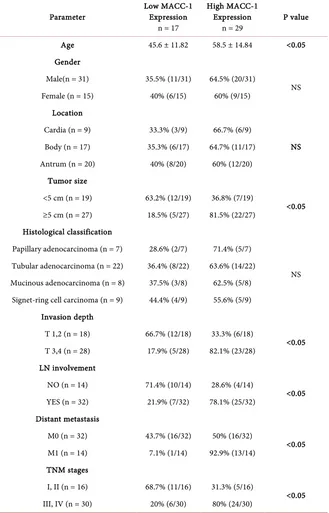
Clinical & pathological characteristics in relation to MACC-1 expression are presented in Table 1. The mean age of the patients was 53.2 ± 11.9. Patients with high MACC-1 expression were significantly older than those with low expres-sion (58.5 ± 14.84 vs 45.6 ± 11.8; p < 0.05) with 67.4% being males and 32.6% females. MACC-1 high-expression was found in 81.5% (22/27) of tumors that were more than 5 cms in size compared to only 36.8% (7/19) of tumors that were <5 cms (p < 0.05). Depth of invasion was found to be significantly associated with high MACC-1 expression as 82.1% (23/28) of T3 and T4 tumors had high
(A) (B)
[image:4.595.211.537.450.644.2](C) (D)
DOI: 10.4236/jct.2019.107050 613 Journal of Cancer Therapy
Table 1. Clinical & pathological characteristics in relation to MACC-1 expression.
Parameter Low MACC-1 Expression n = 17
High MACC-1 Expression
n = 29 P value
Age 45.6 ± 11.82 58.5 ± 14.84 <0.05
Gender
Male(n = 31) 35.5% (11/31) 64.5% (20/31)
NS Female (n = 15) 40% (6/15) 60% (9/15)
Location
Cardia (n = 9) 33.3% (3/9) 66.7% (6/9)
NS Body (n = 17) 35.3% (6/17) 64.7% (11/17)
Antrum (n = 20) 40% (8/20) 60% (12/20) Tumor size
<5 cm (n = 19) 63.2% (12/19) 36.8% (7/19)
<0.05 ≥5 cm (n = 27) 18.5% (5/27) 81.5% (22/27)
Histological classification
Papillary adenocarcinoma (n = 7) 28.6% (2/7) 71.4% (5/7)
NS Tubular adenocarcinoma (n = 22) 36.4% (8/22) 63.6% (14/22)
Mucinous adenocarcinoma (n = 8) 37.5% (3/8) 62.5% (5/8) Signet-ring cell carcinoma (n = 9) 44.4% (4/9) 55.6% (5/9)
Invasion depth
T 1,2 (n = 18) 66.7% (12/18) 33.3% (6/18)
<0.05 T 3,4 (n = 28) 17.9% (5/28) 82.1% (23/28)
LN involvement
NO (n = 14) 71.4% (10/14) 28.6% (4/14)
<0.05 YES (n = 32) 21.9% (7/32) 78.1% (25/32)
Distant metastasis
M0 (n = 32) 43.7% (16/32) 50% (16/32)
<0.05 M1 (n = 14) 7.1% (1/14) 92.9% (13/14)
TNM stages
I, II (n = 16) 68.7% (11/16) 31.3% (5/16)
<0.05 III, IV (n = 30) 20% (6/30) 80% (24/30)
DOI: 10.4236/jct.2019.107050 614 Journal of Cancer Therapy
that of patients with low expression (20%, n = 6/30). In contrast, MACC-1 ex-pression showed no significant relation with gender, tumor location or histolog-ical classification.
Survival analysis
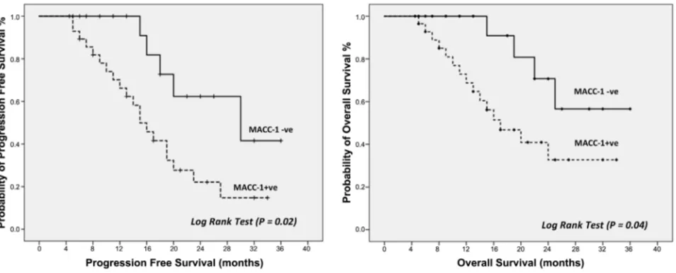
The median progression free survival for patient with low MACC-1 expres-sion was 24 months (95% CI; 11.86 - 31.47) compared to 15 months (95% CI; 10.01 - 17.91) for patient with high MACC-1 expression. The median overall survival for patients with low MACC-1 expression was not reached, while that of patients with high expression was 17 months (95% CI; 10.36 - 21.64).
Progression free and overall survival were significantly higher in patients with low MACC-1 expression compared to patients with high expression (Log Rank test, p = 0.02 and 0.04, consequently) (Figure 2).
4. Discussion
In Egypt, gastric cancer is the 12th most common cancer in both sexes representing
1.6% of the total cancers. It is the 12th leading cause of cancer death representing
2.2% of the total cancer mortality. The median age of GC in the Egyptians is 56 years. The incidence rises with age and 55% of cases occur between 50 and 70 years of age [19].
[image:6.595.63.540.514.707.2]A number of studies have shown that gastric cancer progression, invasion and metastasis are associated with distinct gene expression profiles [1] [20]. In order to identify genes differentially expressed in gastric cancer, gene expression in gastric cancer specimens and healthy tissues were profiled using microarrays. Four hundred thirty-four genes were identified of which, 169 expressed sequence tags (ESTs) that were up-regulated at least 2 folds in primary gastric carcinomas and of these, MACC-1 was found to be one of the most highly upregulated genes [19] [20]. In our study, we verified this microarray data by immune-histo-chemical analysis. We found that gastric cancer specimens expressed high levels of MACC-1,
DOI: 10.4236/jct.2019.107050 615 Journal of Cancer Therapy
while normal gastric mucosa showed low MACC-1 expression. This adds a lot of value because its presence means at least that tissue is not normal. On the other hand, expression of MACC-1 immediately after surgery means incomplete re-moval of cancer. While its presence after means recurrence or new growth. This adds to importance of MACC-1 expression new dimension. These results are not identical to that of Duan et al., (2017), which showed low expression of MACC-1 in few cases of normal gastric tissue adjacent to cancer tissue. Actually, these re-sults weaken the clinical value of MACC-1 expression because negative rere-sults may occur in cancer and positive expression may occur in normal tissue but we could explain these few cases by inaccurate sampling from tissues still not far enough from cancer tissues without clear safety margin. Even if these results were correct still high expression of MACC-1 carry the hope of being valuable marker especially no one single case of normal gastric tissues showed high ex-pression of MACC-1 only very weak exex-pression. Importantly, in our study high MACC-1 expression was significantly correlated with more aggressive clin-ic-pathological features related to size, depth of invasion, lymph node and dis-tant metastases. These findings match with the study done by Jie Ma et al. [3], which showed the same results. Another very valuable point, correlation with severity of the lesions which make value of MACC-1 expression extend to more than a diagnostic tool as in our study, high expression of MACC-1 was not sig-nificantly different among different histological pattern of gastric carcinoma such as papillary, tubular or other forms (p > 0.05) but the situation is much dif-ferent as regard clinical staging. There is very highly significant difference among patients in late stages (III, IV) as almost all the patient showed high ex-pression (92.2%) with exception only one patient in early stages (I, II) showed high expression. Such findings take MACC-1 expression to another dimension to be a severity marker and may be used for follow up or even therapy success marker.
expres-DOI: 10.4236/jct.2019.107050 616 Journal of Cancer Therapy
sion and promotes tumor cell invasion and metastasis [6]. Aberrant activation of the hepatocyte growth factor (HGF)/HGF receptor (MET) signaling pathway is well known documented putative mechanism associated with both malignant transformation and metastatic potential of tumors [3] [5].
5. Conclusion
Our study found that MACC-1 expression is strongly related to gastric cancer stage and both progression free and overall survival, suggesting that MACC-1 promote tumorigenesis and its expression may be used as signal of bad progno-sis in gastric cancer.
Limitation of the study includes limited patient number and lack of long
term follow up that could be solved in future studies in order to increase the ac-curacy of statistics and add another dimension to MACC-1 as prognostic mark-er.
Conflicts of Interest
The authors declare no conflicts of interest regarding the publication of this pa-per.
References
[1] Duan, J., Chen, L., Zhou, M. and Liao, W. (2017) MACC1 Decreases the Chemo-sensitivity of Gastric Cancer Cells to Oxaliplatin by Regulating FASN Expression.
Oncology Reports, 37, 2583-2592.https://doi.org/10.3892/or.2017.5519
[2] Smith, B.R. and Stabile, B.E. (2009) Extreme Aggressiveness and Lethality of Gastric Adenocarcinoma in the Very Young. The Archives of Surgery, 144, 506-510.
https://doi.org/10.1001/archsurg.2009.77
[3] Ma, J., Ma, J., Meng, Q., Zhao, Z.-S. and Xu, W.-J. (2013) Prognostic Value and Clinical Pathology of MACC-1 and c-MET Expression in Gastric Carcinoma. Pa-thology & Oncology Research, 19, 821-832.
https://doi.org/10.1007/s12253-013-9650-0
[4] Okines, A.F., Reynolds, A.R. and Cunningham, D. (2011) Target in Angiogenesis in Esophagogastric Adenocarcinoma. Oncologist, 16, 844-858.
https://doi.org/10.1634/theoncologist.2010-0387
[5] Lai, L. and Gold Schneider, I. (2001) Cutting Edge: Identification of a Hybrid Cyto-kine Consisting of IL-7 and the Beta-Chain of the Hepatocyte Growth Consisting of IL-7 and the Beta-Chain of the Hepatocyte Growth Factor/Scatter Factor. Immu-nology, 167, 3550-3554. https://doi.org/10.4049/jimmunol.167.7.3550
[6] Fock, K.M. (2014) Review Article: The Epidemiology and Prevention of Gastric Cancer. Alimentary Pharmacology & Therapeutics, 40, 250-260.
https://doi.org/10.1111/apt.12814
[7] Mikuriya, Y., Tashiro, H., Kuroda, S.,Nambu, J., Kobayashi, T., Amano, H., Tana-ka, Y. and Ohdan, H. (2015) Fatty Liver Creates a Pro-Metastatic Microenviron-ment for Hepatocellular Carcinoma through Activation of Hepatic Stellate Cells.
International Journal of Cancer, 136, E3-E13.https://doi.org/10.1002/ijc.29096
DOI: 10.4236/jct.2019.107050 617 Journal of Cancer Therapy
11-18.https://doi.org/10.1007/s00109-009-0537-1
[9] Sierra, J.R. and Tsao, M.S. (2011) c-MET as a Potential Therapeutic Target and Biomarker in Cancer. Therapeutic Advances in Medical Oncology, 3, S21-S35.
https://doi.org/10.1177/1758834011422557
[10] Xie, Q.P., Xiang, C., Wang, G., Lei, K.F. and Wang, Y. (2016) MACC1 Upregulation Promotes Gastric Cancer Tumor Cell Metastasis and Predicts a Poor Prognosis.
Journal of Zhejiang University Science B, 17, 361-366.
https://doi.org/10.1631/jzus.B1500236
[11] Wang, L., Wu, Y., Lin, L.,Liu, P., Huang, H., Liao, W., Zheng, D., Zuo, Q., Sun, L., Huang, N., et al. (2013) Metastasis-Associated in Colon Cancer 1 Upregulation Pre-dicts a Poor Prognosis of Gastric Cancer, and Promotes Tumor Cell Proliferation and Invasion. International Journal of Cancer, 133, 1419-1430.
https://doi.org/10.1002/ijc.28140
[12] Wang, G., Fu, Z. and Li, D. (2015) MACC1 Overexpression and Survival in Solid Tumors: A Meta-Analysis. Tumor Biology, 36, 1055-1065.
https://doi.org/10.1007/s13277-014-2736-9
[13] Kokoszyńska, K., Kryński, J., Rychlewski, L., et al. (2009) Unexpected Domain Composition of MACC1 Links MET Signaling and Apoptosis. Acta Biochimica Po-lonica, 56, 317-323. https://doi.org/10.18388/abp.2009_2464
[14] Lang, A.H., Geller-Rhomberg, S., Winder, T., et al. (2012) A Common Variant of the MACC1 Gene Is Significantly Associated with Overall Survival in Colorectal Cancer Patients. BMC Cancer, 12, 20. https://doi.org/10.1186/1471-2407-12-20
[15] Lee, H.S., Cho, S.B., Lee, H.E., et al. (2007) Protein Expression Profiling and Mole-cular Classification of Gastric Cancer by the Tissue Array Method. Clinical Cancer Research, 13, 4154-4163. https://doi.org/10.1158/1078-0432.CCR-07-0173
[16] Mizokami, K., Kakeji, Y., Oda, S., et al. (2006) Clinicopathologic Significance of Hypoxia-Inducible Factor 1 Alpha Overexpression in Gastric Carcinomas. Journal of Surgical Oncology, 94, 149-154. https://doi.org/10.1002/jso.20568
[17] Song, L.B., Liao, W.T., Mai, H.Q., et al. (2006) The Clinical Significance of Twist Expression in Nasopharyngeal Carcinoma. Cancer Letters, 242, 258-265.
https://doi.org/10.1016/j.canlet.2005.11.013
[18] Kolev, Y., Uetake, H., Iida, S., Ishikawa, T., Kawano, T. and Sugihara, K. (2007) Prognostic Significance of VEGF Expression in Correlation with COX-2, Mi-cro-Vessel Density, and Clinicopathological Characteristics in Human Gastric Car-cinoma. Annals of Surgical Oncology, 14, 2738-2747.
https://doi.org/10.1245/s10434-007-9484-7
[19] Ferlay, J., Soerjomataram, I., Ervik, M., Dikshit, R., Eser, S. and Mathers, C. (2015) Cancer Incidence and Mortality Worldwide. International Journal of Cancer, 136, E359-E386.https://doi.org/10.1002/ijc.29210
[20] Xu, Z.Y., Chen, J.S. and Shu, Y.Q. (2010) Gene Expression Profile towards the Pre-diction of Patient Survival of Gastric Cancer. Biomedicine & Pharmacotherapy, 64, 133-139. https://doi.org/10.1016/j.biopha.2009.06.021
[21] Lin, L., Huang, H., Liao, W., Ma, H., Liu, J., Wang, L., Huang, N., Liao, Y. and Liao, W. (2015) MACC1 Supports Human Gastric Cancer Growth under Metabolic Stress by Enhancing the Warburg Effect. Oncogene, 34, 2700-2710.
https://doi.org/10.1038/onc.2014.204
DOI: 10.4236/jct.2019.107050 618 Journal of Cancer Therapy
(2015) Metastasis-Associated in Colon Cancer-1 Upregulates Vascular Endothelial Growth Factor-C/D to Promote Lymphangiogenesis in Human Gastric Cancer.