ISSN Online: 2331-1967 ISSN Print: 2331-1959
DOI: 10.4236/ad.2019.74012 Oct. 11, 2019 257 Archaeological Discovery
Silver and Gold on the Hairs of Holy
Maria-Magdalena, Studied by Scanning Electron
Microscopy and Elemental Analysis
Gérard Lucotte
Institut d’Anthropolgie Moléculaire, Paris, France
Abstract
We have studied by optical microscopy and by SEM-EDX some metallic par-ticles of silver and gold adhering to the Maria-Magdalena hairs. The presence of silver particles is explained by the contact between hairs and the inner side made of silver of the initial reliquary where Maria-Magdalena remains were kept. Presence of gold particles is also explained by the contact between hairs and the gold of the bust-reliquary where hairs of Maria-Magdalena hairs were kept between 1283 and 1793.
Keywords
Maria-Magdalena’s Hair, SEM-EDX Analyses, Silver and Gold Particles, Successive Reliquaries Where Maria-Magdalena’s Remains Were Kept, Calcium Phosphate Deposits, Other Metallic Particles
1. Introduction
Holy Maria-Magdalena (3?-63?) is the most abundantly cited woman in the four Gospels. There were some places (notably Palestine) where Maria-Magdalena could be buried, but according to the French “tradition des Saints de Pro-vence” (Trouillet, 2016) she (and her companions) landed to the present French Mediterranean shores (in a region corresponding to the current part of Les-Saintes-Marie-de-la-mer) and attained further the towns of Marseilles and Aix-en-Provence.
Some relics (cranium, bones and hairs) of the presumed Maria-Magdalena were kept in the Saint-Maximin basilica, where a large lock of Ma-ria-Magdalena’s hair is kept in a dedicated reliquary. We have obtained some hairs from this lock, for scientific purposes (microscopic examinations and How to cite this paper: Lucotte, G. (2019).
Silver and Gold on the Hairs of Holy Ma-ria-Magdalena, Studied by Scanning Elec-tron Microscopy and Elemental Analysis. Archaeological Discovery, 7, 257-282. https://doi.org/10.4236/ad.2019.74012
Received: August 31, 2019 Accepted: October 8, 2019 Published: October 11, 2019
Copyright © 2019 by author(s) and Scientific Research Publishing Inc. This work is licensed under the Creative Commons Attribution International License (CC BY 4.0).
G. Lucotte
DOI: 10.4236/ad.2019.74012 258 Archaeological Discovery
chemical analyses). We have published these last years the mitochondrial DNA haplogroup found by extracting genomic DNA from the bulb of hair number 10 (Lucotte, 2016), the explanation of the brown-red observed colour of the hairs by scanning electron microscopic characterisation of its melanosomes (Lucotte & Thomasset, 2017a), the description of some fennel rests on or at the vicinity of some of these hairs (Lucotte et al., 2018) and marine micro-remains loaded on the hairs (Lucotte et al., 2019).
In the present study, we describe and analyse silver and gold metallic deposits on some of these hairs, by SEM (Scanning Electron Microscopy) and EDX (Energy Dispersive X-ray spectroscopy) analysis.
2. Material and Methods
The material is ten of the lock of Maria-Magdalena’s hairs that were kept in the dedicated reliquary located in the Saint-Maximin basilica. These hairs, num-bered 1 to 10, were loaded on a sterile sticky paper for optical microscopy, SEM and EDX analysis.
All the hairs were examined in confocal stereoscopic micrography. The SEM apparatus used for metallic deposits observations is the FEI model Quanta FEG (an environmental electron microscope apparatus). Elemental analysis of depo-sits was achieved by using EDX, this SEM microscope being equipped with the probe model X-flash 6/30. Both LFD (Large Field Detector) and CBS (Circular Back Scattering) were used, the last one to better detect heavy elements. Each elemental analysis is given in the form of a spectrum, with kiloelectrons/Volts (ke/V) on the abscissa and elemental peaks heights (cps/eV) in ordinates.
3. Results and Discussion
There are numerous particles of silver and of gold on the surface of hair num-bers 1, 2, 3, 4 and 9. As an example Figure 1 shows an optical view of some por-tion of hair number 2 where silver is present under the form of a longitudinal line running along all the hair border, and gold under the form of three local deposits.
3.1. Silver
Figure 2 shows a MEB photograph of some part of the longitudinal line; EDX analysis establishes that it is mainly compounded of silver (of the chloride form, which is the most stable). Two other parts of the line are shown on Figure 3, which are of the same elemental composition. In a fourth part of the line (Figure 4), the height of the chlorine peak is as elevated as that of the silver main peak.
DOI: 10.4236/ad.2019.74012 259 Archaeological Discovery
Figure 1. Optical view (20×) of a portion of hair number 2. S:
silver; G: gold.
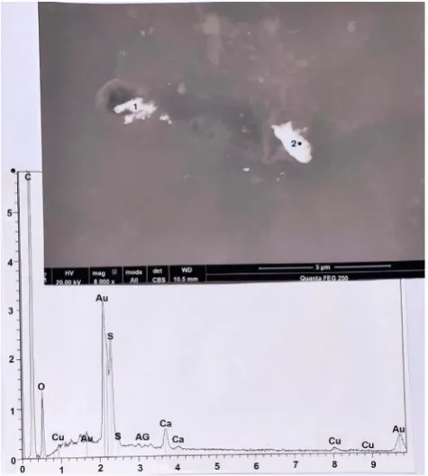
Figure 2. One deposit of silver chloride on hair number 2.
Above: SEM photograph (in CBS, 400×) of the deposit (the black point indicates the location where EDX analysis is rea-lized). Below: spectrum at the black point. C: carbon; O: oxygen; S: sulphur; Cl: chlorine; Ag (three peaks): silver; Ca (two peaks): calcium.
G. Lucotte
[image:4.595.263.485.71.338.2]DOI: 10.4236/ad.2019.74012 260 Archaeological Discovery
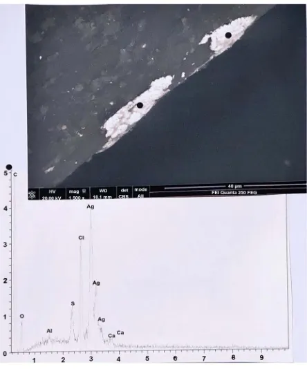
Figure 3. Two other deposits of silver chloride on hair number 2.
Above: SEM photograph (in CBS, 1500×) of the two deposits. Below:
spectrum at the black points.
Figure 4. A fourth deposit of silver chloride on hair number
2. Above: SEM photograph (in CBS, 2500×) of the deposit.
[image:4.595.263.485.388.664.2]DOI: 10.4236/ad.2019.74012 261 Archaeological Discovery
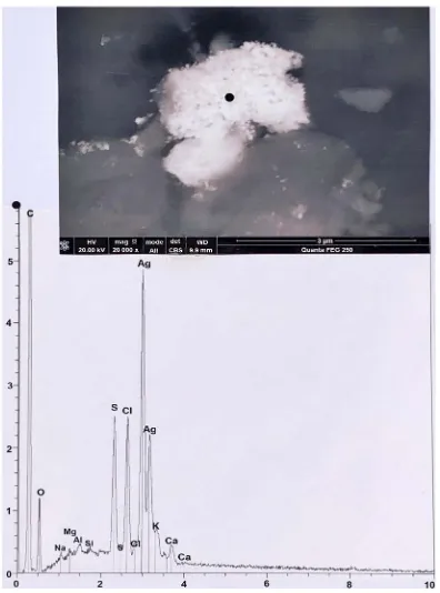
Figure 5. A particle, located on hair number 1 that is a
mix-ture of silver chloride and sulphide. Above: SEM photograph (in CBS, 20000×) of the particle. Below: spectrum at the black point. C: carbon; O: oxygen; Na: sodium; Mg: magne-sium; Al: aluminium; Si: silicium; S (two peaks): sulphur; Cl (two peaks): chlorine; Ag (two peaks): silver; K: potassium; Ca (two peaks): calcium.
Figure 6. Another particle located on hair number 1 that is
[image:5.595.276.469.444.629.2]G. Lucotte
[image:6.595.263.483.73.321.2]DOI: 10.4236/ad.2019.74012 262 Archaeological Discovery
Figure 7. A plaque, located on hair number 1 that is of silver
sulphide with copper. Above: SEM photograph (in CBS, 8000×) of the plaque. Below: spectrum at the black point. C: carbon; O: oxygen; Cu (three little peaks): copper; S (two peaks): sulphur; Ag (four peaks): silver; Ca (two peaks): cal-cium.
Figure 8. A voluminous plaque of pure silver, that is located
[image:6.595.263.486.421.654.2]DOI: 10.4236/ad.2019.74012 263 Archaeological Discovery
3.2. Gold
Figure 9 is a SEM photograph of one of the three particles of gold, depicted on Figure 1, located on hair number 2; it is a multi-lobed particle of about 1.5 µm of maximal length. Its elemental analysis establishes that it is mainly com-pounded of gold (in fact an alloy, with 8.5% of silver and 2.2% of copper). The sulphur content in the sample is, as for all of the gold particles studied here, more elevated than that found in ancient hairs (Lucotte & Thomasset, 2017b); it is deduced that this elemental level in sulphur is mainly due to pollution. The composition of the particle located on hair number 3 (Figure 10) is also that of an alloy of gold, silver and copper. But in the plaque, also located on hair num-ber 3 (Figure 11), the alloy is mainly of gold with traces of copper; it is also the case for a second smaller plaque (Figure 12), located on hair number 1, and for a third plaque (Figure 13), but with silver, located on hair number 1.
Figure 14 shows an example of grains of gold dust, located on hair number 9. Figure 15 shows examples of two little particles of gold (of a gold powder), lo-cated on hair number 3. Figure 16 shows an example of a scale of pure gold, of a relatively great size (of more than 2 µm of maximal length), which is located on hair number 9.
3.3. Copper
There is only one piece of pure copper (Supplementary Figure 1). It is a little plaque of this metal, located on the surface of hair number 3. Most of the cop-per-compounded pieces found are particle of brass: Supplementary Figure 2 shows one of them, also located on hair number 3; there are other plaques of brass, located near hair number 6 and between hairs 9 and 10. Probably they are fragments pulled up to the metal of the reliquary. Supplementary Figure 3 shows a group of little copper-made particles (calibrated at about 5 µm of length, and of a manufactured form), located on some part of hair number 6. Elemental analyses establish that they are particles of copper sulphate; they are powder members of a classical phytosanitary product. Such groups of copper sulphate particles occur also on two other parts of hair number 6, and on hair number 10.
3.4. Other Metallic Particles
3.4.1. LeadThere is only one piece compounded of lead (Supplementary Figure 4), that is located on hair number 9. It is a round (less than 5 µm) particle of composite appearance; elemental analysis establishes that it corresponds to lead phosphate, another phytosanitary product used in the past.
3.4.2. Iron
G. Lucotte
[image:8.595.263.485.70.289.2]DOI: 10.4236/ad.2019.74012 264 Archaeological Discovery
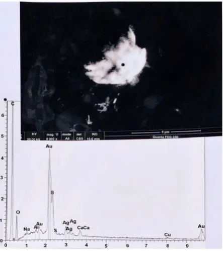
Figure 9. A particle of gold, located on hair number 2.
Above: SEM photograph (in CBS, 20000×) of the particle.
Below: spectrum at the black point. C: carbon, O: oxygen; Cu (three peaks): copper; Mg: magnesium; Al: aluminium; Si: si-licium; Au (two peaks): gold; S: sulphur; Ag: silver; K: potas-sium; Ca (two peaks): calcium. Insert: relative percentages of gold, copper and silver in the alloy.
Figure 10. A particle of gold, located on hair number 3.
[image:8.595.265.487.395.648.2]DOI: 10.4236/ad.2019.74012 265 Archaeological Discovery
Figure 11. A plaque of gold, located on hair number 3.
Above: SEM photograph (in CBS, 30000×) of the plaque. Be-low: spectrum at the black point. C: carbon; O: oxygen; Cu (two peaks): copper; Na: sodium; Al: aluminium; Au (three peaks): gold; Si: silicium; S (two peaks): sulphur; Ca (two peaks): calcium.
Figure 12. A second plaque of gold, located on hair number
1. Above: SEM photograph (in CBS, 24000×) of the plaque.
G. Lucotte
[image:10.595.239.505.73.316.2]DOI: 10.4236/ad.2019.74012 266 Archaeological Discovery
Figure 13. A third plaque of gold, located on hair number 1. Above:
SEM photograph (in CBS, 20000×) of the plaque. Below: spectrum at the black point. C: carbon; O: oxygen; Cu (two little peaks): copper; Au (three peaks): gold; Ag: silver; S (two peaks): sulphur; K: potassium; Ca (two peaks): calcium.
Figure 14. Grains of gold dust, on hair number 9. Above: SEM
[image:10.595.242.505.403.662.2]DOI: 10.4236/ad.2019.74012 267 Archaeological Discovery
Figure 15. Two little particles of gold, on hair number 3. Above:
SEM photography (in CBS, 8000×) of the particles 1 and 2. Below:
spectrum at the black point of 2. C: carbon; O: oxygen; Cu (three peaks): copper; Au (three peaks): gold; S (two peaks): sulphur; Ag: silver; Ca (two peaks): calcium.
Figure 16. A scale of pure gold, located on hair number 9.
Above: SEM photograph (in CBS, 30000×) of the scale. Below:
[image:11.595.249.498.420.650.2]G. Lucotte
DOI: 10.4236/ad.2019.74012 268 Archaeological Discovery
particles are present in number on hairs numbers 1, 2 and 6. Supplementary Figure 6 shows a white-to-electrons particle, located on the hair number 6 bor-der near a transversal alteration of that hair. It is complex in form, compounded of little spherical (showing that it corresponds to some material in fusion) sub-particles. Elemental analysis establishes that it is mainly of pure iron. Such spherical sub-particles, isolated or lumped together, were found on the surfaces of hairs numbers 1, 2 and 3. There are also numerous little particles (of pure or of oxided iron), of various forms, on all hair surfaces; they correspond to com-mon industrial modern pollutants. Supplementary Figure 7 shows a very little (of about 1 µm) particle, located on hair number 1. It is compounded of iron sulphate, a third common phytosanitary product. Such particles, in isolated form or in groups, are found on hair number 2, 3, 5, 9 and 10.
3.4.3. Titanium
Supplementary Figure 8 shows a rod of titanium, located on some part of hair number 3. It is compounded of titanium dioxide (TiO2), the most common me-tallic pollutant after iron. Two other samples of such a rod are found on other parts of hair number 3. Supplementary Figure 9 shows a micro-ball of tita-nium, located on hair number 10 (elemental analysis establishes that titanium is the metallic component of this micro-ball). This unique observation corresponds probably to a droplet of modern white colorant (of titanium), loaded on a bed of calcium carbonate.
3.4.4. Aluminium
Supplementary Figure 10 shows three micro-fragments of aluminium, located at the border of hair number 1; they caused here a local alteration of the hair surface. Elemental analysis establishes that the aluminium is of the oxided form (it corresponds so to a modern aluminium scrop).
3.4.5. Mercury
Supplementary Figure 11 and Supplementary Figure 12 show two cro-drops of mercury, located on hair number 1. Elemental analysis of these mi-cro-drops shows that they are compounded of cinnabar (HgS), a red colorant used since the Middle Age to imitate blood spots (Lucotte et al., 2016). A third micro-drop of cinnabar, located on hair number 3, is shown on Supplementary Figure 13.
3.5. Calcium Phosphate Particles
All of the ten hairs studied have deposits of calcium phosphate on their surfaces. That is important to consider, because hydroxyapatite remains the mineral component found in ancient bones (Lucotte & Thomasset, 2017c).
DOI: 10.4236/ad.2019.74012 269 Archaeological Discovery
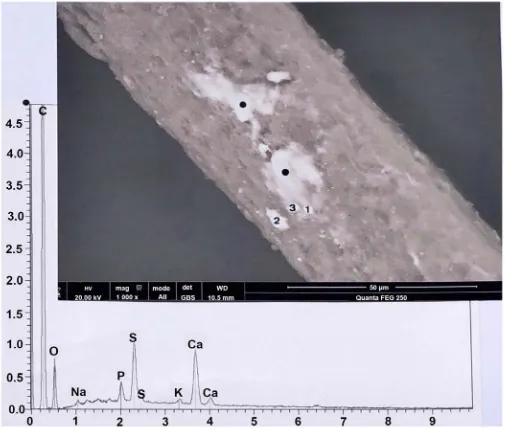
Figure 17. An example of a thin deposit of calcium phosphate,
lo-cated in some part of hair number 3. Above: SEM photograph (in CBS, 1000×) of the deposit (1-3: calcite particles). Below: spectrum at the black points. C: carbon; O: oxygen; Na: sodium; P: phosphor-ous; S (two peaks): sulphur; K: potassium; Ca (two peaks): calcium.
Figure 18. Longitudinal line of six calcium phosphate deposits in
[image:13.595.249.501.372.658.2]G. Lucotte
[image:14.595.249.501.72.399.2]DOI: 10.4236/ad.2019.74012 270 Archaeological Discovery
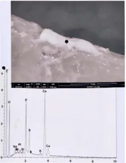
Figure 19. Example of a bone scale (in another part of hair 3).Above:
SEM photograph (in CBS, 2400×) of the bone scale. Below: spectrum at the black point. C: carbon; O: oxygen; Na: sodium; Mg: magne-sium; Al: aluminium; Si: silicium; P: phosphorous; S (two peaks): sulphur; K: potassium; Ca (two peaks): calcium.
In other cases the bone scales appear clearly as loaded on the hair surface (Figure 20). The most extreme situation of osseous scale loading is represented on Figure 21: a voluminous (of more than 50 µm of maximal length) scale of calcium phosphate, very thick and longitudinally fragmented.
In summary, I found on the hairs numerous particles of silver and gold, but also some metallic particles of copper, lead, iron, titanium, aluminium and mer-cury. All the hairs have deposits of calcium phosphate on their surfaces.
DOI: 10.4236/ad.2019.74012 271 Archaeological Discovery
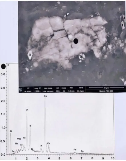
Figure 20. A bone scale, located on hair number 6. Above:
SEM photograph (in CBS, 2000×) of the scale. Below: spectrum at the black point. C: carbon; O: oxygen; Na: sodium; Al: alu-minium; Si: silicium; P: phosphorous; S: sulphur; K: potassium; Ca (two peaks): calcium; Fe (two peaks): iron.
Figure 21. A thick plaque of bone, located on hair number 9.
G. Lucotte
DOI: 10.4236/ad.2019.74012 272 Archaeological Discovery
Figure 22. The ancient (since 1283) bust-reliquary of Maria-Magdalena.
N: golden mask; L: crown of gold; R: bust-reliquary of gold (source: col-lection Lallemant de Betz; Bibliothèque Nationale de France, Paris).
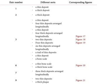
Table 1. Calcium phosphate deposits found on hairs.
Hair number Different sorts Corresponding figures
1 a thin deposit a thick deposit
2 a thick deposit
3
a thin deposit
four thin deposits arranged
longitudinally
a thin deposit
four thick deposits arranged
longitudinally
two thin deposits
Four thin deposits
six thin deposits arranged
longitudinally
a trail of thin deposits
a thin deposit
[image:16.595.206.539.348.635.2] a bone scale
Figure 17 Figure 18 Figure 19
6 a thin bone scale a thick bone scale Figure 20
7 three thick deposits arranged longitudinally
9 two thin deposits a thick plaque Figure 21
Table 2 summarizes all the observations of gold particles on hairs. Probably most of them (particularly those that are of pure gold) are due to close contact between hairs and the gold of the bust-reliquary and that of the crown.
DOI: 10.4236/ad.2019.74012 273 Archaeological Discovery
Table 2. Gold deposits found on hairs.
Hair number Different sorts Corresponding figures
1
a plaque of gold alloy (Au/Ag)
Four little particles of a gold powder
two little particles of a gold powder
two little particles of a gold alloy (Au/Ag/Cu) powder
a second plaque of gold alloy (Au/Ag/Cu)
Figure 12 Figure 13
2
three little particles of a gold alloy (Au/Ag/Cu) powder
a little poly-lobed particle of a gold alloy (Au/Ag/Cu)
six little particles of a gold powder
Figure 9
3
a particle of gold alloy (Au/Ag/Cu)
a plaque of gold alloy (Au/Cu)
two particles of gold alloy (Au/Ag/Cu)
three particles of a gold alloy (Au/Ag/Cu) of a powder
Figure 10 Figure 11 Figure 15
9
a little plaque of pure gold
grains of pure gold dust
a particle of pure gold
Figure 14 Figure 16
Table 3. Silver deposits found on hairs.
Hair number Different sorts Corresponding figures
1
a particle that is a mixture of silver sulphide and chloride
a particle of silver sulphide and gold
a plaque of silver with copper
a granulous particle of silver sulphide
a little particle of silver sulphide
Figure 5 Figure 6 Figure 7
2
a little particle of silver sulphide
about fifteen particles of silver sulphide arranged among the
longitudinal axis of the hair
one plaque of silver chloride on the border of the hair
two plaques of silver chloride on the border of the hair
one plaque of silver chloride on the border of the hair
Figure 2 Figure 3 Figure 4
3
a little plaque of silver sulphide
a particle of silver sulphide/chloride
many little plaques of silver sulphide (with copper traces),
arranged longitudinally
two little plaques of silver sulphide
eight little plaques of silver sulphide arranged longitudinally
a turmoiled little particle of silver sulphide
4 a large plaque of pure silver Figure 8
9 two little plaques of silver sulphide one little pile of particles of silver sulphide, with copper
silver reliquary; possibly some of the silver particles observed on hairs were due to initial contact with the inner side of the reliquary.
4. Conclusion
G. Lucotte
DOI: 10.4236/ad.2019.74012 274 Archaeological Discovery
forms differ: sometimes it is a very thin layer of calcium phosphate loaded on the surface of the hairs; but it can be a thicker deposit, with visible borders. Some-times they consist of plaques of calcium phosphate that mimic a true osseous structure. The trails of calcium observed are often arranged in longitudinal lines along the hair corpus.
Such observations indicate a previous close contact between hairs and some bone. We know that hairs covered cranium in the historical (between 1283 and 1793) bust-reliquary (see Figure 22) where Maria-Magdalena remains were kept. It is during this long time period that the hydroxyapatite of the cranium loaded on hairs.
Because the bust-reliquary was of gold (and also the crown that covered hairs), some of the gold plaques observed on some hairs (particularly those that are of pure gold, like that of Figure 16) can be detached among time from the gold of the bust-reliquary to the hair surface. Generally, gold particles deposited on hairs are made of an alloy of gold with little amounts of silver and copper.
The numerous plaques or silver sulphide (or/and chloride) observed at the surface of some hairs are more difficult to explain; often these plaques are also orientated in longitudinal lines along the hair surface. Because we know that the initial (since 1281) reliquary where the Maria-Magdalena remains were kept was of silver, we suppose that some of the silver plaques observed (particularly those that are of pure silver, like that of Figure 8) were also detached among time from the silver of the inner side of this reliquary.
Other metallic particles observed at the hair surfaces are in fact explained as residues of deposits from the current reliquary, or as traces of phytosanitary products or colorants, or as modern industrial metallic pollutants.
Acknowledgements
We thank F. Racine, the priest of Saint-Maximin-la-Sainte-Baume, who fur-nished Maria-Magdanena’s hairs. Thank you also to T. Thomasset (UTC of Compiègne) for his assistance on the SEM. The present article is a development of some part of the conference I have pronounced at the Saint-Maximin-basilica on the 23rd of July 2016.
Conflicts of Interest
The author declares no conflicts of interest regarding the publication of this pa-per.
References
Franzoni, A. (2016). Sainte Marie-Madeleine et les Saints de Provence dans la Tradition Provençale (Vol. 3). Sainte-Baume: Editions ASTSP.
Lucotte, G. (2016). The Mitochondrial DNA Mitotype of Sainte Marie-Madeleine. Inter-national Journal of Sciences, 5, 10-19. https://doi.org/10.18483/ijSci.1167
DOI: 10.4236/ad.2019.74012 275 Archaeological Discovery Journal of Dermatology and Pigmentation Research, 1, 108-116.
Lucotte, G., & Thomasset, T. (2017b). Scanning Electron Microscopy Characterization and Elemental Analysis of One Hair Located on the Face of the Turin Shroud. Arc-haeological Discovery, 5, 1-21. https://doi.org/10.4236/ad.2017.51001
Lucotte, G., & Thomasset, T. (2017c). An Osseous Remain on the Face of the Turin Shroud. Journal of Anthropology and Archaeology, 5, 20-38.
https://doi.org/10.15640/jaa.v5n1a3
Lucotte, G., D’Hérissart, E., & Thomasset, T. (2019). Marine Micro-Remains on Holy Maria-Magdalena’s Hair, Studied by Scanning Electron Microscopy and Elemental Analysis. Archaeological Discovery, 7, 155-191. https://doi.org/10.4236/ad.2019.73009
Lucotte, G., Derouin, T., & Thomasset, T. (2016). Hematite, Biotite and Cinnabar on the Face of the Turin Shroud: Microscopy and SEM-EDX Analysis. Open Journal of Ap-plied Sciences, 6, 601-625. https://doi.org/10.4236/ojapps.2016.69059
Lucotte, G., Thomasset, T., & Salmon, A. (2018). Fennel (Foeniculum vulgare) Rests on the Holy Maria-Magdalena’s Hairs, Studied by Scanning Microscopy and Elemental Analysis. Archaeological Discovery, 6, 216-270. https://doi.org/10.4236/ad.2018.63012
G. Lucotte
DOI: 10.4236/ad.2019.74012 276 Archaeological Discovery
[image:20.595.256.491.92.298.2]Supplementary Figures
Figure S1. A plaque of copper, located on hair number 3. Above:
SEM photograph (in CBS, 8000×) of the plaque. Below: spectrum at the black point. C: carbon, O: oxygen; Cu (three peaks): cop-per; Al: aluminium; S (two peaks): sulphur; Cl: chlorine; Ca (two peaks): calcium.
Figure S2. A plaque of brass, located on hair number 3. Above:
[image:20.595.263.485.380.658.2]DOI: 10.4236/ad.2019.74012 277 Archaeological Discovery
Figure S3. Particles of copper sulphate, located in one part of hair
number 6. Above: SEM photograph (in CBS, 2000×) of the group of particles (m: mycelium; s: spores). Below: spectrum at the black points. C: carbon; O: oxygen; Cu (three peaks): copper; Al: alumi-nium; S: sulphur; K: potassium; Ca (two peaks): calcium.
Figure S4. A particle of lead phosphate, located on hair number 9.
[image:21.595.248.502.399.641.2]G. Lucotte
[image:22.595.246.502.73.324.2]DOI: 10.4236/ad.2019.74012 278 Archaeological Discovery
Figure S5. A particle of iron oxide, located on the border of hair
number 3. Above: SEM photograph (in CBS, 2000×) of the particle.
Below: spectrum at the black point. C: carbon; O: oxygen; Fe (three peaks): iron; Si: silicium; S (two peaks): sulphur; K: potassium; Ca (two peaks): calcium.
Figure S6. Particles of iron, located at the border of hair number 6.
Above: SEM photograph (in CBS, 2500×) of the group of particles.
[image:22.595.247.501.401.665.2]DOI: 10.4236/ad.2019.74012 279 Archaeological Discovery
Figure S7. A particle of iron sulphate, located on hair number 1.
Above: SEM photograph (in CBS, 5000×) of the particles (4: particle of iron sulphate; 3: particle of copper sulphate; 5: two particles of calcite). Upper spectrum (that of particle 4). C: carbon; O: oxygen; Fe (three peaks): iron; Na: sodium; Mg: magnesium; Al: aluminium; Si: silicium; S (two peaks): sulphur; K: potassium; Ca (two peaks): calcium. Lower spectrum (that of particle 3).C: carbon; O: oxygen; Cu (three peaks): copper; Na: sodium; Mg: magnesium; Al: alumi-nium; S (two peaks): sulphur; K: potassium; Ca (two peaks): calcium; that spectrum corresponds to a particle of copper sulphate.
Figure S8. A rod of titanium, located on hair number 3. Above: SEM
[image:23.595.262.486.422.657.2]G. Lucotte
[image:24.595.252.491.71.390.2]DOI: 10.4236/ad.2019.74012 280 Archaeological Discovery
Figure S9. A micro-ball of titanium, located on hair number 10.
Above: SEM photograph (in CBS, 5000×) of the micro-ball. Below: spectrum at the black point. C: carbon; N: nitrogen; O: oxygen; Na: sodium; Si: silicium; P: phosphorous; S (two peaks): sulphur; Ca (two peaks): calcium; Ti (two peaks): titanium.
Figure S10. Micro-fragments of aluminium, located at the border of
[image:24.595.255.494.466.656.2]DOI: 10.4236/ad.2019.74012 281 Archaeological Discovery
Figure S11. A first micro-drop of mercury, located on hair number 1.
Above: SEM photograph (in CBS, 15000×) of the micro-drop. Below: spec-trum at the black point. C: carbon; O: oxygen; Al: aluminium; Hg (three peaks): mercury; S (two peaks): sulphur; K: potassium; Ca (two peaks): cal-cium.
Figure S12. A second micro-drop of mercury, located on hair number 1.
[image:25.595.235.514.402.658.2]G. Lucotte
[image:26.595.251.496.74.381.2]DOI: 10.4236/ad.2019.74012 282 Archaeological Discovery
Figure S13. A third micro-drop of mercury, located on hair number 3.