Preconcentration techniques for trace explosive sensing
Ross N. Gillanders, James M.E. Glackin, Janja Filipi, Nikola Kezic, Ifor D.W. Samuel, Graham A. Turnbull
PII: S0048-9697(18)35005-8
DOI: https://doi.org/10.1016/j.scitotenv.2018.12.160
Reference: STOTEN 29994
To appear in: Science of the Total Environment
Received date: 30 October 2018 Revised date: 10 December 2018 Accepted date: 10 December 2018
Please cite this article as: Ross N. Gillanders, James M.E. Glackin, Janja Filipi, Nikola Kezic, Ifor D.W. Samuel, Graham A. Turnbull , Preconcentration techniques for trace explosive sensing. Stoten (2018),https://doi.org/10.1016/j.scitotenv.2018.12.160
ACCEPTED MANUSCRIPT
Preconcentration Techniques for Trace Explosive Sensing
Ross N Gillanders a*†, James ME Glackin a†, Janja Filipi b, Nikola Kezic c, Ifor DW Samuel a, Graham A Turnbull a*
a Organic Semiconductor Centre, SUPA, School of Physics & Astronomy, University of St
Andrews, Fife KY16 9SS, Scotland
b Dept. of Ecology, Agronomy and Aquaculture, University of Zadar, Trg kneza Višeslava 9,
23000 Zadar, Croatia
c
HCR-CTRO d.o.o., Sortina 1d, HR-10020 Zagreb, Croatia
*Corresponding authors: [email protected], [email protected]
†These authors contributed equally to this work.
ACCEPTED MANUSCRIPT
Abstract
Trace sensing of explosive vapours is a method in humanitarian demining and Improvised
Explosives Device (IED) detection that has received increasing attention recently, since
accurate, fast, and reliable chemical detection is highly important for threat identification.
However, trace molecule sampling in the field can be extremely difficult due to factors
including weather, locale, and very low vapour pressure of the explosive. Preconcentration
of target molecules onto a substrate can provide a method to collect higher amounts of
analyte for analysis. We used the commercial fluoropolymer Aflas as a preconcentrator
material to sorb explosive molecules to the surface, allowing subsequent detection of the
ACCEPTED MANUSCRIPT
Yellow. The preconcentration effect of Aflas was confirmed and characterised with 2,4-DNT,
prior to field sampling being conducted at a test minefield in Croatia by placing
preconcentration strips in the entrance of the hives, where honeybees have collected
explosive materials during free-flying. In this work we show for the first time a method for
confirmation of landmines combining honeybee colonies containing a preconcentration
material and subsequent monitoring of luminescence quenching.
Highlights
Trace detection of explosive vapours is challenging in real-world environments.
By “preconcentrating” trace vapours onto solid substrates, higher amounts can be
collected for detection.
Our approach combines fluoropolymer preconcentrators, free-flying honeybees for
sampling, and luminescent thin films as the sensing mechanism.
The inexpensive, commercially-available fluoropolymer Aflas has shown good
preconcentration abilities for 2,4-DNT which is typically found in landmines.
Laboratory results and initial field results indicate this method is a very promising
tool for the detection of trace explosive vapours in contaminated land.
Keywords: Nitroaromatic; Apis mellifera carnica; REST sampling; Luminescence quenching; fluoropolymer; honeybee
1. Introduction
Chemical sensing of nitroaromatic compounds can be extremely challenging in the field,
ACCEPTED MANUSCRIPT
scenario is the detection of buried landmines, which remain active and consequently a high
public risk even after decades. While animals can be used for trace vapour mine detection,
alternative techniques that are fast-responding and highly sensitive with low probability for
false detections would be a significant addition to a demining toolkit. Current methods to
detect explosives on minefields include sniffer dogs and Ion Mass Spectrometry (IMS), which
have drawbacks primarily in terms of training, upkeep, cost and behaviour in the former
(Porritt et al., 2015), or cost, lack of selectivity, and reportedly high level of false positives in
the latter (Caygill et al., 2012; Giannoukos et al., 2016).
Remote Explosive Trace Sampling (REST) is a method used in pre-clearance technical surveys
of suspected minefields to ascertain the presence of explosives, and again at the end of the
process for Quality Assurance (QA) (Beyene, 2010; Fjellanger, 2004; Lugo et al., 2017). This
process involves sampling the air above a potential mine into a PVC or polyethylene net,
tagging the sample and sending off-site to specialist centres for trained sniffer dogs to
identify, before sending the information back to the field. However, this process can take up
to several weeks between initial sampling and receiving the result, and requires an
operative to walk on potentially contaminated land. There is therefore a strong interest in
alternative methods both to collect samples across mine-suspected areas without entering
the area, and to detect promptly and sensitively any sampled explosive residues.
Luminescent organic semiconductor films have been attracting a high level of attention in
recent years as explosive vapour sensing films due to their very high sensitivity to
nitroaromatic vapours through an electron transfer process from the polymer to
nitroaromatic molecule giving a light-reducing quenching effect (Ali et al., 2016; Bolse et al.,
ACCEPTED MANUSCRIPT
2007; Toal and Trogler, 2006; Wang et al., 2011b; Yang and Swager, 1998). Instrumentation
based on exploiting these materials has been developed for humanitarian demining
applications (Gillanders, 2017; Wang et al., 2011a) in recent years. The commercial organic
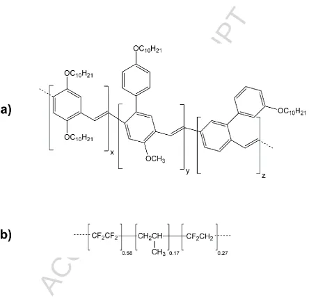
polymer shown in Figure 1 (a) from Merck, Super Yellow, exhibits high sensitivity to
nitroaromatic compounds including 2,4-DNT, a common component in landmine
manufacture and a degradation product of TNT. However, for use in the field, where
uncontrollable environmental factors can disperse the trace vapour plume, a method to
pre-concentrate the target analytes prior to exposure to the sensor would offer an advantage.
Certain polymers have properties that enable them to accumulate explosive vapours to the
surface, based on the hydrogen bond acidity between polymer and target vapour (Abraham,
1993; Houser et al., 2001; McGill et al., 1994). These have been successfully developed for
highly-sensitive Surface Acoustic Wave sensors for hazardous targets including nerve agents
and radionuclides (Grate, 2008), and for HPLC columns applied to environmental monitoring
(Egorov et al., 2006; Grate et al., 2007). These polymers effectively act as a “magnet” for the
explosives, whereby the nitroaromatic compound adsorbs to the surface, and the
application of heat leads to thermal desorption (Camara et al., 2014; Martin et al., 2007;
Serrano et al., 2013; Tiwary et al., 2016; Voiculescu et al., 2006), as shown in Figure 2. The
specially designed materials studied in the literature for this purpose are usually expensive
(approx. $600 for 100 mg at time of writing) which inhibits their ability to be prepared in
large batches for humanitarian demining efforts. A commercial, amorphous random
co-polymer of tetrafluoroethylene (∼56 wt.%), vinylidene fluoride (∼27 wt.%), and propylene
(∼17 wt.%), sold as Aflas and illustrated in Figure 1 (b), has similar structural properties to
ACCEPTED MANUSCRIPT
dyes (Gillanders et al., 2004a; Gillanders et al., 2004b), and is known to have good sorption
kinetics for organic vapours (Wang et al., 1999).
A method to collect these explosives without human activity on the mine-suspected area
would decrease the danger in field sampling. Honeybees foraging for explosives residues
potentially have the advantage of being able to access terrain that may be difficult for
humans or larger animals such as dogs, to safely survey. Honeybees have previously been
used in biosensing applications for targets including radionuclides, pollutants, and
explosives (Barisic D et al., 2002; Girotti et al., 2013; Rodacy et al., 2002; Zarić et al., 2018).
In such an approach, the honeybees’ body hair can electrostatically attract trace particles of
explosives as they free-fly around a contaminated area; after returning to the hive, the inner
environment of the colony may then become rich in explosive molecules for subsequent
detection.
In this work we show for the first time an approach that combines colonies of honeybees to
collect material across an area of landmine contaminated land, with a REST sampling
technique to establish the presence of trace explosives in the field. We show that the
fluoropolymer Aflas can act as an effective preconcentrator material for the accumulation of
nitroaromatic molecules, which can later be released thermally and detected via
luminescence quenching in a semiconducting polymer film. By placing a cartridge of tubes
holding the adsorbent material in the entrance of a hive, bees returning from foraging may
ACCEPTED MANUSCRIPT
be collected and analysed. We compare this with an alternative approach in which air from
[image:8.595.80.525.196.629.2]inside the hive is pumped through a preconcentrator-coated filter for subsequent analysis.
ACCEPTED MANUSCRIPT
Figure 2 –Schematic concept of preconcentrating filter. (a) Air containing the explosive
vapour (shown as blue circles) is drawn through the filter were the molecules of the
explosive adsorb to the polymer surface. (b) Subsequent heating of the preconcentrator
desorbs the explosive molecules for detection by a change in the light emission from a
separate luminescent polymer film.
2. Material and Methods
2.1Polymer sensor and REST filter preparation
Films based on Merck Super Yellow were prepared by spin coating the polymer at 2000 rpm
from toluene solution of a concentration of 6.5 mg/ml onto 1 cm2 cover glasses (Agar
Scientific). Prior to spin coating, the substrates were cleaned ultrasonically for 5 minutes in
toluene, acetone and propan-2-ol, and dried in a dry nitrogen stream prior to being plasma
ACCEPTED MANUSCRIPT
thicknesses, measured with a Veeco Dektak 150 surface profilometer, were found to be in
the region of 100 nm.
Aflas® was purchased from AGC Chemicals Europe Ltd, and was prepared by dissolving in
Tetrahydrofuran (Sigma Aldrich) at a concentration of 155 mg/ml. For characterisation and
air sampling, 10 l of the solution was then drop-cast with a micropipette in a polka-dot grid
pattern of 3x3 spots, with a mass per spot of 1-3 mg, either on silicon wafers (Approx.. 2.5 x
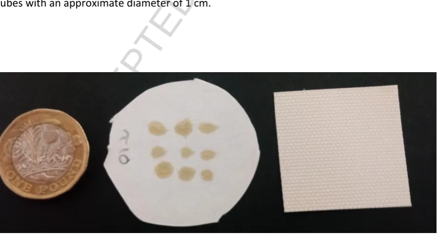
2.5 cm) or standard Whatman filter paper of diameter 4.5 cm, and left to dry prior to use.
An example image of the Aflas-spotted filter is shown in Figure 3. It can be seen that the
uncoated surface of the paper allows for unobstructed air-flow from the air sampler.
For in-situ placement of the preconcentration material in the hive entrance and exit, sheets
of Whatman filter paper were blade-coated with Aflas solution as above and cut into strips
as seen in Figure 3. The strips were placed against strips of acetate sheets and rolled into
[image:10.595.76.525.449.690.2]tubes with an approximate diameter of 1 cm.
Figure 3 – Photograph of a coin for scale (left) with an Aflas-spotted Filter paper (middle)
ACCEPTED MANUSCRIPT
2.2Limit of Detection of Super Yellow sensors
To determine the limit of detection of the super yellow film to nitroaromatic molecules, and
obtain a measure of the typical level of photoluminescence quenching due to a known mass
of explosives, the efficiency of photoluminescence was measured for various concentrations
of 2,4 DNT loaded into the polymer film. The Photoluminescence Quantum Yield (PLQY) of
the Super Yellow films was measured with an integrating sphere (Greenham et al., 1995) in
a Hamamatsu Photonics C9920-02 system with an excitation wavelength of 405 nm. The
PLQY of the film was first measured in its pristine as-spin-coated state. The films were then
contaminated with 2,4-DNT by drop-casting a solution of acetonitrile containing 2,4-DNT
and allowing the solvent to evaporate. The PLQY of the contaminated films, containing
masses of 2,4-DNT between 6 pg and 60 mg, were measured and compared with the initial
values. The error associated with these measurements is 1% of the measured value.
2.3Preconcentration capability of Aflas
To confirm the preconcentrating action of Aflas, the polymer was drop cast onto silicon
wafers which were placed in a custom-built sealed chamber and exposed to 2,4-DNT
vapours generated by flowing nitrogen vapour at a flow rate of 10 Lmin-1 over 1 g of
2,4-DNT powder sealed in a glass tube and held at room temperature at a relative humidity of
40-50%. After exposure to the DNT vapour stream, the samples were placed against a
heating element in a sealed chamber of inner dimensions 53 x 53 x 53 mm, along with the
ACCEPTED MANUSCRIPT
these measurements due to its good thermal conductivity which allowed the Aflas spots to
be heated effectively. To minimise sensor degradation via photo-oxidation in ambient
conditions, the chamber was flushed with clean nitrogen and sealed at the start of each
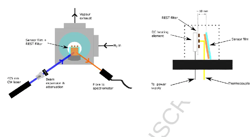
measurement. The sensor was placed 1 cm from the preconcentrator, excited with a 405
nm CW laser diode laser (Photonic Solutions) and its photoluminescence measured over 300
s with a fibre coupled CCD spectrometer taking measurements every 3 s. The
photoluminescence at room temperature was measured for 30 s, then the heater turned on
for approximately 100 s to heat the sample to 100oC. After measurements had been
completed the chamber was flushed again with clean nitrogen to clear the chamber of any
residual explosive vapours.
To confirm the preconcentration action in a simulated minefield environment, the Aflas
polka-dotted onto Whatman filter paper discs were placed over a simulated landmine
containing 1g of DNT in a metal container buried in soil at a depth of 2 cm as described in
(Gillanders, 2017). The relative humidity around the simulated landmine was increased to
70-80% before sampling with a water mist as this has been shown to maximise the amount
of explosive vapour emitted by a landmine (Bach and Mclean, 2003). The paper filter was
placed into a home-made holder attached to an air sampling pump (JS Holdings HF812e).
The filter was held above the simulated landmine, as shown in Figure 5 and air sampled at a
rate of 60 Lmin-1 for 10 minutes. These samples were tested in the same way described for
ACCEPTED MANUSCRIPT
Figure 4 – Experimental set-up for detecting the release of vapours from a heated
preconcentrator film.
Figure 5 – Schematic diagram of the approach to collect vapour and particle samples of explosives above a simulated landmine.
2.4Preconcentrator sampling of trace explosives in the field using honeybee colonies
Honey bee (Apis mellifera carnica) colonies were obtained from a local beekeeper in Zadar
County, Croatia. Three colonies were selected from an area that is not contaminated with
ACCEPTED MANUSCRIPT
modified Liebefeld method (Delaplane et al., 2013). For the field trial colonies were selected
with similar populations of bees, on average 20,000 per colony. We used standard LR hives
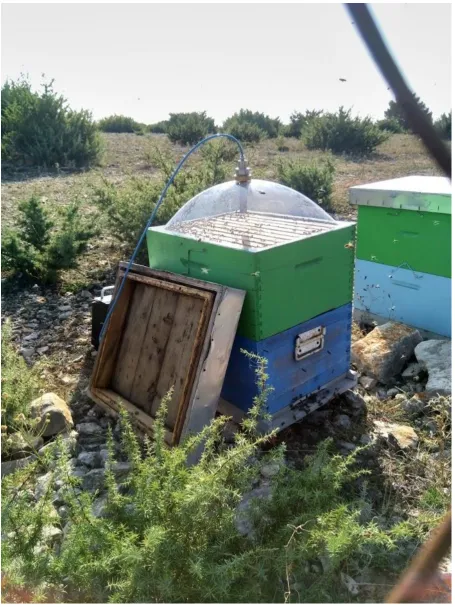
with two supers, a bottom board with entrance, a top board and roof. For the air sampling
the top board and roof were removed to place the cupola on top (figure 8).
The three colonies were located initially on a clean site to collect explosive-free control
samples, before being moved to an explosive-contaminated site to allow sampling of
explosive material in the preconcentrators. Once the colonies were in-situ the bees were
allowed to free-fly, with a sucrose solution of 500 g sugar dissolved in 1.5 L tap water used
to keep the bees within the test area of interest. The control sampling was performed near
Vrsi (“clean site”, Figure 6) and the contaminated colony sampling was conducted on
Benkovac test site (“test site”, Figure 6), in September 2017 and mid-April 2018. The test
site was designed for the testing and validation of mine detectors and mine-clearance
vehicles, with an area of 10,000 m2 and 1,000 deactivated mines buried in a series of test
ACCEPTED MANUSCRIPT
Figure 6 – Sampling site map in Zadar County, Croatia. Map data ©2018 Google.
The main approach tested to collect material gathered by the colonies was to locate
preconcentrators at the entrance to the hives for the bees to mechanically deposit material
onto the Aflas film. Blade-coated filter papers were cut into strips of approximately 3 cm x
10 cm (Figure 7a), placed against a strip of acetate of the same dimensions and rolled into
tubes prior to insertion in plastic cartridges of 4 x 1 cm2. Standard Lexan plates (1 x 1 cm
tube) were cut into 10 cm lengths and used as a cartridge (Figure 7b), which was then
inserted into the entrance of the hives (Figure 7c). To separate bees entering the colony
ACCEPTED MANUSCRIPT
cartridge is protruding from the colony, while for the exit the cartridge was extending into
the colony (figure 7d). Samples were left for periods of one, three days and seven days prior
to collection. At the end of the placement period the papers were removed from the colony,
inserted into glass vials, sealed with Parafilm then placed in airtight sealed bags and stored
ACCEPTED MANUSCRIPT
Figure 7 – (a) A substrate coated with Aflas polymer; (b) Substrates rolled up and inserted
into the adapted entrance tubes; (c) The adapted entrance on the colony; (d) Entrance (left)
and Exit (right) cartridges in the bottom board.
The air from within the bee hives was also sampled after collection of the entrance
cartridges, to compare with the samples of explosive materials deposited on the entrance
and exit cartridges. Uncoated papers were also inserted into the cartridges to confirm
preconcentration efficacy of the Aflas in the field. To sample the hive air, the filters were
inserted into a home-made nozzle and attached to the colony via a specialised cupola
(Figure 8) on the hive, with air being sampled for 10 minutes with a vacuum pump at a
flowrate of 60 Lmin-1. The filters were then immediately placed in glass vial, sealed with
[image:17.595.139.334.369.622.2]ACCEPTED MANUSCRIPT
ACCEPTED MANUSCRIPT
3. Results and Discussion
[image:19.595.102.394.450.740.2]3.1Limit of Detection of Super Yellow with 2,4-DNT
Figure 9 shows the Limit of Detection via change in the PLQY of neat Super Yellow films with
the addition of 2,4-DNT dissolved in acetonitrile. A loss in PLQY efficiency between a pristine
film, with an initial PLQY of 40%, and a contaminated film indicates an increase in the
non-radiative recombination rate, due to photoluminescence quenching by the analyte. At
sub-ng levels the application of the 2,4-DNT solution can be seen to increase the PLQY slightly;
this may be attributable to some swelling of the polymer caused by the solvent reducing
non-radiative interactions between the polymer chains and thereby outweighing any
quenching effect from trace amounts of explosives. Above ng levels however, a large loss of
PLQY can be observed. The results suggest that below 10 ng of explosive material per cm2
on the concentrator is required to quench luminescence at detectable levels.
0.001 0.01 0.1 1 10 100 1000 10000 100000 -20
0 20 40 60 80 100
PLQY dr
op
(%)
ACCEPTED MANUSCRIPT
Figure 9 - Reduction in PLQY of Super Yellow films exposed to different masses of 2,4-DNT in
acetonitrile solution.
3.2 Efficacy of Aflas as a preconcentration material
0 50 100 150 200 250 300
0.4 0.5 0.6 0.7 0.8 0.9 1.0 1.1 DNT exposed Reference Emission in ten sity (a .u. ) t (s)
(a)
500 525 550 575 600 625 650 675
0 50 100 150 200 250 300 350 Coun ts Wavelngth (nm) Pristine
Post exposure (500 ng DNT) on aflas
ACCEPTED MANUSCRIPT
Figure 10 – (a) Photoluminescence emission from the Super Yellow sensor film during the
thermal release of 2,4-DNT vapours from silicon test samples. An Aflas coated silicon
substrate exposed to 2,4-DNT vapour (red line) is compared with a reference uncoated
silicon substrate also previously exposed to 2,4-DNT (black line). The red box indicates the
substrate heating time of 90 s. (b) Emission spectrum from a Super Yellow sensor before and
after exposure to a heated preconcentrator doped with 500 ng of 2,4-DNT.
The Aflas polymer is shown in Figure 10a to be a suitable adsorber for nitroaromatic
vapours. A clean, uncoated silicon reference substrate was exposed to 2,4-DNT vapour and
then heated in the sealed chamber shown in figure 4, while the photoluminescence
emission from a Super Yellow film was measured. A silicon substrate coated with Aflas was
likewise exposed to 2,4-DNT, heated, and the emission from Super Yellow monitored. The
uncoated reference substrate can be seen to cause a decrease in Super Yellow sensor
emission intensity of around 12%, which may in part be due to the elevated temperature
causing some thermal and/or photo-oxidative degradation of the Super Yellow sensing film.
However, the Aflas-coated substrate causes a much greater quenching response of
approximately 45%, showing that explosive vapours have been adsorbed to the Aflas
surface prior to successful thermal desorption. Figure 10b shows the decrease in
fluorescence of Super Yellow after exposure to an Aflas substrate doped with 500 ng
2,4-DNT.
The process was repeated in a simulated real-world environment by sampling air into filter
paper substrates from above a buried simulated landmine. An uncoated filter paper, and an
ACCEPTED MANUSCRIPT
were subsequently heated in the chamber to measure the quenching response of vapours
released onto a Super Yellow sensor film. As shown in Figure 11, the unexposed
Aflas-spotted paper filter causes approximately 10% quenching of the Super Yellow
photoluminescence, attributable to degradation of the sensing polymer. The exposed
uncoated filter paper gives a quenching response of around 27%, which suggests the rough,
fibrous structure of the paper is able to trap some explosive molecules and desorb via
heating. Plain paper has been recently shown to successfully adsorb explosive particles for
photoacoustic sensing (Sharma et al., 2017). However, a clear quenching response of over
70% is shown by the Aflas-spotted filter paper exposed to 2,4-DNT vapour. In addition to the
Aflas polymer sorbing vapours of the 2,4-DNT, the spots on the filter were found to pick up
particles of dust and dirt from the sampling area more effectively than paper alone, further
increasing the effectiveness of the filter.
0 50 100 150 200 250 300
0.2 0.4 0.6 0.8 1.0 1.2 Reference Paper Paper + aflas
ACCEPTED MANUSCRIPT
Figure 11 – Photoluminescence emission from a Super Yellow sensor film during the thermal
release of 2,4-DNT vapours from filter paper samples previously exposed to DNT vapours
from a simulated landmine. Data show the responses to an uncontaminated Aflas-spotted
filter paper (Reference, black line), uncoated filter paper (Paper, red line), and an
Aflas-spotted filter paper (blue line). The red box indicates the substrate heating time of 90 s.
3.3 Preconcentrator sampling of trace explosives in the field using honeybee colonies
Following the initial validation tests of the preconcentrators, samples collected from the test
mine field were subsequently analysed. Preconcentrators taken from the entrance and exit
cartridges in the hives were heated to release sorbed molecules and the photoluminescence
from Super Yellow films was measured as described in section 2.3. The sensing results of a
single hive entrance and exit sampling in September 2017 are shown in Figure 12, while
averaged results from three hives (two hives on the Clean Site) in April 2018 are shown in
Figure 13. The horizontal black line is the benchmark average loss of luminescence from
three separate control measurements (of an unused Aflas filter) due to photo-oxidation of
the Super yellow film in atmospheric conditions. It can be seen in Figure 12 that the highest
quenching response is from the preconcentrator samples collected at the hive entrance
after one day on-site. Weather data from the sampling period shown in Figure 12 show less
favourable conditions in terms of temperature and wind speed on the third and seventh
days thereby reducing bee activity. For instance, the temperature on the first day had a
minimum of 16°C while the other two sampling days had minimums of 12°C and 9.8°C
respectively, which may have reduced bee activity. There was also heavy rainfall of 123.1
ACCEPTED MANUSCRIPT
time, which would have reduced foraging and may have inhibited explosive vapour release
at ground level.
The spread of responses over the trial periods may be attributed to several factors chiefly
including the weather (e.g. during strong winds the bees will remain inside the hive) or
potentially bees finding a reliable pollen (protein) food source after 24-48 hours on-site.
Figure 12 – Response to vapours released after 90 seconds’ heating of the preconcentrator
strips from a single hive entrance in September 2017; sampled both before placement at,
and following 1, 3 and 7 days located on, the test minefield. Bars show the average fraction
of the initial photoluminescence intensity remaining after exposure to molecules released
from the heated preconcentrator, while crosses show individual measurements. Each bar is
an average of three measurements, while the dotted horizontal lines show the standard
0.80 0.85 0.90 0.95 1.00 1.05 1.10
Emission in
ten
sity (a
.u)
Hive Entrance
Hive Exit
ACCEPTED MANUSCRIPT
deviation from the averaged reference. Red bars show results for preconcentrators at the
[image:25.595.136.435.168.449.2]hive entrance while blue bars show results for preconcentrators at the exit.
Figure 13 – Response to vapours released from the preconcentrator strips after 90 seconds
heating - results averaged from three hives on the minefield over three days in April 2018.
The bars show the average fraction of the initial photoluminescence intensity remaining
after exposure to molecules released from the heated preconcentrator, while crosses show
individual measurements. The horizontal line shows the average response from a blank
measurement. “Hive entrance” refers to Aflas-coated papers in the hive entrance, “Hive
Entrance Blank" refers to uncoated papers in the hive entrance; “Hive Exit” refers to
Aflas-coated papers in the hive exit, and “Hive Exit Blank” refers to unAflas-coated papers in the hive
exit. 0.75 0.80 0.85 0.90 0.95 1.00 1.05 1.10 Day 1 Emission in ten sity (a .u)
Clean Day 2 Day 3
Hive Entrance Hive Entrance blank Hive Exit
Hive Exit blank
ACCEPTED MANUSCRIPT
Figure 14 shows the response of the Super Yellow sensor to vapours released from
preconcentrator filters loaded by air-pumping from within a hive. The figure shows
indicative measurements taken over a 3 day period for a hive located on the test minefield.
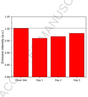
The samples taken from within the colonies show similar levels of luminescence quenching
from the air-sampled preconcentrator as those from the entrance preconcentrators. This
indicates that some quantities of explosive molecules may accumulate in the colony even
after adsorbing to those in the entrance cartridges, and there is potentially enough
explosive material collected inside the colony to be air-sampled and detected.
Figure 14 – Response to vapours released from the preconcentrator air filters after 100
seconds’, sampled from within a hive, following 1, 2 and 3 days located on the test
minefield. Bars show the fraction of the initial photoluminescence intensity remaining after
exposure to molecules released from the heated preconcentrator.
Clean Site Day 1 Day 2 Day 3 0.80
0.85 0.90 0.95 1.00 1.05
Emission in
ten
sity (a
.u.
[image:26.595.141.434.293.614.2]ACCEPTED MANUSCRIPT
4. Conclusions
An inexpensive, commercially-available preconcentrator polymer was used to efficiently
adsorb explosive materials, which could subsequently be released thermally for detection
using a photoluminescence quenching sensor. Preconcentration was tested in the field with
honeybee colonies used as a biological sampling approach to gather residues of buried
explosives over an area. The honeybees were allowed to free-fly around a test site with
known landmine presence, to allow explosive molecules to be picked up electrostatically by
the bees’ body hair. On return to the hive, the bees passed through a tube lined with the
preconcentration polymer to accumulate gathered residues. The preconcentrator was
subsequently heated in a sealed chamber and the released vapours were detected via
luminescence quenching of the conjugated polymer Super Yellow. These results indicate a
preconcentrator at the hive entrance has potential for collecting gathered explosive
materials. This technique was shown for the first time, to the best of our knowledge, and
could provide a pathway to real-time identification of explosives in the field. While the
results are still at an early stage, we expect that improvements in the sampling methods
could provide a robust, inexpensive system to aid humanitarian demining efforts worldwide.
Acknowledgements
This project has received funding from NATO Science for Peace & Security under grant
agreement MYP G5355, the European Union’s Seventh Framework Programme for research,
technological development and demonstration under agreement no 284747, and the EPSRC
ACCEPTED MANUSCRIPT
Appendix A: Supporting InformationData supporting this research can be found at
https://doi.org/10.17630/1b2fa689-d709-4276-8161-ea7918c17716.
References
Abraham MH. SCALES OF SOLUTE HYDROGEN-BONDING - THEIR CONSTRUCTION AND APPLICATION
TO PHYSICOCHEMICAL AND BIOCHEMICAL PROCESSES. Chemical Society Reviews 1993; 22:
73-83.
Ali MA, Shoaee S, Fan SQ, Burn PL, Gentle IR, Meredith P, et al. Detection of Explosive Vapors: The
Roles of Exciton and Molecular Diffusion in Real-Time Sensing. Chemphyschem 2016; 17:
3350-3353.
Bach H, Mclean I. REMOTE EXPLOSIVE SCENT TRACING: GENUINE OR A PAPER TIGER. Journal of Mine
Action 2003; 7: 1-10.
Barisic D, Bromenshenk JJ, Kezic N, Vertacnik A. The Role of Honey Bees in Environmental
Monitoring in Croatia. In: Devillers J, Pham-Delegue M, editors. Honey Bees: Estimating the
Environmental Impact of Chemicals. Taylor & Francis, New York, 2002, pp. 160-185.
Beyene NW. EXTRACTION OF NITROAROMATIC EXPLOSIVE COMPOUNDS FROM SOIL: CRITICAL
REVIEW, 2010.
Bolse N, Eckstein R, Schend M, Habermehl A, Eschenbaum C, Hernandez-Sosa G, et al. A digitally
printed optoelectronic nose for the selective trace detection of nitroaromatic explosive
ACCEPTED MANUSCRIPT
Camara M, James F, Breuil P, Pijolat C, Briand D, de Rooij NF. MEMS-based porous silicon
preconcentrators filled with Carbopack-B for explosives detection. In: Sberveglieri G, Ferrari
V, editors. 28th European Conference on Solid-State Transducers. 87, 2014, pp. 84-87.
Caygill JS, Davis F, Higson SPJ. Current trends in explosive detection techniques. Talanta 2012; 88:
14-29.
Delaplane KS, van der Steen J, Guzman-Novoa E. Standard methods for estimating strength
parameters of Apis mellifera colonies. Journal of Apicultural Research 2013; 52.
Egorov OB, O'Hara MJ, Grate JW. Equilibration-based preconcentrating minicolumn sensors for trace
level monitoring of radionuclides and metal ions in water without consumable reagents.
Analytical Chemistry 2006; 78: 5480-5490.
Fjellanger R. Remote explosive scent tracing - a method for detection of explosive and chemical
substances. Vol 167, 2004.
Giannoukos S, Brkic B, Taylor S, Marshall A, Verbeck GF. Chemical Sniffing Instrumentation for
Security Applications. Chemical Reviews 2016; 116: 8146-8172.
Gillanders RN, Campbell IA, Glackin JME, Samuel IDW, Turnbull GA. Ormosil-coated conjugated
polymers for the detection of explosives in aqueous environments. Talanta 2018; 179:
426-429.
Gillanders RN, Samuel, I.D.W., Turnbull, G.A. A Low-Cost, Portable Optical Explosive-vapour Sensor.
Sensors and Actuators B-Chemical 2017; 245: 334-340.
Gillanders RN, Tedford MC, Crilly PJ, Bailey RT. Erythrosin B encapsulated in a fluoropolymer matrix
for dissolved oxygen optical sensing in aggressive aqueous environments. Journal of
Photochemistry and Photobiology a-Chemistry 2004a; 162: 531-535.
Gillanders RN, Tedford MC, Crilly PJ, Bailey RT. Thin film dissolved oxygen sensor based on platinum
octaethylporphyrin encapsulated in an elastic fluorinated polymer. Analytica Chimica Acta
ACCEPTED MANUSCRIPT
Girotti S, Ghini S, Maiolini E, Bolelli L, Ferri EN. Trace analysis of pollutants by use of honeybees,
immunoassays, and chemiluminescence detection. Analytical and Bioanalytical Chemistry
2013; 405: 555-571.
Grate JW. Hydrogen-bond acidic polymers for chemical vapor sensing. Chemical Reviews 2008; 108:
726-745.
Grate JW, Ozanich R, Hartman JS, O'Hara MJ, Egorov OB, Ieee. Preconcentrating minicolumn sensors
for trace environmental monitoring. 2007 Ieee Sensors, Vols 1-3, 2007, pp. 1357-1360.
Greenham NC, Samuel IDW, Hayes GR, Phillips RT, Kessener Y, Moratti SC, et al. Measurement of
absolute photoluminescence quantum efficiencies in conjugated polymers. Chemical Physics
Letters 1995; 241: 89-96.
Houser EJ, Mlsna TE, Nguyen VK, Chung R, Mowery RL, McGill RA. Rational materials design of
sorbent coatings for explosives: applications with chemical sensors. Talanta 2001; 54:
469-485.
Lugo JH, Zoppi M, Molfino R. Design and Kinematic Modeling of a Screw-Propelled Mobile Robot to
Perform Remote Explosive Scent Tracing Filter Sampling in Forest during Humanitarian
Demining. Advances in Cooperative Robotics 2017: 699-715.
Martin M, Crain M, Walsh K, McGill RA, Houser E, Stepnowski J, et al. Microfabricated vapor
preconcentrator for portable ion mobility spectroscopy. Sensors and Actuators B-Chemical
2007; 126: 447-454.
McGill RA, Abraham MH, Grate JW. CHOOSING POLYMER-COATINGS FOR CHEMICAL SENSORS.
Chemtech 1994; 24: 27-37.
Narayanan A, Varnavski OP, Swager TM, Goodson T, III. Multiphoton fluorescence quenching of
conjugated polymers for TNT detection. Journal of Physical Chemistry C 2008; 112: 881-884.
Porritt F, Shapiro M, Waggoner P, Mitchell E, Thomson T, Nicklin S, et al. Performance decline by
search dogs in repetitive tasks, and mitigation strategies. Applied Animal Behaviour Science
ACCEPTED MANUSCRIPT
Rodacy PJ, Bender SFA, Bromenshenk JJ, Henderson CB, Bender G. The training and deployment of
honeybees to detect explosives and other agents of harm. In: Broach JT, Harmon RS, Dobeck
GJ, editors. Detection and Remediation Technologies for Mines and Minelike Targets Vii, Pts
1 and 2. 4742, 2002, pp. 474-481.
Serrano G, Sukaew T, Zellers ET. Hybrid preconcentrator/focuser module for determinations of
explosive marker compounds with a micro-scale gas chromatograph. Journal of
Chromatography A 2013; 1279: 76-85.
Sharma RC, Kumar S, Gautam S, Gupta S, Srivastava HB. Photoacoustic sensor for trace detection of
post-blast explosive and hazardous molecules. Sensors and Actuators B-Chemical 2017; 243:
59-63.
Shoaee S, Fan SQ, Burn PL, Shaw PE. Photophysics of detection of explosive vapours via
luminescence quenching of thin films: impact of inter-molecular interactions. Physical
Chemistry Chemical Physics 2016; 18: 25861-25868.
Thomas SW, III, Joly GD, Swager TM. Chemical sensors based on amplifying fluorescent conjugated
polymers. Chemical Reviews 2007; 107: 1339-1386.
Tiwary N, Vinchurkar M, Patel M, Nathawat R, Pandey S, Rao VR. Fabrication, Characterization and
Application of ZnO Nanostructure-Based Micro-Preconcentrator for TNT Sensing. Journal of
Microelectromechanical Systems 2016; 25: 968-975.
Toal SJ, Trogler WC. Polymer sensors for nitroaromatic explosives detection. Journal of Materials
Chemistry 2006; 16: 2871-2883.
Voiculescu I, McGill RA, Zaghloul ME, Mott D, Stepnowski J, Stepnowski S, et al.
Micropreconcentrator for enhanced trace detection of explosives and chemical agents. Ieee
Sensors Journal 2006; 6: 1094-1104.
Wang P, Schneider NS, Sung NH. Sorption and diffusion of organic vapors in two fluoroelastomers.
ACCEPTED MANUSCRIPT
Wang Y, Rae BR, Henderson RK, Gong Z, McKendry J, Gu E, et al. Ultra-portable explosives sensor
based on a CMOS fluorescence lifetime analysis micro-system. Aip Advances 2011a; 1.
Wang Y, Turnbull GA, Samuel IDW. Conjugated polymer sensors for explosive vapor detection.
Organic Semiconductors in Sensors and Bioelectronics Iv 2011b; 8118.
Yang JS, Swager TM. Porous shape persistent fluorescent polymer films: An approach to TNT sensory
materials. Journal of the American Chemical Society 1998; 120: 5321-5322.
Zarić NM, Deljanin I, Ilijević K, Stanisavljević L, Ristić M, Gržetić I. Honeybees as sentinels of lead
pollution: Spatio-temporal variations and source appointment using stable isotopes and