ISSN Online: 2164-3199 ISSN Print: 2164-3180
DOI: 10.4236/ojbd.2019.93006 Sep. 17, 2019 47 Open Journal of Blood Diseases
Erythrocyte Senescent Markers by Flow
Cytometry
María Alejandra Ensinck, Melina Eliana Luján Brajovich, Silvia Estela García Borrás,
Carlos Miguel Cotorruelo, Claudia Silvia Biondi
Department of Clinical Biochemestry, Faculty of Biochemical and Pharmaceutical Sciences, National University of Rosario, Rosario, Argentina
Abstract
Background: Mature red blood cells lack protein synthesis and are unable to restore inactivated enzymes, damaged cytoskeleton and membrane proteins. An oxidation breakdown of band 3 is probably part of the mechanism leading to the generation of a senescent cell antigen. This specific signal serves for the clearance of RBCs by inducing the binding of autologous IgG and C3, leading to phagocytosis. In addition, phosphatidilserin molecules appear in the outer membrane and the CD47 expression diminishes. Methods: Erythrocytes of different ages from whole blood were studied by flow cytometry analysing light scatter proprieties, binding of autologous IgG, C3 complement deposits, externalization of phosphatidylserine and CD47 expression. Dot-plot analysis based on forward scatter versus side scatter parameters showed two RBCs populations of different sizes and density. RBCs were further incubated with Alexa 488 IgG, APC-anti-C3, PE-annexin-V and PE-CD47. The comparison of the values obtained for the different variables studied in SeRBC and YRBC populations was carried out by the Student t-test for matched samples or by the Wilcoxon test (after verification of the normality assumption). Results: The percentage of IgG and C3 positive cells was significantly higher in senes-cent red blood cells population. The fraction of annexin-V positive RBCs was also larger in SeRBCs while the CD47 expression was lower in this popula-tion. Conclusions: These results indicate that flow cytometry allow differen-ciation of erythrocytes populations of different ages, turning this tool into an useful alternative option to study erythrocyte aging process. These findings will contribute to a better understanding of the process and mechanisms in-volved in erythrocyte senescence process.
Keywords
Erythrocyte, Senescence, Flow Cytometry How to cite this paper: Ensinck, M.A.,
Brajovich, M.E.L., Borrás, S.E.G., Cotorru-elo, C.M. and Biondi, C.S. (2019) Erythro-cyte Senescent Markers by Flow Cytometry. Open Journal of Blood Diseases, 9, 47-59.
https://doi.org/10.4236/ojbd.2019.93006
Received: August 9, 2019 Accepted: September 14, 2019 Published: September 17, 2019
Copyright © 2019 by author(s) and Scientific Research Publishing Inc. This work is licensed under the Creative Commons Attribution International License (CC BY 4.0).
http://creativecommons.org/licenses/by/4.0/
DOI: 10.4236/ojbd.2019.93006 48 Open Journal of Blood Diseases
1. Introduction
In healthy individuals, the number of erythrocytes exceeds 4 × 10¹²/L in blood stream and has a mean lifespan of 115 days with a variation between 70 and 140 days, and a difference of 15% between individuals. Erythrocyte life is limited by senescence and subsequent removal of aged red blood cells (RBCs) [1] [2] [3].
Under physiological conditions, all individuals have physiologic autologous IgG antibodies to band 3 that bind to senescent (Se) RBCs and initiate their re-moval by macrophage. The accumulation of this specific antibody at the end of erythrocyte life represents an accessible evidence to identify the antigen and subsequently the aging mechanism [4] [5]. The small amount of autologous IgG found in vivo indicates that removal of SeRBCs is an efficient and controlled process. Naturally occurring antibodies (NAbs) that bind to band 3 are impli-cated in the clearance of senescent RBCs. These antibodies have the capacity to stimulate C3b deposition indicating that an effective phagocytosis requires an active complement system, implying its participation in SeRBC clearance [6] [7].
Exposure of phosphatidilserin (PS) in the outer layer membrane has been de-scribed as a marker of apoptosis in nucleated cells [8] [9]. Although apoptosis in RBCs remains controversial, erythrocytes show signs of PS exposure and mem-brane vesicles formation and release [10]. A decrease in phospholipid asymme-try might be one of the physiological aging consequences at membrane level, and in conjunction with other signals, contributes to senescent erythrocytes recogni-tion and removal by phagocytes [11] [12].
The exact nature of the “eat me” signals on RBCs membrane remains undis-covered. It has been generally accepted that CD47 is a “don’t eat me” signal that plays a crucial role in RBCs homeostasis. CD47 is a ubiquitously expressed pro-tein that binds to the inhibitory signal-regulatory propro-tein alpha receptor (SIRPα) present on macrophages and other myeloid cells. The CD47-SIRPα interaction inhibits immune responses such as phagocytosis. Besides functioning as a “don’t eat me” signal, some investigators have shown that a conformational change in the CD47 protein can switch the molecule from a “don’t eat me” to an “eat me” signal. This change in the structure of CD47 can be induced by oxidative stress and promotes trombospondin-1 (TSP-1) binding to CD47, which creates a new binding site for SIRPα. This alternative binding site for SIRPα induces a pro/phagocytic signal for the macrophage. The dual role of CD47-SIRPα in regulating RBCs uptake demonstrates the RBCs clearance complexity [13] [14] [15] [16].
DOI: 10.4236/ojbd.2019.93006 49 Open Journal of Blood Diseases Flow Cytometry (FC) is a very useful tool for cell analysis since it allows the objectively individual study of a large number of cells with high sensitivity and specificity.
The aim of this work was to evaluate IgG, C3 complement, PS and CD47 ex-pression levels on RBCs from different ages by flow cytometry.
2. Materials and Methods
Blood samples (n = 38) were collected from healthy donors into EDTA tubes and stored at 4˚C for less than 24 h until analyzed. 20 of the donors were female and 18 were male. The average age was between 24 and 43 years old.
The study protocol was accepted by the ethical committee of the School of Bi-ochemistry Sciences of Rosario, Argentina and all participants provided written consent according to the principles of the Declaration of Helsinki. The date of approvement by the ethics committee was February 21, 2019 by means of de-canal resolution No. 065/2019.
2.1. Flow Cytometric Analyses
Flow cytometric analyses were performed on a FACSAria II flow cytometer (Becton Dickinson, San Jose, CA, USA). The FACSDiva software was used for acquisition and analysis. The light-scatter and the fluorescence channels were set on a logarithmic scale, a minimum of 100,000 cells were analyzed in each condi-tion.
2.2. Autologous IgG on RBCs Evaluation
The IgG bound to the erythrocytes membrane surface was analysed in fractions of RBCs from different age separated by Percoll gradients and in populations obtained according to the information of light scattering parameters (FSC and SSC) analysed by FC.
The erythrocyte suspensions evaluated were:
- Suspension 1: SeRBCs and young (Y) RBCs populations obtained according to Percoll separation [18].
- Suspension 2: RBCs from whole blood samples.
After 4 washes with 3 ml of phosphate-buffered saline solution (PBS; pH 7.4; 137 mM NaCl, 2.7 mM KCl, 8.1 mM Na2HPO4, and 1.5 mM KH2PO4), suspen-sions were prepared to concentration of 106 cells/mL PBS from each RBCs populations. 50 μL of each suspension were incubated with 50 μL of anti-human IgG labeled with Alexa Fluor 488 (Molecular Probes, Invitrogen, USA) diluted 1:100. After 60 minutes of incubation in the dark, the suspensions were washed once with PBS and resuspended in 0.5 ml of FACS Flow diluent (BD Biosci-ences) [17].
The following controls were used:
DOI: 10.4236/ojbd.2019.93006 50 Open Journal of Blood Diseases The statistical analysis by Wilcoxon test for paired samples showed that the percentage of IgG positive (IgG+) cells do not differ significantly between the populations obtained by both methodologies [18]. The comparable results with the samples without previous separation allowed the study of other senescence markers using whole blood without any separation step, evaluating each marker in the regions defined by FSC and SSC light scattering parameters.
2.3. C3 Complement Analysis
The evaluation of C3 bound to erythrocytes membrane surface was carried out by FC in whole blood samples. RBCs were washed and diluted to a concentration of 106 cells/mL in PBS. 50 μL of RBCs suspension were incubated with 10 μL of anti-C3b (Bioclone, Ortho Diagnostic Systems). After 30 minutes of incubation, a wash with PBS was performed. 50 μL of the allophicocyanin-labeled anti-mouse secondary antibody (APC) (BD Pharmingen USA) was added and incubated 30 min in the dark. The RBCs were washed once with PBS and resuspended in 0.5 ml of FACS Flow diluent (BD Biosciences).
2.4. Flow Cytometric Analyses of Phosphatidylserine Exposure
The evaluation of PS expression on the RBCs external surface is based on its specific binding to annexin V (Annexin V Apotosis Detection Kit I.BD Phar-mingen USA). Suspensions were prepared to a concentration of 106 cells/mL in buffer with calcium (Annexin Binding Buffer). 25 μL of each suspension were incubated with 5 μL of annexin V-phycoerythrin labelled (PE) for 30 minutes in the dark. Subsequently, the samples were washed once with calcium buffer and RBCs were resuspended in 0.5 mL of FACSFlow diluent (BD Biosciences).Negative control: unlabeled RBCs resuspended in calcium buffer.
Positive control: the permeabilization of the erythrocytes was carried out by incubating 50 μL of washed RBCs with 50 μL of 50% ethanol in PBS, 2 minutes at 39˚C. The RBCs were washed and resuspended in Annexin Binding Buffer. 25 μL of this suspension was incubated with 5 μL of annexin V-PE for 30 minutes in the dark. Subsequently, the RBCs were washed with calcium buffer and re-suspended in 0.5 mL of FACSFlow diluent (BD Biosciences). Under these condi-tions, annexin V binds to the exposed PS in the outer layer membrane as well as to the PS present in the inner membrane.
2.5. Assessment of CD47 Expression on RBCs
DOI: 10.4236/ojbd.2019.93006 51 Open Journal of Blood Diseases
2.6. Statistical Analysis
To verify compliance with the assumption of normality required by the different statistical techniques used the Shapiro-Wilk test was applied [18].
The statistical comparison of the results obtained for the different parameters analyzed in SeRBC populations separated by Percoll preformed gradients and studied in whole blood, was carried out using the Wilcoxon test.
The comparison of the values obtained for the different variables studied in SeRBC and YRBC populations was carried out by the Student t-test for matched samples or by the Wilcoxon test (after verification of the normality assumption).
3. Results
3.1. Autologous IgG Evaluation in RBCs
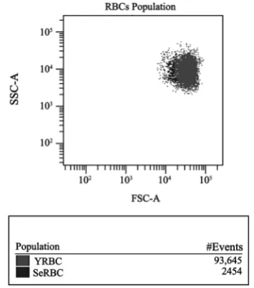
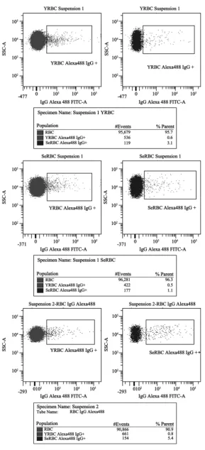
[image:5.595.283.462.491.694.2]Based on data from Kay [19] [20] [21], showing that autologous IgG plays a role in the selective clearance of senescent RBCs, we searched for naturally occurring IgG antibodies (NAbs) directed against RBC proteins. The average values of IgG+ cells percentages in different RBCs suspensions are shown in Table 1. In SeRBCs population, the average values of IgG+ cells percentages were signifi-cantly higher than those observed in YRBCs (p < 0.002) when studying erythro-cyte populations obtained by both methods. The statistical analysis showed that the values of IgG+ cells percentages in SeRBCs and YRBCs populations do not differ significantly between the methodologies used in this work. The compara-ble results in samples without a previous separation step, allowed us to continue the subsequent study of other RBCs senescence markers analysing whole blood, evaluating each marker in regions defined by FSC and SSC light scattering pa-rameters (Figure 1). The analysis strategy used and the results of IgG+ percent-age obtained in suspensions 1 and 2 are shown in Figure 2.
Figure 1. Dot-plot of whole blood with the analysis
DOI: 10.4236/ojbd.2019.93006 52 Open Journal of Blood Diseases
Figure 2. IgG+ RBCs in populations separated by Percoll gradients and
DOI: 10.4236/ojbd.2019.93006 53 Open Journal of Blood Diseases
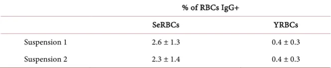
Table 1. IgG+ cells % (mean ± standard deviation). Suspension 1: RBCs separated by
Percoll gradients. Suspension 2: Whole blood.
% of RBCs IgG+
SeRBCs YRBCs
Suspension 1 2.6 ± 1.3 0.4 ± 0.3
Suspension 2 2.3 ± 1.4 0.4 ± 0.3
3.2. C3 Complement Analysis
Naturally occurring auto-antibodies activate the classical complement pathway and stimulate complement amplification and C3b component deposition. There-fore, complement amplification compensates the low affinity of NAbs and gen-erates efficient opsonins. Table 2 describes the median values of C3+ RBCs per-centages of erythrocytes without physical separation. Data corresponding to SeRBCs population was significantly higher than the values obtained from YRBCs (p < 0.0001) (Figure 3).
3.3. Phosphatidylserine Externalization Measurement
We also assessed phosphatidylserine exposure on the outer leaflet plasma mem-brane, which acts as a signal triggering erythrophagocytosis. We observed difference in the phosphatidylserine externalization level between SeRBCs and YRBCs (Figure 4). Table 3 shows the percentages of RBCs with increased PS on the outer side membrane, based on annexin V binding affinity. The median value of annexin V positive cells (AV+) percentage was significantly higher in SeRBC compared to YRBCs (p < 0.001).
3.4. Assessment of CD47 Expression on RBCs
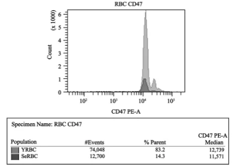
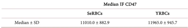
Recent studies have demonstrated that CD47 membrane antigen is an important self-recognition marker involved in protecting the circulating RBCs from phagocytosis [13]. Comparative analyses of membrane CD47 expression in SeRBCs versus YRBCs showed a general decrease in MFI values in senescent erythrocytes population (Figure 5). The median of CD47 MFI was significantly lower in SeRBCs than in YRBCs (p < 0.001), as shown in Table 4.
4. Discussion
Erythrocyte senescence is associated with cell shrinkage, plasma membrane vesiculization, progressive shape change from discocytes to spherocytes, cy-toskeletal and membrane proteins alteration. This process is also characterized by changes in the plasma membrane phospholipids asymmetry that lead to the externalization of PS, which may represent one of the signals that would allow SeRBCs macrophages ingestion [22] [23] [24].
DOI: 10.4236/ojbd.2019.93006 54 Open Journal of Blood Diseases immediate clearance [2] [12] [25] [26]. However, separation techniques used are laborious and require a considerable amount of sample.
Figure 3. Dot-plot of whole blood with the analysis strategy used to evaluate C3+
[image:8.595.261.488.328.498.2]RBCs in populations from different ages.
Figure 4. Dot-plot of whole blood with the analysis strategy used to evaluate PS
expression by binding to Annexin V+ in RBCs populations from different ages.
[image:8.595.256.487.544.708.2]DOI: 10.4236/ojbd.2019.93006 55 Open Journal of Blood Diseases
Table 2. C3+ RBCs % in SeRBCs and YRBCs populations.
% of RBCs C3+
SeRBCs YRBCs
[image:9.595.209.538.182.232.2]Median ± SD 2.2 ± 1.4 0.2 ± 0.6
Table 3. AV+ RBCs % in populations of erythrocytes from different ages, evaluated
through PS binding affinity to annexin V.
% of RBCs Annexin V+
SeRBCs YRBCs
Median ± SD 1.1 ± 0.5 0.15 ± 0.1
Table 4. CD47 expression in RBCs populations from suspension 2.
Median IF CD47
SeRBCs YRBCs
Median ± SD 11010.0 ± 882.9 11965.0 ± 945.7
The evaluation of erythrocyte senescence markers by flow cytometry was per-formed using information obtained from light scattering parameters and fluo-rescence intensity measurements. The forward scatter parameter corresponds to the scattered light collected between 0˚ and 10˚ and is related to the size of cells, while the side scatter value refers to the scattered light collected at 90˚ and is re-lated to the internal cells structure. Given that FSC represents cell size and SSC is related to the internal cellular complexity, when a sample of RBCs without prior separation step is acquired, we consider that the population of SeRBCs is in the area of lower FSC and greater dispersion of SSC, while the YRBCs fraction would be located in the region of higher FSC and more homogeneous SSC.
We have evaluated the usefulness of FC in the study of senescence erythro-cyte, comparing the IgG percentages bound to SeRBCs and YRBCs fractions separated with Percoll and in samples without previous separation. The compa-rable values allowed us to investigate changes that take place during red blood cells aging, using a small amount of sample, avoiding separation method ma-nipulations and decreasing the work time. In addition, the analysis strategy used in the results evaluation and the large number of cells studied allowed by FC, support this methodology election to continue our study of senescence markers in aged erythrocytes.
[image:9.595.207.539.262.312.2]proc-DOI: 10.4236/ojbd.2019.93006 56 Open Journal of Blood Diseases esses seem to be responsible for premature RBCs clearance in haemoglobi-nopathies, membrane protein deficiencies, Down’s syndrome, Alzheimer’s dis-ease, etc [29] [30]. Free radical oxidation maybe an important factor underlying the formation of the senescent signal. There is evidence connecting the RBC oxidation levels in vivo to the breakdown of Band 3 and to the autologous IgG binding [31]. The senescent neo-antigen appearance induces the binding of both autologous IgGs and C3 complement fraction to the membrane, triggering erythrophagocytosis [32] [33]. NAbs activate classical complement pathway which yields a proportional number of deposited C3b [29].
In this study we observed that the C3b positive cells percentage is significantly different in the two regions: low FSC and high SSC (SeRBCs) over the area of the highest FSC and lowest SSC (YRBCs). These findings demonstrate a significant increase in C3b bound in SeRBCs populations. The C3b deposition indicates the participation of this complement component as an additional factor in the SeRBCs removal, making these cells more susceptible to erythrophagocytosis.
In healthy cells, phosphatidylserine is normally found on the inner leaflet RBCs membrane. However, in apoptosis a large increase of exposed PS amounts is seen. This increase of PS expression is proposed to be an “eat me” signal for an apoptotic cell phagocyte recognition, resulting in a non-inflammatory clearance of dying cells [34]. For a long time, it has been proposed that apoptotic cells that expressing PS can be cleared from circulation via macrophages by recognizing them through specific PS-receptors [8]. The analysis of PS expression in the erythrocyte membrane, evaluated by its annexin V binding affinity, showed an increased PS percentage in the region assigned to SeRBCs. This increase would allow its recognition by macrophages, contributing to their elimination. So, it could be considered as a signal that would lead to programmed cell death.
In erythrocytes, CD47 is part of the Rh complex and interacts with Band 3 and Protein 4.2 [35]. CD47 expression has been evaluated in a number of studies, but its role in RBCs human membrane has not been fully clarified. RBCs are cleared as a result of the accumulation of “eat me” signals on membrane via macro-phage-dependent phagocytosis.
The conformational changes in CD47 expression serve as a switch from an erythrocyte phagocytosis inhibitory signal to a stimulating one. In CD47 expres-sion FC investigation, we observed lower CD47 expresexpres-sion intensity in the re-gion assigned to SeRBCs. Considering that CD47 is part of the Band 3 macro complex, the modifications in these proteins, mainly in Band 3, would have an indirect effect in CD47 conformation, triggering an inhibitory phagocytosis sig-nal [36].
Further studies should be carried out by the proposed methodology to analyze other cell markers involved in the RBC senescence process.
5. Conclusions
DOI: 10.4236/ojbd.2019.93006 57 Open Journal of Blood Diseases by immune system. Our results indicate that FC allows differentiation of RBCs populations with different sizes and densities. The aging markers studied al-lowed us to characterize cells of different ages. Therefore, this methodology could be an alternative tool to study erythrocyte aging using small sample quan-tities.
The identification of the process involved in RBCs aging constitutes a major challenge in current medicine research and transfusion. In this way, FC would contribute to the study of RBC progressive structural and biochemical alterna-tions that occur during storage (RBC storage lesion). These findings would pro-vide new insights into RBCs homeostasis.
Ethics Approval and Consent to Participate
The study protocol was accepted by the ethical committee of the School of Bio-chemistry Sciences of Rosario, Argentina and all participants provided written consent according to the principles of the Declaration of Helsinki.
Authors’ Contributions
MAE participated in the design of the study, carried out the flow cytometry stu-dies and drafted the manuscript. MLB participate in the experimental assay and helped to revise the manuscript. SGB helped to revise the manuscript. CC par-ticipated in the design of the study, performed the statistical analysis. CB con-ceived of the study, and participated in its design and coordination and helped to draft the manuscript. All authors read and approved the final manuscript.
Conflicts of Interest
The authors declare no conflicts of interest regarding the publication of this pa-per.
References
[1] Franco, R. (2012) Measurement of Red Cell Lifespan and Aging. Transfusion Medi-cine and Hemotherapy, 39, 302-307.https://doi.org/10.1159/000342232
[2] Cohen, R.M., Franco, R., Khera, P.K., Smith, E.P., Lindsell, C.J., Ciraolo, P.J., Palas-cak, M.B. and Joiner, C.H. (2008) Red Cell Life Span Heterogeneity in Hematologi-cally Normal People Is Sufficient to Alter HbA1c. Blood, 112, 4284-4291.
https://doi.org/10.1182/blood-2008-04-154112
[3] Mock, D.M., Matthews, N.I., Zhu, S., Strauss, R.G., Schmidt, R.L., Nalbant, D., Cress, G.A. and Widness, J.A. (2011) Red Blood Cell (RBC) Survival Determined in Humans Using RBCs Labeled at Multiple Biotin Densities. Transfusion, 51, 1047-1057.https://doi.org/10.1111/j.1537-2995.2010.02926.x
[4] Kay, M. (2005) Immunoregulation of Cellular Lifespan. Annals of the New York Academy of Sciences, 1057, 85-111.
https://doi.org/10.1111/j.1537-2995.2010.02926.x
[5] Lutz, H.U. and Bogdanova, A. (2013) Mechanism Staging Senescent Red Blood Cells for Clearance in Healthy Humans. Frontiers in Physiology, 4, 387.
DOI: 10.4236/ojbd.2019.93006 58 Open Journal of Blood Diseases
[6] Lutz, H.U. (2012) Naturally Occurring Anti-Band 3 Antibodies in Clearance of Se-nescent and Oxidatively Stressed Human Red Blood Cells. Transfusion Medicine and Hemotherapy,39, 321-327.https://doi.org/10.1159/000342171
[7] Franco, R., et al. (2013) Changes in the Properties of Normal Human Red Blood Cells during in Vivo Aging. American Journal of Hematology, 88, 44-51.
https://doi.org/10.1002/ajh.23344
[8] Nguyen, D.B., Wagner-Britz, L., Maia, S., Steffen, P., Wagner, C., Kaestner, L. and Bernhardt, I. (2011) Regulation of Phosphatidylserine Exposure in Red Blood Cells.
Cellular Physiology and Biochemistry, 28, 847-856. https://doi.org/10.1159/000335798
[9] Zwaal, R.F., Comfurius, P. and Bevers, E.M. (2005) Surface Exposure of Phosphati-dylserine in Pathological Cells. Cellular and Molecular Life Sciences, 62, 971-988.
https://doi.org/10.1007/s00018-005-4527-3
[10] Lang, F., Gulbins, E., Lerche, H., Huber, S.M., Kempe, D.S. and Foller, M. (2008) Eryptosis, a Window to Systemic Disease. Cellular Physiology and Biochemistry, 22, 373-380. https://doi.org/10.1159/000185448
[11] Lang, F., Gulbins, E., Lang, P.A., Zappulla, D. and Foller, M. (2010) Ceramide in Suicidal Death of Erythrocytes. Cellular Physiology and Biochemistry, 26, 21-28. https://doi.org/10.1159/000315102
[12] Oldenborg, P.A., Zheleznyak, A., Fang, Y.F., Lagenaur, C.F., Gresham, H.D. and Lindberg, F.P. (2000) Role of CD47 as a Marker of Self on Red Blood Cells. Science, 288, 2051-2054.https://doi.org/10.1126/science.288.5473.2051
[13] Oldenborg, P.A., Gresham, H.D. and Lindberg, F.P. (2001) CD47-Signal Regulatory Protein Alpha (SIRPAlpha) Regulates Fcgamma and Complement Receptor-Mediated Phagocytosis. The Journal of Experimental Medicine, 193, 855-862.
https://doi.org/10.1084/jem.193.7.855
[14] Barclay, A.N. and van den Berg, T.K. (2014) The Interaction between Signal Regu-latory Protein Alpha (SIRPAlpha) and CD47: Structure, Function, and Therapeutic Target. Annual Review of Immunology, 32, 25-50.
[15] Burger, P., Hilarius-Stokman, P., de Korte, D., van den Berg, T.K. and van Bruggen, R. (2012) CD47 Functions as a Molecular Switch for Erythrocyte Phagocytosis.
Blood, 119, 5512-5521.https://doi.org/10.1182/blood-2011-10-386805
[16] Piomelli, S. and Seaman, C. (1993) Mechanism of Red Blood Cell Aging: Relation-ship of Cell Density and Cell Age. American Journal of Hematology, 42, 46-52. https://doi.org/10.1002/ajh.2830420110
[17] Lutz, H.U., Stammler, P., Fasler, S., Ingold, M. and Fehr, J. (1992) Density Separa-tion of Human Red Blood Cells on Self Forming Percoll R Gradients: CorrelaSepara-tion with Cell Age. Biochimica et Biophysica Acta, 1116, 1-10.
https://doi.org/10.1016/0304-4165(92)90120-J
[18] Steel, R.G.D., Torrie, J.H. and Dickey, D.A. (1997) Principles and Procedures of Sta-tistics. A Biometrical Approach. 3rd Edition, McGraw Hill, New York.
[19] Kay, M.M. (1975) Mechanism of Removal of Senescent Cells by Human Macro-phages in Situ. Proceedings of the National Academy of Sciences of the United States of America, 72, 3521-3525.https://doi.org/10.1073/pnas.72.9.3521
[20] Lutz, H.U. (2012) Naturally Occurring Anti-Band 3 Antibodies in Clearance of Se-nescent and Oxidatively Stressed Human Red Blood Cells. Transfusion Medicine and Hemotherapy, 39, 321-327.https://doi.org/10.1159/000342171
DOI: 10.4236/ojbd.2019.93006 59 Open Journal of Blood Diseases
Signalling in Mature Red Cells: From Basic Science to Transfusion Practice. Blood Transfusion, 8, s39-s47.
[22] Arashiki, N., Kimata, N., Manno, S., Mohandas, N. and Takakuwa, Y. (2013) Mem-brane Peroxidation and Methemoglobin Formation Are Both Necessary for Band 3 Clustering: Mechanistic Insights into Human Erythrocyte Senescence. Biochemis-try, 52, 5760-5769. https://doi.org/10.1021/bi400405p
[23] Arese, P., Turrini, F. and Schwarzer, E. (2005) Band 3/Complement-Mediated Rec-ognition and Removal of Normally Senescent and Pathological Human Erythro-cytes. Cellular Physiology and Biochemistry,16, 133-146.
https://doi.org/10.1159/000089839
[24] Mohandas, N. and Gallagher, P.G. (2008) Red Cell Membrane: Past, Present, and Future. Blood, 112, 3939-3948.https://doi.org/10.1182/blood-2008-07-161166
[25] Tanner, M.J.A. (2002) Band 3 Anion Exchanger and Its Involvement in Erythrocyte and Kidney Disorders. Current Opinion in Hematology, 9, 133-139.
https://doi.org/10.1097/00062752-200203000-00009
[26] Lutz, H.U. (2004) Innate Immune and Non-Immune Mediators of Erythrocyte Clearance. Cellular and Molecular Biology, 50, 107-116.
[27] Kay, M. (2005) Immunoregulation of Cellular Life Span. Annals of the New York Academy of Sciences, 1057, 85-111. https://doi.org/10.1196/annals.1356.005
[28] Hornig, R. and Lutz, H.U. (2000) Band 3 Protein Clustering on Human Erythro-cytes Promotes Binding of Naturally Occurring Anti-Band 3 and Anti-Spectrin An-tibodies. Experimental Gerontology, 35, 1025-1044.
https://doi.org/10.1016/S0531-5565(00)00126-1
[29] Pantaleo, A., Giribaldi, G., Mannu, F., et al. (2008) Naturally Occurring Anti-Band 3 Antibodies and Red Blood Cell Removal under Physiological and Pathological Con-ditions. Autoimmunity Reviews, 7, 457-462.
https://doi.org/10.1016/j.autrev.2008.03.017
[30] Kay, M.M., Bosman, G.J., Shapiro, S.S., et al. (1986) Oxidation as a Possible Mecha-nism of Cellular Aging: Vitamin E Deficiency Causes Premature Aging and IgG Binding to RBCs. Proceedings of the National Academy of Sciences, 83, 2463-2467.
https://doi.org/10.1073/pnas.83.8.2463
[31] Kay, M.M., Goodman, S.R., Sorensen, K., et al. (1983) Senescent Cell Antigen Is Immunologically Related to Band 3. Proceedings of the National Academy of Sci-ences,80, 1631-1635.https://doi.org/10.1073/pnas.80.6.1631
[32] Lutz, H.U., Stammler, P. and Fasler, S. (1993) Preferential Formation of C3b-IgG Complexes in Vitro and in Vivo from Nascent C3b and Naturally Occurring Anti Band 3 Antibodies. The Journal of Biological Chemistry, 268, 17418-17426.
[33] Boas, F.E., Forman, L. and Beutler, E. (1998) Phosphatidylserine Exposure and Red Cell Viability in Red Cell Aging and in Haemolytic Anemia. Proceedings of the Na-tional Academy of Sciences,95, 3077-3081.https://doi.org/10.1073/pnas.95.6.3077
[34] Bruce, L.J., Beckmann, R., Ribeiro, M.L., Peters, L.L., Chasis, J.A., Delaunay, J., Mohandas, N., Anstee, D.J. and Tanner, M.J. (2003) A Band 3-Based Macrocomplex of Integral and Peripheral Proteins in the RBC Membrane. Blood, 101, 4180-4188.
https://doi.org/10.1182/blood-2002-09-2824
[35] de Back, D.Z., Kostova, E.B., van Kraaij, M., van den Berg, T.K. and van Bruggen, R. (2014) Of Macrophages and Red Blood Cells: A Complex Love Story. Frontiers in Physiology, 5, 9.https://doi.org/10.3389/fphys.2014.00009