REACTIVE LYMPHDENITIS: AN
Pathologist, Apollo BGS Hospital, Mysore Karnataka, India
ARTICLE INFO ABSTRACT
Background and Objectives:
lymphadenopathy, biopsies of lymph nodes have drastically reduced in number. However some clinical scenario warrants further evaluation of lymph nodes, especially as lymphomas are a heterogenous gro
lymphadenitis where the architecture and distribution of cells played a key role in diagnosis. Methods:
Biopsies were obtained in nine cases of reactive lymphadenitis.
lymphadenitis diagnosed on histopathology were also diagnosed on cytology. 5 cases reported as reactive on cytology showed features of tub
Hodgkin’s lymphoma, metastatic deposits of squamous cell carcinoma and Castleman’s disease (one each) on histological examination.
reactive lymphad
diagnosis can not be excluded. Architecture of the lymph node is key in the diagnosis of most lymphomas.
patient, should always be interpreted with the clinical picture in mind. It does not rule out serious underlying disorder.
Copyright © 2018, Tejaswini Gudibande. This is an open use, distribution, and reproduction in any medium, provided
INTRODUCTION
With the establishment of FNA procedure for diagnosis of lymphadenopathy, biopsies of lymph nodes have drastically reduced in number. However some clinical scenario warrants further evaluation of lymph nodes, especially a
are a heterogenous group of disorders. One of the most problematic areas in cytology is the distinction between a reactive and neoplastic lymphoid proliferation
2001). The diagnosis of a malignant lymphoma is straightforward when bizarre or highly pleomorphiclymphoid cells are present in the aspiration smears (Yao
Otherwise, the cytologic diagnosis of malignant lymphomas often is based on the presence of a relatively monomorphic lymphoid population whereas a polymorphous lymphoid population typically is observed in reactive lymphoid processes (Yao et al., 2001). Potential misdiagnoses may occur when certain lymphomas present with an apparently heterogeneous cellular pattern (Yao et al., 2001
Organizational context: A total of 491 patients were
diagnosed with reactive lymphadenitis during a study period. FNA diagnosis of other types of lymph node lesions were excluded. Above table highlights the marked reduction in the number of lymph node biopsies performed at our hospital a
ISSN: 0975-833X
Article History: Received 28th March, 2018
Received in revised form 15th April, 2018
Accepted 19th May, 2018
Published online 30th June, 2018
Citation: Dr. Tejaswini Gudibande, 2018. “Reactive lymphdenitis: An engima Key words:
Lymph Nodes,
FNAC, Reactive Lymphadenitis, Histological Correlation.
*Corresponding author
RESEARCH ARTICLE
REACTIVE LYMPHDENITIS: AN ENIGMA
*
Dr. Tejaswini Gudibande
Pathologist, Apollo BGS Hospital, Mysore Karnataka, India
ABSTRACT
Background and Objectives: With the establishment of FNA procedure for diagnosis of lymphadenopathy, biopsies of lymph nodes have drastically reduced in number. However some clinical scenario warrants further evaluation of lymph nodes, especially as lymphomas are a heterogenous group of disorders. This case series includes five cases diagnosed on FNAC as reactive lymphadenitis where the architecture and distribution of cells played a key role in diagnosis. Methods: A total of 491 patients were diagnosed with reactive lymphadenitis
Biopsies were obtained in nine cases of reactive lymphadenitis.
lymphadenitis diagnosed on histopathology were also diagnosed on cytology. 5 cases reported as reactive on cytology showed features of tubercular lymphadenitis, Hodgkin’s lymphoma, Non Hodgkin’s lymphoma, metastatic deposits of squamous cell carcinoma and Castleman’s disease (one each) on histological examination. Interpretation: Positive predictive value of FNA diagnosis of reactive lymphadenitis was 77.27%. That leaves a huge number of cases where possibilities of other diagnosis can not be excluded. Architecture of the lymph node is key in the diagnosis of most lymphomas. Conclusion: Reactive lymphadenitis / a negative cytology, although r
patient, should always be interpreted with the clinical picture in mind. It does not rule out serious underlying disorder.
open access article distributed under the Creative Commons Attribution provided the original work is properly cited.
With the establishment of FNA procedure for diagnosis of lymphadenopathy, biopsies of lymph nodes have drastically clinical scenario warrants further evaluation of lymph nodes, especially as lymphomas group of disorders. One of the most problematic areas in cytology is the distinction between a reactive and neoplastic lymphoid proliferation (Yao et al., The diagnosis of a malignant lymphoma is straightforward when bizarre or highly pleomorphiclymphoid Yao et al., 2001). Otherwise, the cytologic diagnosis of malignant lymphomas presence of a relatively monomorphic lymphoid population whereas a polymorphous lymphoid population typically is observed in reactive lymphoid . Potential misdiagnoses may occur when certain lymphomas present with an apparently
2001).
A total of 491 patients were diagnosed with reactive lymphadenitis during a study period. FNA diagnosis of other types of lymph node lesions were the marked reduction in the number of lymph node biopsies performed at our hospital a
decade after increased application of FNA. obtained in nine cases of reactive lymphadenitis.
Personal content
Case 1: A 55 year old female complained of
left sub-mandibular region since 4
fever, a weight loss of about 25%and right sided parotid swelling. The sub-mandibular swelling measured 4×4 cm, was firm, nodular, non tender and immobile. Sonography of neck show enlarged sub-mandibular glands; multiple enlarged lymph nodes noted in level I, II and III on left side. Thyroid was enlarged, both lobes showed multiple hypo nodules. An opinion of lymphoma was given. investigations were normal and HIV, HB
A clinical diagnosis of Hodgkin’s lymphoma was made. yielded a polymorphous population of lymphoid cells, tingible body macrophages, immunoblasts and plasma cells; and was
reported as reactive lymphadenitis.
immunostaining were advised because of the high clinical suspicion of malignancy. Histological examination showed effaced architecture. Lymph node was replaced by
polymorphous cell population
eosinophils, histiocytes, and plasma cells, and neut along with large atypical cells resembling centroblasts and immunoblasts.
International Journal of Current Research
Vol. 10, Issue, 06, pp.70565-70569, June, 2018
Reactive lymphdenitis: An engima”, International Journal of Current Research
Available online at http://www.journalcra.com
Pathologist, Apollo BGS Hospital, Mysore Karnataka, India
With the establishment of FNA procedure for diagnosis of lymphadenopathy, biopsies of lymph nodes have drastically reduced in number. However some clinical scenario warrants further evaluation of lymph nodes, especially as lymphomas are a up of disorders. This case series includes five cases diagnosed on FNAC as reactive lymphadenitis where the architecture and distribution of cells played a key role in diagnosis. A total of 491 patients were diagnosed with reactive lymphadenitis during a study period. Biopsies were obtained in nine cases of reactive lymphadenitis. Result: Four cases of reactive lymphadenitis diagnosed on histopathology were also diagnosed on cytology. 5 cases reported as ercular lymphadenitis, Hodgkin’s lymphoma, Non Hodgkin’s lymphoma, metastatic deposits of squamous cell carcinoma and Castleman’s disease (one Positive predictive value of FNA diagnosis of enitis was 77.27%. That leaves a huge number of cases where possibilities of other diagnosis can not be excluded. Architecture of the lymph node is key in the diagnosis of most Reactive lymphadenitis / a negative cytology, although reassuring for the patient, should always be interpreted with the clinical picture in mind. It does not rule out serious
ribution License, which permits unrestricted
decade after increased application of FNA. Biopsies were obtained in nine cases of reactive lymphadenitis.
A 55 year old female complained of swelling in the mandibular region since 4-5 months with high grade fever, a weight loss of about 25%and right sided parotid mandibular swelling measured 4×4 cm, was firm, nodular, non tender and immobile. Sonography of neck mandibular glands; multiple enlarged lymph nodes noted in level I, II and III on left side. Thyroid was enlarged, both lobes showed multiple hypo-echoic An opinion of lymphoma was given. Routine investigations were normal and HIV, HBsAg was non reactive. A clinical diagnosis of Hodgkin’s lymphoma was made. FNA yielded a polymorphous population of lymphoid cells, tingible body macrophages, immunoblasts and plasma cells; and was
reported as reactive lymphadenitis. Biopsy and
ing were advised because of the high clinical Histological examination showed effaced architecture. Lymph node was replaced by
polymorphous cell population including lymphocytes,
eosinophils, histiocytes, and plasma cells, and neutrophils, along with large atypical cells resembling centroblasts and
INTERNATIONAL JOURNAL OF CURRENT RESEARCH
Table 1. Comparison of Number Of Biopsies In Our Institute A Decade Ago
Time period 2000 – 02 2010 – 12
[image:2.595.68.544.92.505.2]Number of lymph node biopsies received in the department 311 22
Table 2 . Cyto – Histological Coorelation A
Sl no.
AG
E
SEX
Cytology diagnosis Biopsy diagnosis Y
/ N
Final Diagnosis (IHC ETC.)
1. 55 F Reactive Lymphadenitis Hodgkin’s Disease N NHL – DLBCL
2. 45 F Reactive Lymphadenitis Castleman’s Disease Y
3. 45 F Reactive Lymphadenitis Reactive Lymphadenitis Y
4. 45 F Reactive Lymphadenitis Reactive Lymphadenitis N Small Lymphocytic Lymphoma 5. 45 M Reactive Lymphadenitis Reactive Lymphadenitis Y
[image:2.595.85.521.539.774.2]6. 50 M Reactive Lymphadenitis Reactive Lymphadenitis Y 7. 53 M Reactive Lymphadenitis Tubercular Lymphadenitis N 8. 58 M Reactive Lymphadenitis Moderately Differentiated Squamous Cell ca N 9. 50 F Reactive Lymphadenitis Reactive Lymphadenitis Y
Figure 1. Gray zone lymphoma
In the presence of an appropriate cellular background and some large cells (bilobed and multilobed) with large inclusion-like nucleoli, a histopathological diagnosis of Hodgkin’s
Lymphoma – Mixed Cellularity was made.
Immunophenotyping was ordered to confirm the diagnosis. Immunohistochemistry revealed immunoreactivity for LCA, CD 20. CD 3, CD 30, Alk 1 and EMA negative. CD 30 stained some large cells, Mib1 was 90%. High grade NHL – Diffuse Large B Cell type.
Case 2: A 45 year old lady was admitted with pain abdomen
since 2 days and 2 episodes of vomiting. She was on Category I ATT since past 2 months because of suspected abdominal Koch’s and tubercular lymphadenitis. Patient had bilateral cervical lymphadenopathy which was referred for cytology. She also had persistent ascites and intermittent melena. CXR was done to find a pulmonary focus. Permeative pattern of
osteolysis in the entire left 5th rib with mild expansion was
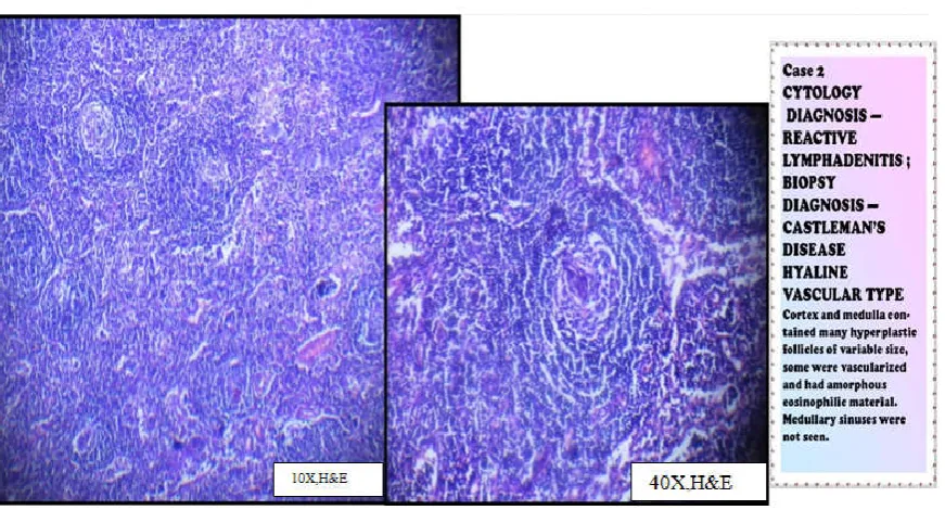
found incidentally. The rib showed cortical erosion with associated soft tissue component. Lungs were clear. Left basal pleura were thickened. Left pleural effusion, mediastinal and axillary lymphadenopathy was also seen. She was non reactive for HIV and HBsAg. FNA of posterior cervical lymph node measuring 5×6 cm was done. Smears were highly cellular. A mixed population of small and large lymphoid cells including follicular centre cells and tingible-body macrophages were seen. Few immunoblastsand plasma cells were seen. There were no epithelioid cells, giant cells or necrosis. A diagnosis of reactive hyperplasia was given. As a conclusive diagnosis in line with varied clinical symptoms was not reached, biopsy correlation was suggested. Histological examination showed cortex and medulla containing many hyperplastic follicles of variable size, some of which were vascularised with hyalinised
capillariesand had amorphous eosinophilic material. Germinal centres had eosinophilic material within the cell aggregates. Parafollicular zone contained branching capillaries, mature lymphocytes and plasma cells. Medullary sinuses were not seen. These findings confirmed CD, hyaline vascular type.
Similar cases have been recorded by Agarwalet al2and Sudha
et al. (2010).
Case 4: A 45yr old lady presented with left upper cervical
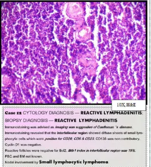
[image:3.595.155.452.60.388.2]lymphadenopathy. She had mild to moderatefever on and off. No history of night sweats or weight loss. Patient had undergone lymph node excision 8 years ago which was reported as reactive lymphadenitis. On examination, left upper cervical lymph nodes measured 5×5 cm. They were mildly tender, firm, nodular, matted. USG neck showed multiple level II, III and IV lymph nodes. Multiple nodules were noted in both lobes of thyroid. FNA showed a reactive lymphadenitis. CT scan also showed multiple enlarged lymph nodes in the left side of the neck along IJV, largest showing intense enlargement. ? Castleman’sdisease. Biopsy correlation was suggested for definite diagnosis. The smaller lymph node was excised initially as the larger node was close to the IJV. Histopathological examination showed that thenode was fragmented. Follicles with reactive centres, scattered thick walled vessels and numerous vesicles with high endothelial cells were noted. No granulomas or any evidence of metastasis were seen. Features were supportive of clinical suspicion of Castleman’s disease. As the patient was not willing to undergo excision of the larger lymph node, we decided to study the lymph node with immunemarkers. The Immunostaining revealed that the interfollicular region showed diffuse sheets of small lymphocytic cells which were positive for CD20, CD5 & CD23. CD138 was non contributory.
Cyclin D1 was negative. Reactive follicles were negative for Bcl2; Mib1 index in interfollicular region was 15%; PBC and BM not known. These features were diagnostic of Nodal involvement by Small lymphocytic lymphoma.
Problem: As policemen of the body’s immunity, lymph nodes
enlarge or react to a plethora of insult to the body. It ranges from viral, immunologic, bacterial to metastatic or circulating tumour cells. Whereas a non hematologic tumour deposit in lymph node is easier to diagnose in view of known history of primary, a hematologic or lymphoid neoplasm is a con artist. It mimics the problem of terrorism infiltrating civilian life.A few cases in the present case series have highlighted these problems.
Lessons for the Field
1.Hodgkin’s disease vs. reactive lymphadenitis: one should consider the diagnosis of Hodgkin’s disease when examining a reactive – appearing aspirate of a clinically suspicious lymph node. Reed – Sternberg cells and variants can be few in number and may be overlooked. Careful examination of all
smears is mandatory (Ioachim, 2008). 2.HLvs NHL is a
primary diagnosis usually followed by further classification
into subtypes and variants.4 Differentiating HL from NHL is
particularly important since it has direct implications for the prognosis and treatment of patients (Ioachim, 2008). The histologic pictures of their classic forms are sufficiently characteristic to allow for confident differential diagnosis in most cases (Ioachim, 2008). Nonetheless, it was noted that 20% to 30% of patients with HL treated with the otherwise successful standard therapy do not respond to treatment and finally die of their disease (Ioachim, 2008). The new methodologies, allowing for immunophenotypic and genetic analysis of the neoplasms, revealed the existence of lymphomas that did not fit exactly in the categories previously defined because of a variety of overlapping features (Ioachim, 2008). Such cases with mixed features, a few of which had been reported since the early 1990s , were the subject of two workshops and several studies dedicated to these entities, presently referred to as gray-zone lymphomas (Ioachim, 2008). Lymphomas that exhibit features overlapping with those of the classical type of HL belong to three groups: (a) nodular lymphocyte-predominant HL (NLPHL) and T-cell rich large B-cell lymphoma (TCRLBCL); (b) anaplastic large-cell lymphomas (ALCL; ALK+ and ALK-) and peripheral T-cell lymphoma (PTCL); and (c) primary mediastinal large-cell B-cell lymphoma (PMLBCL) and diffuse large B-B-cell lymphoma
(DLBCL) (Ioachim, 2008).
In NLPHL, the neoplastic cells (L&H cells) are of B-cell type; however, they are clearly different from H/R-S cells
of classical HL (Ioachim, 2008).
T-cell rich large B-cell lymphoma may show cells of
H/R-S type, but they are of B-cell type and express BCL-2. Eosinophils are not in the background, as in classical
or HL, and EBV gene products are not identified. 4
The cells of ALCL tumors, even when similar to H/R-S
cells commonly express the T-cell antigens and, in most cases, the ALK fusion gene, which is always negative in HL. The ALK–ALCL lymphomas can be distinguished by the lack of PAX5 antigen, which is usually dimly positive in H/R-S cells (Ioachim, 2008).
Peripheral T-cell lymphomas express T-cell markers as
well as cytotoxic molecules, such as granzyme B,
whereas HLs are of B-cell origin and do not express T-cell or cytotoxic markers (Ioachim, 2008).
The third group of large B-cell lymphomas constitutes the
most common overlapping gray zone with HLs since it is now established that the latter also originate in the B cells
of the lymphoid follicles (Ioachim, 2008).The large
B-cell lymphomas arising primarily in the mediastinum originate in the B cells of the thymic medulla and, by mediastinal location and histologic similarity, may create difficult problems of differential diagnosis with HL
(Ioachim, 2008). These gray-zone lymphomas share
features of DLBCL and of classical HL, with expression of CD30, CD15, as well as the pan–B-cell antigens CD20 and CD79a, suggesting that they represent a transition between the two tumor types (Ioachim, 2008).
Gray-zone lymphomas generally exhibit a more aggressive behavior than their HL counterparts, thus underlining the importance of their differential diagnosis (Ioachim, 2008). They require distinct treatments, and the addition of the anti–
B-cell rituximab antibody to the full combination
chemotherapy has been suggested.
Atypical lymphoproliferative disorders: The atypical
lymphoproliferative disorders include Castleman’s disease, Angioimmunoblastic lymphadenopathy with dysproteinemia, Lymphomatoidgranulomatosis, and Lymphomatoid papulosis (Brown et al., 2006). These diseases are distinct from the benign lymphoproliferative disorders in that they have significant potential for or have already acquired a malignant
phenotype (Brown, 2006).Castleman’s disease is an unusual
form of benign lymph node hyperplasia which form a differential diagnosis to lymphoma, tuberculosis, sarcoidosis,
silicosis, histoplasmosis etc. in anterior mediastinum (Gregson,
2003); although most common in the middle and posterior
compartments, in the anterior mediastinum the lesion tends to
be lobulated (Eisenberg, 1997).
Solution
Reactive lymphadenitis / a negative cytology, although
reassuring for the patient, should always be interpreted with the clinical picture in mind. It does not rule out serious underlying disorder. A greater awareness among the primary care physician or surgeon who orders for FNA study of a clinically significant lymph node is the requirement of the day. It should always be taken with a pinch of salt. As lymphoid lesions often have a quick clinical course, decisions for appropriate investigations have to be quick.
Patients should also be made aware of the varied types
of lymphoproliferative lesions and advances in their diagnostic tools. A informed decision has to be made keeping these factors. Often lymph node is an organ which is unknown to the non medical fraternity, unless they have experience with oncology patients among family members. Public awareness is the need of the hour just as in any field of medicine.
Unresolved questions
The pros and cons of surgical excision of inaccessible lymph
nodes, especially in the mediastinum, periaortic,
Reactive lymphadenitis can occur in one lymph node concurrent to occurrence of more serious lesions elsewhere.
REFERENCES
Agarwal D, Bansal P, Rani B, Sharma S, Chawla S, Bharat V et al. 2010. Evaluation of etiology of lymphadenopathy in different age groups using Fine Needle Aspiration Cytology: A retrospective study. The Internet Journal of Pathology. 2010; 10(2)
Brown RJ, 2006. Skarin TA in The Lymphomas. Cancellos GP, Lister TA, Young B 2nd Ed. Saunders Elsevier.
Eisenberg RL. 1997. Clinical Imaging, an atlas of differential
diagnosis. 3rd Ed. Lippincott – Raven.
Gregson RHS in Textbook of Radiology & Imaging. Sutton D. 7th Ed. Churchill Livingstone. 2003.
Ioachim HL, Medeiros LJ. 2008. Ioachim’s Lymph Node
Pathology. 4th edition.Wolter Kluwer: Lippincott Williams
and Wilkins; March.
Sudha A, Vivekanand N. 2010. Cytologic picture of Castleman’s disease. A report of two cases.Journal of Cytology. Oct; 27(4): 152 – 154.
Yao JL, Cangiarella JF, Cohen JM, Chhieng DC. 2001.
Fine-Needle Aspiration Biopsy of Peripheral T-Cell
Lymphomas: A Cytologic and Immunophenotypic Study of
33 Cases. Cancer cytopathol. Apr25; 93(2): 151-159