ISSN Online: 2327-509X ISSN Print: 2327-5081
DOI: 10.4236/jbm.2019.711007 Nov. 8, 2019 76 Journal of Biosciences and Medicines
Penetration of Topical Glucosamine Sulfate into
the Synovial Fluid of Patients with Knee
Osteoarthritis: A Nonrandomized, Open-Label,
Single Dose, Bioavailability Study
Matthew Kong
1, Khadijah Binte Hashim
1, Phoebe Lin
1, Gaëlle Coestesquis
2, Aiyun Xu
2,
Frederick Lebes
2, Choon Meng Ting
31School of Applied Science, Temasek Polytechnic, Singapore 2Lynk Biotechnologies Pte. Ltd., Singapore
3T & T Family Health Clinic & Surgery, Singapore
Abstract
Objective: This study aims to show that a proprietary topical cream can de-liver glucosamine through the skin into the synovial fluid of osteoarthritic patients. This cream contains 10% w/w glucosamine sulfate. It also aims to determine the endogenous level of glucosamine in the synovial fluid of these patients. Therapeutic effectiveness of glucosamine is not addressed in this study. Design: This phase IV, open-label, nonrandomized study enrolled 240 patients. Participants from the Test group received a single dose treat-ment (2 g of cream), and synovial fluid samples were collected 1 - 3 hours post-treatment. Patients from the Control group were not subjected to any treatment but their synovial fluid was also sampled to establish a glucosa-mine concentration baseline for Time-0 (T0). Glucosaglucosa-mine concentrations were determined by HPLC analysis. Results: The mean glucosamine con-centration in the synovial fluid of patients from the Test group (100.56 ng/ml, 95% CI 66.36 - 134.76, n = 117) was higher than in the Control group (17.83 ng/ml, 95% CI 7.42 - 28.24, n = 117) resulting in a significant between-group difference (p < 0.0001). While the gender of the subjects did not appear to affect the results, a significant difference was observed with age variation. Conclusion: The results suggest that glucosamine can be topically delivered across the human skin into the synovial fluid using a proper vehicle. This suggests that other water-soluble molecules could si-milarly be delivered transdermally, alleviating the need for oral delivery in cases where oral administration is difficult, or when harmful side effects could ensue.
How to cite this paper: Kong, M., Hashim, K.B., Lin, P., Coestesquis, G., Xu, A.Y., Lebes, F. and Ting, C.M. (2019) Penetra-tion of Topical Glucosamine Sulfate into the Synovial Fluid of Patients with Knee Osteoarthritis: A Nonrandomized, Open- Label, Single Dose, Bioavailability Study. Journal of Biosciences and Medicines, 7, 76-90.
https://doi.org/10.4236/jbm.2019.711007
Received: October 4, 2019 Accepted: November 5, 2019 Published: November 8, 2019
Copyright © 2019 by author(s) and Scientific Research Publishing Inc. This work is licensed under the Creative Commons Attribution International License (CC BY 4.0).
DOI: 10.4236/jbm.2019.711007 77 Journal of Biosciences and Medicines
Keywords
Osteoarthritis of the Knee, Transdermal Delivery, Glucosamine, Synovial Fluid, Joints
1. Introduction
Osteoarthritis (OA) is a highly prevalent joint disease characterized by joint car-tilage degeneration, a condition that affects 9.6% of men and 18% of women over 60 years old worldwide [1].
Our joints are cushioned by cartilages and lubricated by synovial fluid such that we can move any joint freely without pain. The principal lubricating sub-stances in our cartilage and synovial fluid are proteoglycans and glycosami-noglycans (GAGs). Glucosamine, the main ingredient needed to produce GAGs, is naturally produced by our body, but the body loses its capacity to produce suf-ficient glucosamine with age and causes thinning of the cartilage, leading to joint degeneration [2].
Contradictory research studies on glucosamine were published [2], and OA treatment guidelines developed by scientific organizations are often debated. Organizations such as the American College of Rheumatology (ACR) or the Os-teoarthritis Research Society International (OARSI) recommend against the use of glucosamine for the treatment of OA. According to the OARSI Guidelines, glucosamine is not an efficacious disease-modifying drug for OA, and also does not guarantee outcomes as a pain reliever. However, it is important to note that transdermal delivery or skin application of glucosamine was not considered in the construct of the OARSI Guidelines. However, other organizations such as the European League Against Rheumatism (EULAR) and the European Society or Clinical and Economic Aspects of Osteoporosis and Osteoarthritis (ESCEO) recommend symptomatic slow-acting drugs such as glucosamine sulfate for the background therapy of knee OA. This was based on high-quality evidence show-ing that glucosamine was superior to placebos in the treatment of pain and func-tional impairment [3].
For targeted drug administration, it is widely established that topical admini-stration offers numerous advantages over oral delivery [3]. The benefits of topi-cal administration include maximizing the bioavailability of the drug, optimizing therapeutic efficacy, and minimizing side effects [4]. Topical delivery avoids the occurrence of the hepatic first-pass effect, and also has advantages over both in-travenous and intramuscular routes as it is a painless and noninvasive method of drug delivery [3].
ad-DOI: 10.4236/jbm.2019.711007 78 Journal of Biosciences and Medicines ministration was superior to the oral route in improving stiffness and function of the joint [2]. In a different study by Cohen et al., 63 patients suffering from OA of the knee were randomly divided into 2 groups. The group receiving topi-cal glucosamine and chondroitin showed significant pain relief compared to the placebo group [5]. These papers have demonstrated the statistical relationship between the application of topical glucosamine and positive OA outcomes, but have not demonstrated that the applied glucosamine has indeed crossed the skin to reach the diseased joint.
Hence, this study is designed to produce the evidence that glucosamine did actually reach the diseased area after topical application. The aim of the present nonrandomized, open-label, single dose study was to investigate the feasibility of penetration of a 10% glucosamine cream into the synovial fluid of patients with joint effusions and intended arthrocentesis (synovial fluid aspiration). A second outcome was to determine the endogenous level (or baseline) of glucosamine in synovial fluid of osteoarthritic knees for the group with no treatment adminis-tered. The hypothesis of the study was that higher concentrations of glucosa-mine would be found in the synovial fluid of patients using the cream.
Whether the topical glucosamine dosage was sufficient to be translated into clinical effects was not addressed in this study and could be an objective for fu-ture work. To our knowledge, this is the first study describing the bioavailability of glucosamine in human synovial fluid following the application of a topical cream containing glucosamine.
2. Methods
2.1. Ethical Considerations
The study protocol and related materials were approved by the Institutional Re-view Board of Temasek Polytechnic, Singapore. The study was conducted in conformance with the current revision of the Declaration of Helsinki, and with current Good Clinical Practice, Singapore Guidelines. Written informed consent was obtained from all participants before commencement of the study. The study was registered at clinicaltrials.gov (NCT03743896) and with the Singapore Health Sciences Authority (CTC1600169). This report adheres to the Transpar-ent Reporting of Evaluations with Non-randomised Designs (TREND) state-ment, which complement the widely adopted CONsolidated Standards Of Re-porting Trials (CONSORT) statement [6].
2.2. Patient Eligibility and Study Design
DOI: 10.4236/jbm.2019.711007 79 Journal of Biosciences and Medicines 2 and 3) prior to recruitment. Patients with any form of personal glucosamine supplement or pain relief treatment taken during the last 24 hours were excluded from this study. Additional exclusion criteria included known allergy to shell-fish, glucosamine, and capsaicin, as well as a history of skin sensitivity, skin ab-normality at application site and women who were nursing. Participants were grouped into Control (untreated) and Test (treated) groups.
The outpatient clinic, located in Singapore, specialises in arthrocentesis and viscosupplementation. The medical doctor in charge of the clinic has been per-forming these procedures since 1991 [7]. He is also the author of a handbook about viscosupplementation for OA knees, and in this book, he describes the ways patients are diagnosed with knee OA as well as the procedures for synovial fluid extraction and viscosupplementation [8].
2.3. Study Intervention and Allocation
TGC® Plus Capsaicin, a transdermal glucosamine cream containing 10% w/w glucosamine sulfate, is commercially available for joint pain relief, and manu-factured by Lynk Biotechnologies Pte. Ltd., Singapore. It is listed as a medicinal product exempted from registration by the Singapore Health Sciences Authority. All patients taking part in the study were offered a free 45 g tube of TGC® Plus Capsaicin. The joint fluid aspiration cost was waived for patients of the Test group.
Participants were given full discretion to decide which of the two groups they wished to be assigned to. Test group volunteers were given treatment by the clinic’s nurses of a topical application of the cream (2 g single dose treatment) on the affected knee 1 - 3 hours before the extraction of the synovial fluid by the medical practitioner. The single dose of 2 g (200 mg glucosamine) was decided due to surface area limitations. A larger amount would have been impractical for topical application. The synovial fluid was collected after 1 to 3 hours, based on results from previous internal studies. In these studies, the maximum glucosa-mine concentration in mice plasma was obtained within this time range. The Control group volunteers were not given any treatment and proceeded directly for the extraction of fluid. This fluid, which is normally discarded by the clinic, was collected for both groups.
Since the primary endpoint is to determine the concentration of glucosamine present in the synovial fluid, there was no possibility of subjective bias or pla-cebo effect. Hence no randomisation or patient’s blinding was necessary. How-ever, blind analyses were conducted independently by third party testing lab (Temasek Polytechnic, Singapore). Synovial fluid samples were only labelled with each participant’s identification code.
2.4. Materials
DOI: 10.4236/jbm.2019.711007 80 Journal of Biosciences and Medicines glacial acetic acid (ACS grade, 99.0%) were obtained from Merck Millipore.
2.5. Sample Preparation and Analyses
The synovial fluid samples were centrifuged at 4˚C, 1400×g for 15 minutes im-mediately after collection. The cell-free supernatant was harvested and stored at −80˚C pending assay. The concentration of glucosamine in each synovial fluid sample was determined by a validated high-performance liquid chromatogra-phy-ultraviolet visible spectrometry (HPLC-UV) method. After thawing at room temperature, synovial fluid sample (200 μl) was pipetted into a 1.5-ml centrifuge tube and acetonitrile (400 μl) was subsequently added for protein precipitation. Methanol (200 μl) was then added and the resultant mixture was vortexed and centrifuged at 14,000 × g for 2 minutes. The supernatant was recovered and dried using a freeze dryer (Scanvac, Labogene). To the dried residue, 0.1 ml of derivatiz-ing reagent, 1-naphthyl isothiocyanate in methanol:acetonitrile:triethylamine (1:1:0.2) mixture at 50 mg/ml was added. The mixture was vortexed and left at ambient conditions for 30 minutes. The reaction was quenched by adding 0.2 ml of acetic acid solution (1.5% v/v). The excess derivatizing reagent and its degra-dation products were partitioned into an organic phase by the addition of 1.0 ml of methylene chloride. The upper aqueous layer was transferred to another 2-ml centrifuge tube and the extraction process was repeated three times. 150 - 170 µl of the upper aqueous layer was filtered using a 0.45 µm syringe filter (Thermo Scientific) and transferred into vials for HPLC analyses.
The instrument and the optimised parameters used are as follows: Agilent 1290 Liquid Chromatography system with autosampler and UV detector; Col-umn type: Two Inertsil ODS-3 (4.0 mm × 250 mm, 3 µm) connected in se-quence; Column temperature: 27˚C; Flow rate: 0.3 ml/min; Injection volume: 10 µl; Detection wavelength: 224 nm; HPLC elution: Binary gradient elution with mobile phase A (80:20 ultrapure water:acetonitrile with 0.04% v/v acetic acid and 0.04% v/v triethylamine) and mobile phase B (Acetonitrile); 0 - 65 min (100% A), 65 - 85 min (100% B), 85 - 100 min (100% A).
Due to potential matrix effects in analysing synovial fluid, the standard addi-tion method was used to quantify the concentraaddi-tion of glucosamine. The stan-dard addition method was carried out by spiking each synovial fluid sample separately with two different concentrations of standard solutions of glucosa-mine hydrochloride to achieve additional concentrations of glucosaglucosa-mine of 250 ng/ml and 500 ng/ml, respectively.
2.6. Sample Size Estimation
aspira-DOI: 10.4236/jbm.2019.711007 81 Journal of Biosciences and Medicines tion, potential trial participants were readily available and synovial fluid samples are usually discarded. Therefore, there were no ethical concerns by having a high number of participants.
2.7. Statistical Analysis and Outliers
Analyses were conducted using GraphPad Software © 2018. Outcomes were as-sessed using a univariate, unpaired Student t-test. Two-tailed P-values < 0.01 were considered statistically significant. The Fisher’s test was used for compari-son of baseline categorical data.
The Z-score method was used to identify outliers in both the Control group and the Test group dataset. Samples with a Z-value greater than 3 were taken to be outliers and excluded from the concentration calculation.
3. Results
3.1. Participants Disposition and Baseline Characteristics
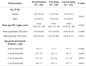
From May 2016 to March 2017, 240 patients undergoing arthrocentesis were se-lected for the study, with 120 participants in each group. The participants con-sisted of 58 (24.17%) men and 182 (75.83%) women, with mean age of 64.24 ± 8.58 years old (mean ± SD). There were no notable differences in participant demographic between Control and Test groups (Table 1).
[image:6.595.212.538.457.707.2]The patient enrolment process and sample analysis process are summarized in the enrolment chart (Figure 1). There was no follow up necessary and no devia-tion from the protocol.
Table 1. Demographic details of study participants—sex, mean age, and age group.
Characteristics All participants (N = 240) Test group (N = 120) Control group (N = 120) P-value
Sex, N (%)
Female 182 (75.83) 87 (72.50) 95 (79.17)
0.2912 Male 58 (24.17) 33 (27.50) 25 (20.83)
Mean age (SD, range), years (8.58, 39 - 80) 64.24 (8.37, 39 - 80) 63.61 (8.78, 39 - 80) 64.88 0.2526
Mean age female (SD), years 64.54 (8.87) 64.13 (8.54) 64.92 (9.19) 0.5498 Mean age male (SD), years 63.29 (7.62) 62.24 (7.88) 64.68 (7.17) 0.2301
Age group participants, N female + male
39 to 50 years old 14 + 6 5 + 5 9 + 1 0.1409 51 to 60 years old 47 + 10 28 + 3 19 + 7 0.1602 61 to 70 years old 72 + 33 35 + 23 37 + 10 0.0574
71 to 80 years old 49 + 9 19 + 2 30 + 7 0.4647
DOI: 10.4236/jbm.2019.711007 82 Journal of Biosciences and Medicines
Figure 1. Enrolment chart. 120 participants were selected for each group. All synovial
fluid samples were included in the main analysis.
A total of 240 samples of synovial fluid were collected. The Control group, consisting of 120 samples, were not subjected to any treatment prior to arthro-centesis. The Test group, with a total of 120 samples, were treated with a single dose (2 g) of topical glucosamine cream 1 to 3 hours prior to arthrocentesis.
3.2. Overall Results—Test Group vs Control Group
In Table 2, the HPLC results show that the mean concentration of glucosamine of the Test group was 100.56 ng/ml (95% CI 66.36 - 134.76, n = 117) and is sta-tistically higher (P < 0.0001) than that of the corresponding value for the Control group (17.83 ng/ml, 95% CI 7.42 - 28.24; n = 117). Between 1 and 3 hours post-treatment, the mean concentration of glucosamine measured from the Test group was more than five times than that of the Control group, demonstrating a significant difference between samples from the two groups.
DOI: 10.4236/jbm.2019.711007 83 Journal of Biosciences and Medicines
Figure 2. Distribution of glucosamine concentrations (in ng/ml) in synovial fluid samples of subjects from the Control group and
[image:8.595.210.541.339.398.2]the Test group. The comparison shows higher concentrations of glucosamine present in samples of Test group subjects compared to samples of Control group subjects.
Table 2. Mean glucosamine concentrations of test and control groups.
Overall Results Glucosamine concentration (ng/ml) P-value Mean (95% CI)
Control (n = 117) 17.83 (7.42 - 28.24)
<0.0001 Test (n = 117) 100.56 (66.36 - 134.76)
Abbreviation: CI = Confidence Interval.
(82%) have no detectable glucosamine, while only 48 samples in the Test group (41%) have no detectable glucosamine. In addition, 18 samples of the Test group (15.3%) had a measured glucosamine concentration of >200 ng/ml, compared to only 2 samples of the Control group (1.7%) surpassing this concentration. This difference in detectable levels of glucosamine between the two groups implies that a sufficient amount of glucosamine was absorbed through the skin and de-livered to the subject’s synovial fluid within one to three hours after the applica-tion of topical glucosamine.
3.3. Results—By Sex
Table 3(a) shows the glucosamine concentrations in synovial fluid samples from subjects in both groups, categorised by sex. There was a significant difference between Test and Control groups for both male and female subjects. However, the difference between male (136.94 ng/ml, 95% CI 61.48 - 212.40; n = 33) and female (86.26 ng/ml, 95% CI 48.33 - 124.20; n = 84) Test Groups was not statis-tically significant (P = 0.1878).
3.4. Results—By Age Group
DOI: 10.4236/jbm.2019.711007 84 Journal of Biosciences and Medicines data indicates a significant difference between Test and Control groups for the older age group (P < 0.0001), but not for the younger age group (P = 0.0896).
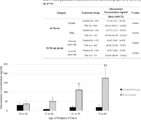
[image:9.595.63.539.287.696.2]When subjects were further categorised into specific age groups with a nar-rower 10-year interval, glucosamine concentrations for Test and Control sub-jects in the age groups of 39 - 50 years old (P = 0.8044) and 51 - 60 years old (P = 0.0879) were not statistically significant. However, significant differences were observed among subjects in the advanced age group of 61 - 70 years old (P = 0.0014) and 71 - 80 years old (P = 0.0019) showing more pronounced differences in glucosamine concentration. While the glucosamine concentration remains relatively constant for all the Control group participants, a different trend of data among participants of the Test group suggests that glucosamine concentration in the synovial fluid, following application of topical glucosamine, increases with age (Figure 3).
Table 3. Mean glucosamine concentrations of test and control groups: (a) by sex, (b) by
age group.
Category Treatment Group
Glucosamine
Concentration (ng/ml) P-value Mean (95% CI)
(a) By sex
Female Control (n = 93) 17.34 (5.13 - 29.55) 0.0004 Test (n = 84) 86.26 (48.33 - 124.20)
Male Control (n = 24) 19.73 (−0.17 - 39.63) 0.0106 Test (n = 33) 136.94 (61.48 - 212.41)
(b) By age group
39 to 60 years old
Control (n = 33) 14.82 (3.60 - 26.03)
0.0896 Test (n = 40) 46.96 (13.94 - 79.97)
61 to 80 years old
Control (n = 84) 19.01 (5.08 - 32.95)
<0.0001 Test (n = 77) 128.40 (80.00 - 176.81)
Figure 3. Distribution of glucosamine concentrations in synovial fluid samples of Control and Test group subjects by 10-year age
DOI: 10.4236/jbm.2019.711007 85 Journal of Biosciences and Medicines
3.5. Safety
There were no allergic or adverse reactions reported during or after the applica-tion of the glucosamine cream by participants of the Test group.
4. Discussion
4.1. Overview
The results of the study present empirical evidence of glucosamine bioavailabil-ity and transdermal delivery into human synovial fluid after application of a topical glucosamine cream on knees of osteoarthritic patients.
Observations show that the mean glucosamine concentration in the synovial fluid of subjects from the Test group was significantly higher than the glucosa-mine concentration in the synovial fluid of subjects from the Control group. These results are corroborated by another study assessing the glucosamine con-centration in synovial fluid after 14-day treatment of 1500 mg oral glucosamine [9]. This study reported a median glucosamine concentration of 777 ng/ml for synovial fluid samples collected 3 hours after the last dose of the 2-week treat-ment. Our study shows that with a single dose of cream (containing 200 mg glucosamine) applied on skin, a mean glucosamine concentration of 100.56 ng/ml can be detected. Correlations of absorbed glucosamine levels in synovial fluid to demographic information of the patients, such as age and sex, were also studied.
4.2. Endogenous Glucosamine Level
When subjects in the Control group were categorised by sex or age, it was ob-served that the mean glucosamine concentrations were within a consistent range. It can be seen that the baseline (endogenous) glucosamine concentration in synovial fluid does not vary across sex and age. In this study, the glucosamine concentration from subjects in the Control group (17.83 ng/ml, 95% CI 7.42 - 28.24; n = 117) regardless of sex or age is similar to the results from two previous studies, where the endogenous glucosamine concentrations in synovial fluid were reported to be 36.5 ng/ml [9] and 32.3 ng/ml [10].
4.3. Impact of Sex on Glucosamine Absorption
DOI: 10.4236/jbm.2019.711007 86 Journal of Biosciences and Medicines that the degree or rate of absorption were not dependant on gender [11] [12]. Similar to these studies, this transdermal study of topical glucosamine applica-tion also showed that it was not selective and independent of sex.
4.4. Impact of Age on Glucosamine Absorption
A possible correlation with participants’ age was also considered in this study. The approach employed was grouping the subjects into two age categories of 39 - 60 years old and 61 - 80 years old, and analysing the glucosamine concentra-tion of each subject within each age category. The glucosamine level of younger Test group patients did not significantly increase following treatment (P = 0.0896). However, the older Test group patients were found to have a signifi-cantly higher level of glucosamine (P < 0.0001) compared to the control group. As shown in Figure 3, an increasing trend in glucosamine levels, as a function of age, became more prominent when age groupings were modified into more spe-cific age categories of 10-year intervals. The results suggested that the rate of transdermal glucosamine transport increases with age and that transdermal de-livery of glucosamine might have higher rates in people of advanced age than their younger counterparts. More data would be needed to confirm this trend as the population of younger patients was limited in this study.
A direct correlation of transdermal delivery rate with skin ageing was postu-lated in this work. It has been reported that skin thins progressively at an accel-erating rate with age [13], and the thinning of skin tissue may allow better transdermal absorption of glucosamine. Konda and colleagues reported that po-tential differences in skin from individuals of varying age, pharmacokinetics with transdermal delivery may be altered. Other factors such as active ingredient physicochemical characteristics and formulation components, determine whether a specific drug will have pharmacokinetic differences across age groups [14]. This can be the basis for further studies of how transdermal glucosamine trans-port is affected by age, and shows the potential for age-specific glucosamine cream products with optimised transport efficiencies.
4.5. Transdermal Glucosamine Absorption
DOI: 10.4236/jbm.2019.711007 87 Journal of Biosciences and Medicines the synovial membrane. The synovial matrix provides the permeable pathway through which exchange of molecules occurs, but also offers sufficient outflow resistance to retain large solutes of synovial fluid within the joint cavity [17].
It is scientifically established that water-soluble compounds like glucosamine cannot readily permeate through the lipophilic layers of the skin. The results of this study demonstrate the potential that if suitable vehicles are used to enable the transport of glucosamine, as in the case of TGC® Plus Capsaicin, transdermal delivery might also be possible for other water-soluble compounds.
4.6. Elimination of Outliers
Only six outliers were identified (three in each group) and excluded from the main results. The HPLC analysis were repeated and it was confirmed that the results markedly deviated from other observations. Even if the outliers are in-cluded in the calculations, the average concentration of glucosamine of the Test group is still significantly higher (145.84 ng/ml, 95% CI 80.26 - 211.41; n = 120) than the corresponding value for the Control group (35.92 ng/ml, 95% CI 12.76 - 59.08, n = 120); at P = 0.002.
4.7. Study Limitations
The study design deliberately involved a large sample size to improve the confi-dence level of results obtained. The Control group acted as a proxy for a Time-0 (T0) sample to establish a baseline of glucosamine level in the synovial fluid of subjects in the Test group. Ideally, a T0 sample should have been used as a con-trol. As arthrocentesis is a relatively painful procedure, this study was designed, for ethical reasons, to avoid subjecting the patients to an additional extraction in order to obtain T0 data. Instead we have collected a significant number of sam-ples from a control group that is already seeking arthrocentesis treatment as a proxy for T0 data, hence avoiding any additional pain.
As this study was not a pharmacokinetic study, the Tmax (time at which the highest concentration is observed) was unknown. As the synovial fluid samples for the Test group were collected one to three hours after the application of the glucosamine cream, the concentrations reported here are likely to be underesti-mated and could have been higher if the Tmax had been determined beforehand.
col-DOI: 10.4236/jbm.2019.711007 88 Journal of Biosciences and Medicines lected for each patient and this does not change the overall conclusions of the study. Assuming that the synovial fluid volume extracted is proportional to the total synovial fluid originally present, the absolute amount of glucosamine pre-sent in each sample was still significantly higher (P = 0.0093) for the Test group, compared to the Control group. Further research work will be necessary to measure the total volume of synovial fluid and get a more accurate profile of glucosamine level in synovial fluid.
5. Conclusions and Recommendations for Further Studies
This clinical study presented empirical evidence of transdermal delivery of glu-cosamine resulting in its bioavailability in the synovial fluid of test patients with OA of the knee. This study reinforces results from a prior unpublished pharma-cokinetic study where glucosamine was detected in mice plasma 1 - 3 hours after topical glucosamine treatment. Whether the amount of glucosamine delivered is sufficient to be translated into therapeutic effect remains to be elucidated and could be the subject for future studies.This study demonstrated that with a proper vehicle, a water-soluble molecule such as glucosamine can be delivered through human skin, and further studies may be conducted to investigate the potential transdermal delivery of other compounds using the same vehicle. The transdermal delivery route represents a highly attractive alternative to oral delivery, especially for demographic groups such as the elderly. The rate and mechanism of glucosamine absorption is not documented in this work, and further research is recommended on specific chemical properties of cream formulations that may control the glucosamine transdermal transport steps from a cream matrix, through skin layers.
Acknowledgements
The authors wish to thank Siti Norasikin for her assistance with numerous HPLC analyses, and Chua Yek Tann and Sheng Ping for their contributions to earlier analytical work.
Conflicts of Interest
Ting, C.M. has declared no conflicts of interest. Temasek Polytechnic, School of Applied Science was engaged by Lynk Biotechnologies Pte. Ltd. for data acquisi-tion, analysis and interpretation.
References
[1] WHO (2019) Chronic Rheumatic Conditions: Osteoarthritis.
http://www.who.int/chp/topics/rheumatic/en
[2] Hammad, Y.H., Hala, R.M. and Sobhy, M.M. (2015) Clinical and Biochemical Study of the Comparative Efficacy of Topical versus Oral Glucosamine/Chondroitin Sul-fate on Osteoarthritis of the Knee. The Egyptian Rheumatologist, 37, 85-91. https://doi.org/10.1016/j.ejr.2014.06.007
DOI: 10.4236/jbm.2019.711007 89 Journal of Biosciences and Medicines
(2017) Skin Delivery of Glucosamine and Chondroitin Sulphates—A Perspective on the Conservative Treatment for Osteoarthritis of the Knee. Journal of Biosciences and Medicines, 5, 11-20.https://doi.org/10.4236/jbm.2017.54002
[4] Onigbinde, A.T., Talabi, A.E. and Shehu, R.A. (2011) Acute Effects of Combination of Glucosamine Sulphate Iontophoresis with Exercise on Fasting Plasma Glucose of Participants with Knee Osteoarthritis. Hong Kong Physiotherapy Journal, 29, 79-85.
https://doi.org/10.1016/j.hkpj.2011.06.003
[5] Cohen, M., Wolfe, R., Mai, T. and Lewis, D. (2003) A Randomized, Double Blind, Controlled Trial of a Topical Cream Containing Glucosamine Sulfate, Chondroitin Sulfate, and Camphor for Osteoarthritis of the Knee. The Journal of Rheumatology, 30, 523-528.
[6] CDC (2018) Transparent Reporting of Evaluations with Non-Randomised Designs (TREND). https://www.cdc.gov/trendstatement/index.html
[7] YTL Community (2008) Suffering Worn-out, Osteo-Arthritic Knees? Cheah Ui-Hoon Finds out How to Avoid Knee Replacement Surgery.
http://www.ytlcommunity.com/commnews/shownews.asp?newsid=37518
[8] Ting, C.M. (2007) Advancing Visco Supplementation—A Handbook on Viscosup-plementation for OA Knees. Armour Publishing Pte Ltd., Singapore.
[9] Persiani, S., Rotini, R., Trisolino, G., Rovati, L.C., Locatelli, M., Paganini, D., et al.
(2007) Synovial and Plasma Glucosamine Concentrations in Osteoarthritic Patients Following Oral Crystalline Glucosamine Sulphate at Therapeutic Dose. Osteoarthri-tis Cartilage, 15, 764-772. https://doi.org/10.1016/j.joca.2007.01.019
[10] Pastorini, E., Rotini, R., Guardigli, M., Vecchiotti, S., Persiani, S., Trisolino, G., et al.
(2009) Development and Validation of a HPLC-ES-MS/MS Method for the Deter-mination of Glucosamine in Human Synovial Fluid. Journal of Pharmaceutical and Biomedical Analysis,50, 1009-1014. https://doi.org/10.1016/j.jpba.2009.07.008
[11] Roy, S.D. and Flynn, G.L. (1990) Transdermal Delivery of Narcotic Analgesics: pH, Anatomical, and Subject Influences on Cutaneous Permeability of Fentanyl and Sufentanil. Pharmaceutical Research, 7, 842-847.
https://doi.org/10.1023/A:1015912932416
[12] So, J., Ahn, J., Lee, T.H., Park, K.H., Paik, M.K., Jeong, M., et al. (2014) Comparison of International Guidelines of Dermal Absorption Tests Used in Pesticides Expo-sure Assessment for Operators. Toxicological Research, 30, 251-260.
https://doi.org/10.5487/TR.2014.30.4.251
[13] Farage, M.A., Miller, K.W., Elsner, P. and Maibach, H.I. (2013) Characteristics of the Aging Skin. Advances in Wound Care, 2, 5-10.
https://doi.org/10.1089/wound.2011.0356
[14] Konda, S., Meier-Davis, S.R., Cayme, B., Shudo, J. and Maibach, H.I. (2012) Age- Related Percutaneous Penetration Part 2: Effect of Age on Dermatopharmacokinet-ics and Overview of Transdermal Products. Skin Therapy Letter, 17, 5-7.
[15] Anderson, J.W., Nicolosi, R.J. and Borzelleca, J.F. (2005) Glucosamine Effects in Humans: A Review of Effects on Glucose Metabolism, Side Effects, Safety Consid-erations and Efficacy. Food and Chemical Toxicology, 43, 187-201.
https://doi.org/10.1016/j.fct.2004.11.006
[16] Uldry, M., Ibberson, M., Hosokawa, M. and Thorens, B. (2002) GLUT2 Is a High Affinity Glucosamine Transporter. FEBS Letters, 524, 199-203.
https://doi.org/10.1016/S0014-5793(02)03058-2
DOI: 10.4236/jbm.2019.711007 90 Journal of Biosciences and Medicines
https://doi.org/10.2478/intox-2013-0019
[18] Geborek, P., Saxne, T., Heinegard, D. and Wollheim, F.A. (1988) Measurement of Synovial Fluid Volume Using Albumin Dilution upon Intraarticular Saline Injec-tion. The Journal of Rheumatology, 15, 91-94.
[19] Rekonen, A., Oka, M. and Kuikka, J. (1973) Measurement of Synovial Fluid Volume by a Radioisotope Method. Scandinavian Journal of Rheumatology, 2, 33-35.
https://doi.org/10.1080/03009747309097148
[20] Kraus, V.B., Stabler, T.V., Kong, J., Varju, G. and McDaniel, G. (2007) Measure-ment of Synovial Fluid Volume Using Urea. Osteoarthritis Cartilage, 15, 1217-1220.