2-Hydroxyimino-1-phenylethanone
thiosemicarbazone monohydrate
Nursabah Sarıkavaklı,aI:knur Babahan,aErtan S¸ahinband Tuncer Ho¨kelekc*
aAdnan Menderes University, Department of Chemistry, 09010 Aydın, Turkey, b
Atatu¨rk University, Department of Chemistry, 22240 Erzurum, Turkey, and
cHacettepe University, Department of Physics, 06800 Beytepe, Ankara, Turkey
Correspondence e-mail: merzifon@hacettepe.edu.tr
Received 14 February 2008; accepted 20 February 2008
Key indicators: single-crystal X-ray study;T= 294 K; mean(C–C) = 0.004 A˚; Rfactor = 0.062;wRfactor = 0.155; data-to-parameter ratio = 18.7.
In the title thiosemicarbazone derivative, C9H10N4OSH2O,
intramolecular N—H N hydrogen bonds result in the formation of two nearly coplanar five- and six-membered rings, which are also almost coplanar with the adjacent phenyl ring. The oxime group has anEconfiguration and is involved in intermolecular O—H O hydrogen bonding as a donor. In the crystal structure, intramolecular O—H S and N—H N and intermolecular O—H O and N—H S hydrogen bonds generate edge-fused R2
2
(8) and R4 1
(11) ring motifs. The hydrogen-bonded motifs are linked to each other to form a three-dimensional supramolecular network.
Related literature
For general backgroud, see: Lukevicset al.(1995); Liberta & West (1992); Hagenbach & Gysin (1952); Joneset al.(1965); Brockman & Thomson (1956); Klaymanet al.(1979); Petering & van Giesen (1966); Sevagapandian et al. (2000); Forman (1964); Holanet al.(1984); Balsamoet al.(1990); Marsmanet al.(1999); Karleet al.(1996); Etteret al.(1990); Chertanovaet al.(1994); Bernstein et al.(1995). For related structures, see: Sarıkavaklıet al.(2007); O¨ zel Gu¨ven et al.(2007); Ho¨kelek, Batı et al. (2001); Ho¨kelek, Zu¨lfikarog˘lu & Batı (2001); Bu¨yu¨kgu¨ngo¨ret al.(2003); Ho¨keleket al.(2004a,b); Ho¨kelek
et al.(2004). For the synthesis, see: El-Shazlyet al.(2005). For bond-length data, see: Allenet al.(1987).
Experimental
Crystal data
C9H10N4OSH2O
Mr= 240.29
Monoclinic,C2=c a= 28.5615 (3) A˚ b= 4.6805 (3) A˚ c= 22.0977 (4) A˚
= 127.24 (2)
V= 2351.8 (6) A˚3
Z= 8
MoKradiation
= 0.27 mm1
T= 294 (2) K 0.300.200.15 mm
Data collection
Rigaku R-AXIS RAPID-S diffractometer
Absorption correction: multi-scan (Blessing, 1995)
Tmin= 0.940,Tmax= 0.960
31269 measured reflections 3607 independent reflections 2146 reflections withI> 2(I) Rint= 0.090
Refinement
R[F2> 2(F2)] = 0.062
wR(F2) = 0.154
S= 1.04 3607 reflections 193 parameters 8 restraints
H atoms treated by a mixture of independent and constrained refinement
max= 0.14 e A˚
3
min=0.31 e A˚
[image:1.610.312.566.355.432.2]3
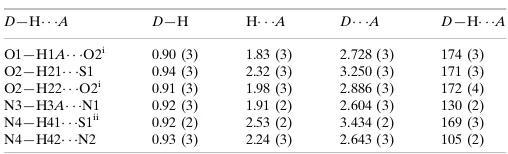
Table 1
Hydrogen-bond geometry (A˚ ,).
D—H A D—H H A D A D—H A
O1—H1A O2i
0.90 (3) 1.83 (3) 2.728 (3) 174 (3) O2—H21 S1 0.94 (3) 2.32 (3) 3.250 (3) 171 (3) O2—H22 O2i 0.91 (3) 1.98 (3) 2.886 (3) 172 (4) N3—H3A N1 0.92 (3) 1.91 (2) 2.604 (3) 130 (2) N4—H41 S1ii
0.92 (2) 2.53 (2) 3.434 (2) 169 (3) N4—H42 N2 0.93 (3) 2.24 (3) 2.643 (3) 105 (2)
Symmetry codes: (i)xþ1 2;yþ
1 2;zþ
3
2; (ii)x;yþ1;zþ1.
Data collection:CrystalClear(Rigaku/MSC, 2005); cell refinement:
CrystalClear; data reduction:CrystalClear; program(s) used to solve structure:SHELXS97(Sheldrick, 2008); program(s) used to refine structure: SHELXL97 (Sheldrick, 2008); molecular graphics:
ORTEP-3 for Windows(Farrugia, 1997) andPLATON(Spek, 2003); software used to prepare material for publication:WinGX(Farrugia, 1999).
The authors are indebted to the Department of Chemistry, Atatu¨rk University, Erzurum, Turkey, for the use of the X-ray diffractometer purchased under grant No. 2003/219 of the University Research Fund.
Supplementary data and figures for this paper are available from the IUCr electronic archives (Reference: XU2403).
References
Allen, F. H., Kennard, O., Watson, D. G., Brammer, L., Orpen, A. G. & Taylor, R. (1987).J. Chem. Soc. Perkin Trans. 2, pp. S1–19.
Balsamo, A., Macchia, B., Martinelli, A., Orlandini, E., Rossello, A., Macchia, F., Bocelli, G. & Domiano, P. (1990).Eur. J. Med. Chem.25, 227–233. Bernstein, J., Davis, R. E., Shimoni, L. & Chang, N.-L. (1995).Angew. Chem.
Int. Ed. Engl.34, 1555–1573.
Blessing, R. H. (1995).Acta Cryst.A51, 33–38.
Brockman, R. W. & Thomson, J. R. (1956).Cancer Res.16, 167–170. Bu¨yu¨kgu¨ngo¨r, O., Ho¨kelek, T., Tas¸, M. & Batı, H. (2003).Acta Cryst.E59,
o883–o885.
organic compounds
Acta Cryst.(2008). E64, o623–o624 doi:10.1107/S1600536808004947 Sarıkavaklıet al.
o623
Acta Crystallographica Section E
Structure Reports
Online
716.
Shazly, R. M., Al-Hazmi, G. A. A., Ghazy, S. E., Shahawi, M. S. & El-Asmy, A. A. (2005).Spectrochim. Acta,A61, 243–252.
Etter, M. C., MacDonald, J. C. & Bernstein, J. (1990).Acta Cryst.B46, 256–262. Farrugia, L. J. (1997).J. Appl. Cryst.30, 565.
Farrugia, L. J. (1999).J. Appl. Cryst.32, 837–838. Forman, S. E. (1964).J. Org. Chem.29, 3323–3327.
Hagenbach, R. E. & Gysin, H. (1952).Experientia,8, 184–185.
Ho¨kelek, T., Batı, H., Bekdemir, Y. & Ku¨tu¨k, H. (2001).Acta Cryst.E57, o663– o665.
Ho¨kelek, T., Bu¨yu¨kgu¨ngo¨r, O., Tas¸, M. & Batı, H. (2004a).Acta Cryst.E60, o109–o111.
Ho¨kelek, T., Bu¨yu¨kgu¨ngo¨r, O., Tas¸, M. & Batı, H. (2004b).Acta Cryst.E60, o406–o408.
Ho¨kelek, T., Tas¸, M. & Batı, H. (2004).Cryst. Res. Technol.39, 363–367. Ho¨kelek, T., Zu¨lfikarog˘lu, A. & Batı, H. (2001).Acta Cryst.E57, o1247–o1249. Holan, G., Johnson, W. M. P., Rihs, K. & Virgona, C. T. (1984).Pestic. Sci.15,
361–368.
Jones, D. H., Slack, R., Squires, S. & Woolridge, K. R. H. (1965).J. Med. Chem. 8, 676–680.
7128–7133.
Klayman, D. L., Bartoserich, J. F., Griffin, T. S., Manson, C. J. & Scovill, J. P. (1979).J. Med. Chem.22, 885–893.
Liberta, A. E. & West, D. X. (1992).Biometals,5, 121–125.
Lukevics, E., Jansone, D., Rubina, K., Abele, E., Germane, S., Leite, L., Shymaska, M. & Popelis, J. (1995).Eur. J. Med. Chem.30, 983–986. Marsman, A. W., Leussing, E. D., Zwikker, J. W. & Jenneskens, L. W. (1999).
Chem. Mater.11, 1484–1491.
O¨ zel Gu¨ven, O¨., Erdog˘an, T., C¸aylak, N. & Ho¨kelek, T. (2007).Acta Cryst.E63, o3463–o3464.
Petering, H. G. & van Giesen, G. J. (1966).The Biochemistry of Copper, pp. 197–208. New York: Harriman.
Rigaku/MSC (2005).CrystalClear. Rigaku/MSC, The Woodlands, Texas, USA. Sarıkavaklı, N., S¸ahin, E. & Ho¨kelek, T. (2007).Acta Cryst.E63, o3601. Sevagapandian, S., Rjagopal, G., Nehru, K. & Athappan, P. (2000).Transition
Met. Chem.25, 388–393.
supporting information
sup-1
Acta Cryst. (2008). E64, o623–o624
supporting information
Acta Cryst. (2008). E64, o623–o624 [doi:10.1107/S1600536808004947]
2-Hydroxyimino-1-phenylethanone thiosemicarbazone monohydrate
Nursabah Sar
ı
kavakl
ı
,
İ
knur Babahan, Ertan
Ş
ahin and Tuncer H
ö
kelek
S1. Comment
Thiosemicarbazones are derivatives of carbonyl compounds and they have a wide range of biological activities,
depending on the parent aldehyde or ketone (Lukevics et al., 1995; Liberta & West, 1992). Some of the
thio-semicarbazone derivatives have antitumour (Hagenbach & Gysin, 1952), antiviral (Jones et al., 1965), antileukaemic
(Brockman & Thomson, 1956) and antimalarial (Klayman et al., 1979) activities. Thus, some of them have been used as
drugs and have the ability to form complexes (Petering & van Giesen, 1966).
Oxime and dioxime derivatives are very important compounds in the chemical industry and medicine (Sevagapandian et
al., 2000). They have a broad pharmacological activity spectrum, encompassing antibacterial, antidepressant and
antifungal activities (Forman, 1964; Holan et al., 1984; Balsamo et al., 1990). The oxime (–C=N—OH) moiety is
potentially ambidentate, with possibilities of coordination through nitrogen and/or oxygen atoms. It is a functional group
that has not been extensively explored in crystal engineering. In the solid state, oximes are usually associated via O—
H···N hydrogen bonds of length 2.8 Å.
Oxime groups possess stronger hydrogen-bonding capabilities than alcohols, phenols, and carboxylic acids (Marsman et
al., 1999), in which intermolecular hydrogen bonding combines moderate strength and directionality (Karle et al., 1996)
in linking molecules to form supramolecular structures; this has received considerable attention with respect to
directional noncovalent intermolecular interactions (Etter et al., 1990).
The structures of some oxime and dioxime derivatives have been determined in our laboratory, including those of
2,3-dimethylquinoxaline-dimethyl-glyoxime (1/1), [(II) Hökelek, Batı et al., 2001], 1-(2,6-dimethylphenylamino)-
propane-1,2-dione dioxime, [(III) (Hökelek, Zülfikaroğlu & Batı, 2001), N-hydroxy-2-oxo-2,N′-diphenylacetamidine,
[(IV) (Büyükgüngör et al., 2003], N-(3,4-dichlorophenyl)-N′-hydroxy-2-oxo-2-phenylacetamidine, [(V) Hökelek et al.,
2004], N-hydroxy-N′-(1-naphthyl)-2-phenylacetamidin-2-one [(VI) Hökelek et al., 2004a], N
-(3-chloro-4-methylphenyl)-N′-hydroxy-2 -oxo-2-phenylacetamidine [(VII) Hökelek et al., 2004b], 2-(1H-benzimidazol -1-yl)-1-phenylethanone
oxime [(VIII) Özel Güven et al., 2007] and (1Z,2E)-1-(3,5-dimethyl-1H-pyrazole-1-yl)ethane-1,2-dione dioxime [(IX)
Sarıkavaklı et al., 2007]. The structure determination of the title compound, (I), a thiosemicarbazone derivative with one
2-hydroxyimino -1-phenyl-ethanone, one thiosemicarbazone moieties and one uncoordinated water molecule, was carried
out in order to investigate the strength of the hydrogen bonding capability of the oxime and thiosemicarbazone groups
and to compare the geometry of the oxime moiety with the previously reported ones.
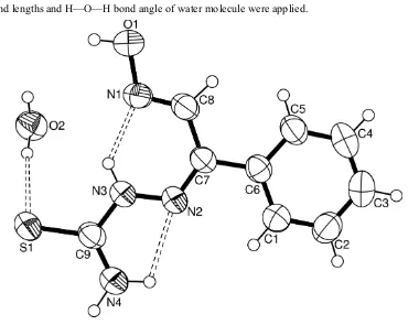
In the molecule of the title compound, (I), (Fig. 1) the bond lengths (Allen et al., 1987) and angles are generally within
normal ranges. Ring A (C1—C6) is, of course, planar. The intramolecular N—H···N hydrogen bonds (Table 1) result in
the formation of two more planar five- and six-membered rings B (C9/N2—N4/H42) and C (C7/C8/N1—N3/H3A). The
compared with the corresponding values in compounds (II)-(VII) (Table 2). The oxime moiety has an E configuration [C7
—C8—N1—O1 177.1 (2)°; Chertanova et al., 1994]. In this configuration, the oxime groups are involved as donors in O
—H···O intermolecular hydrogen bondings (Table 1).
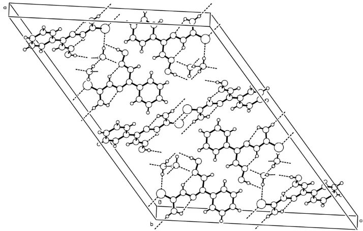
In the crystal structure, intramolecular O—H···S and N—H···N and intermolecular O—H···O and N—H···S hydrogen
bonds (Table 1) generate edge-fused R22(8) and R41(11) ring motifs (Fig. 2) (Bernstein et al., 1995). The hydrogen bonded
motifs are linked to each other to form a three dimensional network (Fig. 3). The intra- and intermolecular hydrogen
bonds seem to be effective in the stabilization of the crystal structure.
S2. Experimental
The title compound was prepared according to the literature method (El-Shazly et al., 2005). 2-Isonitrosoacetophenone
(149 mg, 1 mmol) was reacted with thiosemicarbazide (91 mg, 1 mmol) in ethanol-water mixture (1:1) by refluxing for
24 h. Then, a few drops of glacial acetic acid were added. The formed precipitate was filtered and recrystallized from
ethanol to obtain yellow crystals (yield: 155 mg, 70%).
S3. Refinement
H atoms were located in difference syntheses and refined isotropically [O—H = 0.903 (18)–0.940 (17) Å; Uiso(H) =
0.100 (10)–0.133 (14) Å2, N—H = 0.915 (17)–0.930 (17) Å; U
iso(H) = 0.077 (8)–0.084 (9) Å2 and C—H = 0.92 (3)–
0.97 (3) Å; Uiso(H) = 0.071 (8)–0.092 (9) Å2]. The restrains on the O—H (for OH) and N—H (for NH and NH2) bonds
[image:4.610.116.496.352.647.2]and O—H bond lengths and H—O—H bond angle of water molecule were applied.
Figure 1
The molecular structure of the title molecule with the atom-numbering scheme. Displacement ellipsoids are drawn at the
supporting information
sup-3
[image:5.610.127.483.74.371.2]Acta Cryst. (2008). E64, o623–o624 Figure 2
A part of the crystal structure of (I), showing the formation of R22(8) and R41(11) ring motifs. Hydrogen bonds are shown
as dashed lines.
Figure 3
[image:5.610.125.482.424.652.2]Crystal data
C9H10N4OS·H2O Mr = 240.29
Monoclinic, C2/c
Hall symbol: -C 2yc
a = 28.5615 (3) Å
b = 4.6805 (3) Å
c = 22.0977 (4) Å
β = 127.24 (2)°
V = 2351.8 (6) Å3 Z = 8
F(000) = 1008
Dx = 1.357 Mg m−3 Melting point: 443 K
Mo Kα radiation, λ = 0.71073 Å Cell parameters from 2893 reflections
θ = 2.3–30.5°
µ = 0.27 mm−1 T = 294 K Prism, yellow
0.30 × 0.20 × 0.15 mm
Data collection
Rigaku R-AXIS RAPID-S diffractometer
Radiation source: fine-focus sealed tube Graphite monochromator
ω scans
Absorption correction: multi-scan (Blessing, 1995)
Tmin = 0.940, Tmax = 0.960
31269 measured reflections 3607 independent reflections 2146 reflections with I > 2σ(I)
Rint = 0.090
θmax = 30.5°, θmin = 2.3°
h = −40→40
k = −6→5
l = −31→31
Refinement
Refinement on F2 Least-squares matrix: full
R[F2 > 2σ(F2)] = 0.062 wR(F2) = 0.154 S = 1.04 3607 reflections 193 parameters 8 restraints
Primary atom site location: structure-invariant direct methods
Secondary atom site location: difference Fourier map
Hydrogen site location: inferred from neighbouring sites
H atoms treated by a mixture of independent and constrained refinement
w = 1/[σ2(F
o2) + (0.0474P)2 + 0.9027P] where P = (Fo2 + 2Fc2)/3
(Δ/σ)max < 0.001 Δρmax = 0.14 e Å−3 Δρmin = −0.31 e Å−3
Special details
Geometry. All e.s.d.'s (except the e.s.d. in the dihedral angle between two l.s. planes) are estimated using the full covariance matrix. The cell e.s.d.'s are taken into account individually in the estimation of e.s.d.'s in distances, angles and torsion angles; correlations between e.s.d.'s in cell parameters are only used when they are defined by crystal symmetry. An approximate (isotropic) treatment of cell e.s.d.'s is used for estimating e.s.d.'s involving l.s. planes.
Refinement. Refinement of F2 against ALL reflections. The weighted R-factor wR and goodness of fit S are based on F2, conventional R-factors R are based on F, with F set to zero for negative F2. The threshold expression of F2 > σ(F2) is used only for calculating R-factors(gt) etc. and is not relevant to the choice of reflections for refinement. R-factors based on F2 are statistically about twice as large as those based on F, and R- factors based on ALL data will be even larger.
Fractional atomic coordinates and isotropic or equivalent isotropic displacement parameters (Å2)
x y z Uiso*/Ueq
S1 0.08587 (3) 0.69943 (16) 0.58554 (3) 0.0693 (2)
supporting information
sup-5
Acta Cryst. (2008). E64, o623–o624
O2 0.22848 (8) 0.6577 (4) 0.70204 (11) 0.0720 (5)
H21 0.1878 (8) 0.692 (7) 0.6673 (16) 0.133 (14)*
H22 0.2445 (13) 0.817 (5) 0.7313 (16) 0.113 (12)*
N1 0.19953 (8) 0.7460 (4) 0.83853 (10) 0.0576 (5)
N2 0.09978 (8) 0.3607 (4) 0.75639 (10) 0.0577 (5)
N3 0.10929 (9) 0.5206 (5) 0.71329 (10) 0.0619 (5)
H3A 0.1412 (9) 0.641 (5) 0.7347 (14) 0.077 (8)*
N4 0.02448 (9) 0.3422 (5) 0.60650 (11) 0.0654 (6)
H41 −0.0011 (10) 0.316 (5) 0.5550 (10) 0.076 (8)*
H42 0.0210 (12) 0.238 (5) 0.6394 (14) 0.084 (9)*
C1 0.07415 (11) 0.0295 (6) 0.83508 (14) 0.0644 (6)
H1 0.0517 (11) 0.012 (5) 0.7826 (15) 0.074 (8)*
C2 0.06113 (12) −0.1405 (6) 0.87378 (17) 0.0721 (7)
H2 0.0280 (11) −0.257 (5) 0.8455 (15) 0.071 (8)*
C3 0.09577 (13) −0.1362 (6) 0.95201 (17) 0.0724 (7)
H3 0.0859 (12) −0.245 (6) 0.9780 (16) 0.083 (9)*
C4 0.14393 (14) 0.0388 (6) 0.99041 (16) 0.0742 (7)
H4 0.1666 (12) 0.036 (6) 1.0426 (16) 0.092 (9)*
C5 0.15737 (13) 0.2112 (6) 0.95211 (14) 0.0639 (6)
H5 0.1913 (13) 0.330 (6) 0.9810 (17) 0.089 (9)*
C6 0.12248 (9) 0.2111 (5) 0.87338 (12) 0.0521 (5)
C7 0.13487 (9) 0.3927 (5) 0.82959 (11) 0.0516 (5)
C8 0.18351 (10) 0.5985 (5) 0.87101 (13) 0.0580 (6)
H8 0.2005 (11) 0.621 (5) 0.9241 (15) 0.080 (8)*
C9 0.07134 (10) 0.5057 (5) 0.63645 (11) 0.0556 (5)
Atomic displacement parameters (Å2)
U11 U22 U33 U12 U13 U23
S1 0.0632 (4) 0.0943 (5) 0.0452 (3) −0.0096 (3) 0.0301 (3) 0.0036 (3)
O1 0.0703 (11) 0.0827 (12) 0.0573 (10) −0.0268 (9) 0.0324 (9) −0.0116 (9)
O2 0.0630 (11) 0.0814 (13) 0.0698 (12) 0.0040 (10) 0.0393 (10) −0.0022 (10)
N1 0.0518 (10) 0.0649 (12) 0.0491 (10) −0.0074 (9) 0.0269 (9) −0.0074 (8)
N2 0.0588 (11) 0.0704 (12) 0.0439 (10) −0.0070 (9) 0.0311 (9) −0.0025 (8)
N3 0.0611 (12) 0.0780 (14) 0.0419 (9) −0.0147 (10) 0.0287 (9) −0.0037 (9)
N4 0.0628 (12) 0.0832 (15) 0.0449 (11) −0.0118 (11) 0.0299 (10) −0.0060 (10)
C1 0.0567 (13) 0.0777 (17) 0.0544 (14) −0.0031 (12) 0.0313 (12) 0.0037 (12)
C2 0.0633 (15) 0.0771 (18) 0.0776 (18) −0.0037 (14) 0.0436 (15) 0.0078 (14)
C3 0.0841 (19) 0.0761 (18) 0.0785 (19) 0.0119 (15) 0.0603 (17) 0.0164 (14)
C4 0.093 (2) 0.0816 (18) 0.0567 (15) 0.0102 (16) 0.0496 (15) 0.0081 (14)
C5 0.0737 (16) 0.0693 (15) 0.0500 (13) −0.0021 (13) 0.0381 (12) −0.0023 (11)
C6 0.0534 (12) 0.0566 (12) 0.0491 (11) 0.0061 (10) 0.0325 (10) 0.0012 (9)
C7 0.0503 (11) 0.0596 (13) 0.0429 (11) 0.0026 (10) 0.0272 (9) −0.0009 (9)
C8 0.0563 (13) 0.0668 (14) 0.0435 (11) −0.0018 (11) 0.0263 (10) −0.0028 (10)
S1—C9 1.681 (2) C2—H2 0.93 (3)
O1—H1A 0.903 (18) C3—C2 1.378 (4)
O2—H21 0.940 (17) C3—C4 1.368 (4)
O2—H22 0.907 (18) C3—H3 0.93 (3)
N1—O1 1.385 (2) C4—H4 0.92 (3)
N1—C8 1.264 (3) C5—C4 1.381 (4)
N2—N3 1.362 (3) C5—H5 0.95 (3)
N2—C7 1.297 (3) C6—C1 1.389 (3)
N3—H3A 0.923 (17) C6—C5 1.386 (3)
N4—C9 1.320 (3) C6—C7 1.483 (3)
N4—H41 0.915 (17) C7—C8 1.469 (3)
N4—H42 0.930 (17) C8—H8 0.97 (3)
C1—C2 1.374 (3) C9—N3 1.355 (3)
C1—H1 0.93 (2)
N1—O1—H1A 104.6 (19) C3—C4—C5 121.2 (3)
H21—O2—H22 106 (3) C3—C4—H4 116.6 (19)
C8—N1—O1 112.88 (19) C5—C4—H4 122.2 (19)
C7—N2—N3 118.58 (19) C4—C5—C6 120.9 (3)
C9—N3—N2 120.1 (2) C4—C5—H5 118.5 (18)
C9—N3—H3A 117.9 (16) C6—C5—H5 120.6 (18)
N2—N3—H3A 122.0 (17) C1—C6—C7 119.6 (2)
C9—N4—H41 120.3 (17) C5—C6—C1 117.3 (2)
C9—N4—H42 117.9 (17) C5—C6—C7 123.0 (2)
H41—N4—H42 121 (2) N2—C7—C8 125.4 (2)
C2—C1—C6 121.2 (2) N2—C7—C6 116.00 (19)
C2—C1—H1 118.9 (16) C8—C7—C6 118.60 (18)
C6—C1—H1 119.7 (16) N1—C8—C7 122.4 (2)
C1—C2—C3 120.9 (3) N1—C8—H8 122.8 (16)
C1—C2—H2 118.0 (16) C7—C8—H8 114.7 (16)
C3—C2—H2 121.1 (16) N4—C9—N3 117.2 (2)
C4—C3—C2 118.4 (3) N4—C9—S1 124.28 (17)
C4—C3—H3 120.7 (18) N3—C9—S1 118.46 (18)
C2—C3—H3 120.8 (18)
O1—N1—C8—C7 177.1 (2) C1—C6—C5—C4 −0.6 (4)
C7—N2—N3—C9 −175.9 (2) C7—C6—C5—C4 179.8 (2)
N3—N2—C7—C8 2.8 (3) C5—C6—C7—N2 177.2 (2)
N3—N2—C7—C6 −179.56 (19) C1—C6—C7—N2 −2.4 (3)
C6—C1—C2—C3 −0.1 (4) C5—C6—C7—C8 −5.0 (3)
C4—C3—C2—C1 −0.8 (4) C1—C6—C7—C8 175.4 (2)
C2—C3—C4—C5 1.0 (4) N2—C7—C8—N1 −7.6 (4)
C6—C5—C4—C3 −0.3 (4) C6—C7—C8—N1 174.8 (2)
C5—C6—C1—C2 0.8 (4) S1—C9—N3—N2 −178.23 (16)
supporting information
sup-7
Acta Cryst. (2008). E64, o623–o624 Hydrogen-bond geometry (Å, º)
D—H···A D—H H···A D···A D—H···A
O1—H1A···O2i 0.90 (3) 1.83 (3) 2.728 (3) 174 (3)
O2—H21···S1 0.94 (3) 2.32 (3) 3.250 (3) 171 (3)
O2—H22···O2i 0.91 (3) 1.98 (3) 2.886 (3) 172 (4)
N3—H3A···N1 0.92 (3) 1.91 (2) 2.604 (3) 130 (2)
N4—H41···S1ii 0.92 (2) 2.53 (2) 3.434 (2) 169 (3)
N4—H42···N2 0.93 (3) 2.24 (3) 2.643 (3) 105 (2)