Myosin 5c is a class V myosin that functions in secretory granule trafficking
Damon Todd Jacobs
A dissertation submitted to the faculty of the University of North Carolina at Chapel Hill in partial fulfillment of the requirements for the degree of Doctor of Philosophy in the
School of Medicine (Department of Cell and Molecular Physiology)
Chapel Hill 2008
Approved by:
Carol A. Otey, Ph.D.
Richard E. Cheney, Ph.D.
C. William Davis, Ph.D.
Ann Erickson, Ph.D.
James M. Anderson, MD, Ph.D.
Abstract:
DAMON JACOBS: Myosin Vc is Class V Myosin that Functions in Secretory Granule Trafficking
(Under the direction of Richard E. Cheney, Ph.D.)
Myosin motor proteins are a super family of mechanoenzymes that carry out diverse functions in plants and animals. Class V myosins power the movement of membranous organelles along tracks composed of actin filaments. Myosin Vc (Myo5c) is the third and final member of the myosin V family to be discovered. The mRNA distribution of
Myo5c indicated that it is preferentially expressed in epithelial cells and glandular tissues. Initial studies in HeLa cells indicated that Myo5c was associated with an endocytic recycling compartment. Kinetic studies showed that Myo5c is a non- processive vertebrate class V myosin. These studies revealed that, unlike Myo5a and Myo5b, Myo5c is a low duty ratio motor and the rate limiting step of the ATPase cycle is in a prehydrolysis state. More recently, we began to explore the functions of Myo5c in exocrine secretion. Tissue surveys and immunoblots of rat tissues showed that Myo5c is expressed most abundantly in acinar cells and localizes to the apical domain. Our studies utilized exocrine–derived MCF-7 cells to reveal the first endogenous localization of Myo5c in human cells. Myo5c clearly labeled distinct membrane compartments consisting of puncta and long, slender tubules. Our generation of a full-length, GFP- tagged Myo5c expression construct showed that in living cells Myo5c-associated puncta and tubules exhibit differing localization, dynamics, and dependence upon the
cytoskeleton. Our key results in MCF-7 cells demonstrated that Myo5c is tightly
associated with secretory granules and that expression of a dominant-negative Myo5c tail disrupts the distribution of secretory granules. Furthermore, in acinar cells of the
lacrimal gland, Myo5c tail partially inhibited carbachol stimulated secretion. Together
these results strongly indicate that Myo5c is a unique vertebrate class V myosin with
important functions in exocrine secretion.
Dedication
This work documents a great personal achievement and is dedicated to my American Indian ancestors who did not have opportunities or were denied opportunities for education. This dissertation is also dedicated to our daughter, Sophia Zavarine Jumping
Eagle Jacobs, who was born in our home in Chapel Hill, North Carolina.
“Sophia, may all your dreams come true”
Acknowledgements:
First and foremost, I wish to acknowledge my graduate advisor, Dr. Richard E Cheney, whose guidance and mentorship through my graduate studies was invaluable. I wish to thank Dr. Cheney for demanding a rigorous approach toward scientific investigation and his commitment toward education. I appreciate the many great discussions over good coffee, covering topics such as cell biology, wildland firefighting, and the natural world.
I wish to acknowledge my family, particularly Mom, Carmen Marie Jacobs, for understanding and support during my graduate studies.
I wish to thank the Thompson family in Fort Hall, Idaho for their lifelong friendship and support during all endeavors of my life, you guys are the best!
I wish to acknowledge Dr. Debra Kendall for a note on the back page of a Bluebook test booklet that sparked my initial interest in pursuing graduate studies. I also wish to thank Dr. Shere Byrd, for guidance while attending Fort Lewis College and for providing my first opportunity to perform biological research.
I wish to thank Dr. Sharon Milgram and Jan McCormick of the Cell and Molecular
Physiology department at the University of North Carolina-Chapel Hill.
Most of all, I wish to acknowledge and thank Diane Marie Gercke, for her unwavering
support and commitment to our family. Diane, my partner in life, has been a great source
of inspiration and motivation, and has provided invaluable perspective, patience, and
understanding during my graduate studies.
Preface:
This dissertation is the culmination of several years of intense study focused on unconventional myosins. My attention was directed primarily toward investigating the fundamental physiology and cell biology of myosin Vc (Myo5c). The first paper reporting the sequence and discovery of this molecular motor had just been published (Rodriguez and Cheney, 2002) when I began working on this project. Although Myo5a had been the subject of intensive research, almost nothing was known concerning the function(s) of Myo5c. This study of Myo5c allowed me to explore the mechanisms of actin-based membrane transport in vertebrate cells and tissues. In addition to Myo5c, I also performed studies with myosin X (Myo10) that revealed a novel form of motility within filopodia. A brief summary of my accomplishments are listed below:
1. As a first step toward determining the function(s) of Myo5c and identifying the organelle(s) that it is associated with, I began a systematic analysis of Myo5c localization in rat tissues. This led to the discovery of the striking localization at the apical domain of acinar cells in exocrine glands such as the lacrimal gland.
These results were presented at a Gordon Research Conference (Salivary Glands
and Exocrine Secretion) where I won a student poster award for this work. This
presentation of my results also led to a collaboration with the Hamm-Alvarez lab
at the University of Southern California to investigate the role of Myo5c in lacrimal gland acinar cells.
2. In the introduction (Chapter 1) I provide a detailed background on class V
myosins and the functions they perform in organelle trafficking. This chapter was commissioned by Nature Reviews Molecular Cell Biology and will serve as the basis of a review article title “Class V Myosins: Motors for organelle transport”.
It is currently undergoing revision by Dr. Richard Cheney.
3. Chapter 2 is a data chapter that contains the manuscript, Marchelletta et al., 2008,
“The Class V Myosin Motor, Myosin 5c, Localizes to Mature Secretory Vesicles
and Facilitates Exocytosis in Lacrimal Acini.” This manuscript was published in
the American Journal of Physiology-Cell Physiology. This collaboration with the
Hamm-Alvarez lab at USC is a study on the role of Myo5c in secretion from
isolated rabbit lacrimal glands. I am second author on this publication and my
contributions to this study included the initial localization of Myo5c in a lacrimal
gland and showing the presence of all three class V myosins (Myo5a, Myo5b, and
Myo5c) by immunoblot. In addition, I generated a green fluorescent protein
(GFP) tagged, full-length human Myo5c cDNA, and provided other constructs
and Myo5c-specific antibodies, all of which made this study possible. I also
contributed to the intellectual conception of the study, provided drafts of a portion
of the introduction, results, and discussion, and provided comments on several
draft manuscripts.
4. In chapter three, I present a manuscript of my core work on the localization, dynamics, and functions of Myo5c. This manuscript is titled “Myosin Vc is a class V myosin that functions in secretory granule trafficking”, and is currently in revision at Molecular Biology of the Cell. This study utilized an exocrine-derived cell line (MCF-7) that expresses an abundance of Myo5c protein, which allowed the first immunolocalization of Myo5c in a cell line. Furthermore, in this study a clonal line that stably expresses Myo5c at near physiological levels was generated and utilized in numerous experiments. Although the MCF-7 cells do not exhibit classic epithelial polarity, they provided a model system in which to obtain high- resolution, time-lapse images of secretory granule trafficking using TIRF
microscopy. I am the first author on this manuscript and I am deeply involved in the intellectual conception of the study. I also designed and carried out all experiments, and I wrote the initial draft of the manuscript. Drs. Julie Donaldson and Roberto Weigert, at the NIH, are collaborators on this manuscript. This collaboration originally focused on the endocytic recycling of MHC class I molecules in HeLa cells, however, when many endocytic recycling assays did not consistently support a role for Myo5c in recycling, the focus shifted to the
localization and dynamics of Myo5c on secretory granules in MCF-7 cells.
5. The concluding chapter (Chapter 4) will discuss results from previous chapters
and present original ideas on the functions of Myo5c in secretory granule
trafficking. Potential regulatory mechanisms of class V myosins associating with cargo will also be discussed.
6. Appendix Chapter 1 is a manuscript published in the Journal of Biological Chemistry (Takagi et al., 2008) that determined the kinetic parameters of human Myo5c. This paper is important because it demonstrated that, in contrast to Myo5a and Myo5b, Myo5c is a non-processive vertebrate class V myosin. My primary contribution to this study consisted of generating a GFP-tagged Myo5c- HMM construct that made this study possible.
7. Appendix Chapter 2 is a manuscript on the discovery of a novel form of motility in filopodia. This manuscript represents the first direct visualization of the dynamics of single molecules of Myosin 10 in living cells. We also provide evidence that Myo5a can also undergo similar movements at the single molecule level. My contributions to this study included setting up the TIRF imaging system that made these studies possible and acquiring the first data revealing this novel form of motility.
Additionally, during my graduate studies I was actively involved with minority issues and recruitment activities sponsored by the Graduate School. These activities included serving as the president of the First Nations Graduate Circle student organization, organizing American Indian recruitment events and American Indian research
conferences at UNC-CH, serving on the Provost’s Committee on Native American Issues
(PCNAI) at UNC-CH, serving on the search committee for the inaugural director of the
American Indian Center at UNC-CH, and serving on the American Indian Center
Advisory Committee. These activities allowed me to take advantage of valuable
professional development opportunities offered at the University of North Carolina-
Chapel Hill.
Table of Contents
CHAPTER 1: INTRODUCTION ... 1
CLASS V MYOSINS: MOTORS FOR ORGANELLE TRANSPORT ... 1
Abstract... 3
Introduction... 4
Structure of class V myosins... 5
Functions of class V myosins ... 8
Biophysical and biochemical properties of class V myosins... 22
Regulatory mechanisms of class V myosins... 24
Conclusions... 40
References... 42
CHAPTER 2: THE CLASS V MYOSIN MOTOR, MYOSIN 5C, LOCALIZES TO MATURE SECRETORY VESICLES, AND FACILITATES EXOCYTOSIS IN LACRIMAL ACINI... 53
Abstract... 55
Introduction... 56
Materials and Methods... 60
Results... 70
Discussion... 108
Text Footnotes ... 115
Acknowledgements... 116
References... 117
CHAPTER 3: MYOSIN VC IS A CLASS V MYOSIN THAT FUNCTIONS IN
SECRETORY GRANULE TRAFFICKING... 122
Abstract... 124
Introduction... 125
Materials and Methods... 129
Results... 137
Discussion... 181
Acknowledgements... 190
Supplemental Methods... 197
References... 198
CHAPTER 4: CONCLUSIONS AND FUTURE DIRECTIONS ... 206
Class V myosins are actin-based molecular motors that transport organelles... 207
How is organelle specificity achieved?... 207
Myo5c is a class V myosin for exocrine secretion ... 211
Myo5c, non-processivity, and exocrine secretion... 219
References... 231
APPENDICES ... 237
APPENDIX 1: MYOSIN VC IS A LOW DUTY RATIO, NON-PROCESSIVE MOLECULAR MOTOR ... 238
Abstract... 240
Introduction... 242
Materials and Methods... 246
Results... 251
Discussion... 282
Acknowledgments... 290
References... 291
APPENDIX 2: SINGLE MOLECULE IMAGING OF MYOSIN-X REVEALS A NOVEL FORM OF MOTILITY IN FILOPODIA... 298
Abstract... 300
Introduction... 301
Materials and Methods... 304
Results... 307
Discussion... 329
Acknowledgements... 331
References... 336
List of Tables
Table 1-1: Class V myosins associate with several organelles... 11 Table 2-1. Bulk Protein release in LGAC transduced with GFP or GFP-Myo5c-tail. ... 89 Table A1-1: Kinetic parameters of the MVc-S1 ATPase cycle with
corresponding values for myosins Va, Vb and Drosophila myosin V... 256
List of Figures
Figure 1-1: Structure of the vertebrate class V myosins... 6
Figure 1-2: Current models of class V myosin organelle transport functions. ... 9
Figure 1-3: Crystal structure of the Myo2p globular tail... 26
Figure 1-4: Diagram of docking complexes for different organelles. ... 30
Figure 1-5: Rab proteins label distinct organelles. ... 32
Figure 1-6: General mechanisms of regulation of Class V myosins. ... 38
Figure 2-1. Localization of endogenous Myo5c and actin filaments in LG and LGAC... 71
Figure 2-2: Myo5c and Rab3D co-localized in LGAC... 74
Figure 2-3: GFP-Myo5c-tail associates with mature SVs. ... 79
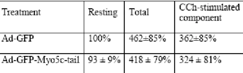
Figure 2-4: Expression of GFP-Myo5c-tail suppresses CCh-stimulated syncollin-GFP release in LGAC. ... 82
Figure 2-5: Expression of GFP-Myo5c-tail suppresses CCh-stimulated SC release in LGAC... 86
Figure 2-6: Electron Microscopy micrographs of Myo5c and GFP-Myo5c- tail enriched around mSV in resting and CCh-stimulated LGAC. ... 90
Figure 2-7: GFP-Myo5c-full is localized to mSVs... 94
Figure 2-8: GFP-Myo5c-full and GFP-Myo5c-tail both localize to pIgAR/SC enriched mSVs beneath the APM. ... 96
Figure 2-9: GFP-Myo5c-tail-enriched mSVs exhibit reduced diameter and infrequent association with actin-coated structures relative to GFP-Myo5c- full-enriched mSVs in stimulated LGAC. ... 100
Figure 2-10. GFP-Myo5c-tail-enriched mSVs exhibit reduced diameter in the presence and absence of CCh, and reduced association with actin-coated structures in CCh-stimulated LGAC. ... 104
Supplemental Figure 2-1: Myo5c and endogenous Rab3D are co-localized
on mSVs in LGAC... 106
Figure 3-1. Endogenous Myo5c localizes to small puncta and slender tubules. ... 138 Figure 3-2. Dynamics of GFP-Myo5c associated puncta and tubules... 143 Movie 3-1: Timelapse movie illustrating the dynamics of Myo5c puncta. ... 145 Movie 3-2: Timelapse movie demonstrating that Myo5c associated tubules move rapidly and microtubule-dependently whereas Myo5c puncta move slowly and microtubule-independently... 147 Figure 3-3. The localization of Myo5c is distinct from that of Myo5a and Myo5b. ... 150 Movie 3-3: The localization and dynamics of Myo5c are distinct from those of Myo5b. ... 152 Figure 3-4. Distribution of Myo5c relative to selected membrane compartments. ... 155 Figure 3-5. Myo5c localizes on secretory granules. ... 158 Movie 3-4: mCherry-Myo5c localizes on chromogranin A-GFP labeled
secretory granules. ... 160 Movie 3-5: mCherry-Myo5b does not localize on chromogranin A-GFP
labeled secretory granules... 163 Movie 3-6: Myo5c associated secretory granules can be triggered to undergo secretion... 168 Figure 3-6: Myo5c labeled secretory granules can be triggered to
undergo secretion... 172 Movie 3-7: Myo5c remains associated with secretory granule membranes
following exocytosis. ... 174 Movie 3-8: Myo5c labeled secretory granules are not perturbed by exogenous
markers of secretory granules. ... 176 Figure 3-7. Dominant negative Myo5c disrupts the distribution of
secretory granules. ... 179
Figure 3-8: Summary Diagram: Myo5c is associated with dynamic tubules
and regulated secretory granules... 183
Supplemental Figure 1. Myo5c is expressed in exocrine secretory tissues. ... 191
Supplemental Figure 2: Class V myosin constructs used to investigate the
cellular functions of Myo5c. ... 193
Supplemental Figure 3: Dominant-negative Myo5c specifically associates with secretory granules and disrupts their distribution in MCF-7 cells... 195
Movie 4-1: Myo5c localizes to vacuole-like structures in a temporally- regulated manner... 227
Figure 4-1: Myo5c is enriched in regions of protrusive activity in MCF-7 cells. ... 229
Figure A1-1: SDS polyacrylamide electrophoretogram of MVc-S1 ... 252
Figure A1-2: Steady-state actin-activated ATPase activity of MVc-S1... 254
Figure A1-3: Interaction of MVc-S1 with actin in the absence of nucleotide and in the presence of ADP. ... 259
Figure A1-4: Interaction of ATP with acto-MVc-S1... 262
Figure A1-5: MANT-ADP release from acto-MVc-S1. ... 265
Figure A1-6: Acto-MVc-S1 ADP affinity. ... 267
Figure A1-7: Transient kinetics of ATP hydrolysis by MVc-S1 monitored by quenched-flow... 270
Figure A1-8: Pi release from MVc-S1 and acto-MVc-S1 monitored by MDCC-PBP fluorescence. ... 273
Figure A1-9: in vitro actin gliding assay of eGFP-MVc-HMM. ... 276
Figure A1-10: Dependence of steady-state parameters (ATPase activity, duty ratio) of MVc-S1 on (A) actin and (B) background ADP concentration. ... 280
Figure A1- 11: Phylogenetic analysis of various myosin Vs from different organisms. ... 287
Movie A2-1. TIRF microscopy of single GFP molecules adsorbed on a coverslip. ... 308
Movie A2-2. TIRF microscopy of GFP-Myo10 in HeLa cell filopodia... 310
Movie A2-3. TIRF microscopy reveals fast forward movements of faint
particles of GFP-Myo10 in a filopodium... 312
Figure A2-1. TIRF microscopy reveals fast forward movements of faint particles of GFP-Myo10 in living cells... 314 Figure A2-2. Faint particles of GFP-Myo10 exhibit characteristics of single molecules... 318 Figure A2-3. Fast forward movements require the Myo10 motor domain... 321 Figure A2-4. Fast forward movements are inhibited by latrunculin B. ... 325 Figure A2-5. Faint particles of GFP-Myo5a can also move forward and rearward in living cells... 327 Figure A2-S1. Dynamics of GFP-Myo10 in living HeLa cells imaged with TIRF... 332 Figure A2-S2. Tip localization of different Myo10 constructs and TIRF
imaging of a forced dimer and a motor domain point mutant. ... 334
List of Abbreviations Ad- adenovirus
APM- apical plasma membrane ATP- Adenosine Triphosphate BSA- bovine serum albumin Ca++ - calcium
CgA- Chromogranin A
DIC- differential interference contrast DNA- deoxyribonucleic acid
DN- dominant negative EM- electron microscopy ER- endoplasmic reticulun F-actin- Filamentous actin GFP- green fluorescent protein HA- hemagglutinin epitope Kd- Kilodalton
LG-lacrimal gland
LGAC- lacrimal gland acinar cells
MANT-ATP- 2'-(or-3')-O-(N-methylanthraniloyl) adenosine 5'-triphosphate, trisodium salt;
MANT-ADP- 2'-(or-3')-O-(N-methylanthraniloyl)adenosine 5'-diphosphate, disodium salt;
MOPS,
N-morpholino-propanesulfonic acid mRNA- messenger RNAmSV- mature secretory vesicle nm - nanometer
NPY- neuropeptide -Y
pIgAR- polymeric immunoglobulin A receptor SC- secretory component
SEM- standard error of the mean sIgA- secretory immunoglobulin A
TIRF- total internal reflection fluorescence
m- micrometer
M- micromolar
CHAPTER 1: INTRODUCTION
CLASS V MYOSINS: MOTORS FOR ORGANELLE TRANSPORT
Class V myosins: Motors for organelle transport
Damon T Jacobs and Richard E Cheney
University of North Carolina-Chapel Hill, Cell and Molecular Physiology department 5314 Medical Biomolecular Research Building Chapel Hill, NC 27599-7545
Corresponding author:
Richard E Cheney, Professor of Cell and Molecular Physiology cheneyr@med.unc.edu
919-966-0331
Abstract
Myosins are actin-based molecular motors that are responsible for carrying out a
variety of biological processes such as muscle contraction, mRNA transport, and cell
signaling. Class V myosins are an evolutionarily conserved class of myosin with key
functions in organelle transport. This family of motor proteins is essential for normal
physiology, and mutations to class V myosins can cause lethal diseases. Studies of class
V myosins have led to important insights on membrane trafficking, molecular motor
function, and the determinants of organelle/cargo specificity.
Introduction
Class V myosins are among the most ancient and broadly distributed class of myosin (Berg et al., 2001; Odronitz and Kollmar, 2007). Class V myosins are also leading candidates to carry out actin-based organelle transport (Berg et al., 2001). In the budding yeast, Saccharomyces cereviseae, there are two class V myosins (Myo2p and Myo4p) that function in polarized secretion, organelle inheritance, and mRNA transport (Valiathan and Weisman, 2008). In mammals, the three class V myosins, myosin Va (Myo5a), myosin Vb (Myo5b), and myosin Vc (Myo5c), appear to be differentially expressed in a tissue specific manner (Rodriguez and Cheney, 2002). In humans, defects in class V myosins can cause serious diseases such as Griscelli Syndrome Type I
(Pastural et al., 1997) and microvillar inclusion disease (Muller et al., 2008). These disorders are a direct result of trafficking defects of class V myosin-associated organelles.
Studies in isolated melanocytes (pigment producing cells) showed that Myo5a stably
associates with melanosomes (pigment granules) by forming a tripartite protein docking
complex on the surface of the organelle (Wu et al., 2002b). These studies also revealed
that melanosomes undergo long-range, microtubule-based transport from the cell center
and are retained in the actin-rich cell periphery by Myo5a, leading to a dual-filament
transport model for organelle transport (Wu et al., 1998). Since the discoveries of the
association of Myo2p with secretory vesicles in yeast (Johnston et al., 1991) and Myo5a
with melanosomes in vertebrates (Provance et al., 1996), the number of organelles
associated with class V myosins has increased substantially. This list now includes
neuronal secretory vesicles (Prekeris and Terrian, 1997; Bridgman, 1999), endoplasmic
reticulum (Takagishi et al., 1996; Estrada et al., 2003), and several plasma membrane
receptors (Roland et al., 2007), among others. Recent structural studies have afforded much progress in determining the basis for cargo selection and regulation of motor activity (Liu et al., 2006; Pashkova et al., 2006). Here we will review recent progress and key ideas on the roles of class V myosins in membrane trafficking and will
emphasize recent research in budding yeast and mammals.
Structure of class V myosins
Vertebrate class V myosins are motor proteins composed of two identical heavy chains with six light chains per heavy chain (Espreafico et al., 1992) (Figure 1-1). At the N-terminus of each heavy chain there is a highly conserved myosin motor domain, which is then followed by a region that contains six tandem repeats of a light chain binding motif, and finally a tail domain at the C-terminus that can be divided into a proximal tail and a distal globular tail. The head or motor domain hydrolyzes ATP and binds to F- actin during the catalytic cycle to produce force (Cheney et al., 1993). The neck or light chain binding domain consists of six tandem repeats of a conserved consensus sequence (
IQxxxRGxxxR), termed “IQ motif” that binds to calmodulin or calmodulin-like light chains with high affinity (Cheney and Mooseker, 1992). This region of the molecule also serves as a rigid lever arm that is important for force transduction and step size
(Sakamoto et al., 2003). The proximal tail is predicted to consist largely of coiled-coil
and mediates dimerization of the myosin V heavy chains (Cheney et al., 1993). The
globular tail of class V myosins is required for cargo attachment and interacts with a
docking complex on the surface of the organelle (Wu et al., 2002b; Ishikawa et al.,
Figure 1-1: Structure of the vertebrate class V myosins. a. The predicted structure of
Myo5a consists of a conserved head domain, a neck domain containing six IQ motifs
(light chain binding motifs), followed by a region of predicted coiled coil and a globular
tail. Each molecule consists of two identical heavy chains and six accessory light
chains/heavy chain. b. Domain alignment of mouse Myo5a, rat Myo5b, and human
Myo5c shows that vertebrate class V myosins are highly homologous and amino acid
sequence alignment (not shown) predicts ~ 50% overall identity between family
members. Class V myosin domains are conserved within the family and the structures
are predicted to be highly homologous. (Figure adapted from Cheney and Rodriguez,
2002).
Head
(Motor domain)
Neck
(Regulatory light chain binding sites)
Proximal Tail
(Dimerization Domain)
Globular Tail
(Cargo Binding Domains)
~61% aa identity
~49%
Myo5c
~50%
754 902 1109 1149 1351
1742 aa 203 kDa human (AF272390)
Myo5b (myr 6)
764 912 1093 1140 1227 1339 1438
1846 aa 214 kDa rat (NM_017083)
head neck exon Fglobular tail
Myo5a (dilute)
764 911 1108 1156 1235 1347 1471
1880aa
~215 kDa human (Y07759)
Myo5a
Exon F
a. b.
are important for providing cargo specificity (Pashkova et al., 2005) (discussed below).
The general model of vertebrate class V myosin function consists of a two-headed motor transporting an organelle in a hand-over-hand fashion along an actin filament (Liu et al., 2006).
Functions of class V myosins
Class V myosins are broadly expressed and have important functions in vertebrates. Messenger RNA expression data indicates that, although most tissues express all three class V myosins, one family member appears to predominate over the others (Rodriguez and Cheney, 2002). Yeast have been utilized extensively as a model system where much of our knowledge of class V myosins has been demonstrated.
Functional studies in both yeast and mammals show that class V myosins perform transport and tethering tasks and that many functions are conserved across species.
(Figure 1-2) These studies show that each class V myosin appears to associate with a specific set of organelles in a cell type specific manner (See table 1). The two class V myosins in budding yeast perform such tasks as transporting membranous organelles and mRNAs along actin cables (Estrada et al., 2003). Since most studies in yeast have focused on the budding yeast, we will limit our discussion to Myo2p and Myo4p.
Yeast
Myo2p transports secretory vesicles in yeast.
Myo2p was the first class V myosin to be discovered and was initially shown to
transport secretory vesicles to sites of active cell growth, such as the tip of a growing bud
(Johnston et al., 1991; Govindan et al., 1995). (Figure 1-2a) Mutants of Myo2p exhibit
an accumulation of secretory vesicles within the mother cell during bud formation,
Figure 1-2: Current models of class V myosin organelle transport functions. a. In yeast class V myosins transport secretory vesicles and organelles along actin cables into the bud tip during cell division. b. Myo5a functions to tether or transport melanosomes in the actin-rich dendritic extensions of melanocytes. c. Myo5a is implicated in the
recruitment and anchoring of smooth endoplasmic reticulum in dendritic spines of
Purkinje neurons in mice. d. In neuroendocrine cells, Myo5a maintains a normal
distribution of dense-core secretory granules.
Transport/tethering of melanosomes
Anchoring of calcium stores/SER in dendritic spines
Transport of secretory vesicles and cellular organelles in yeast
a. b.
c. d. Distributing secretory granules in
neuroendocrine cells Actin Cables
(-)
(+) F-actin
Microtubules
(+) (-)
(+)
(+)
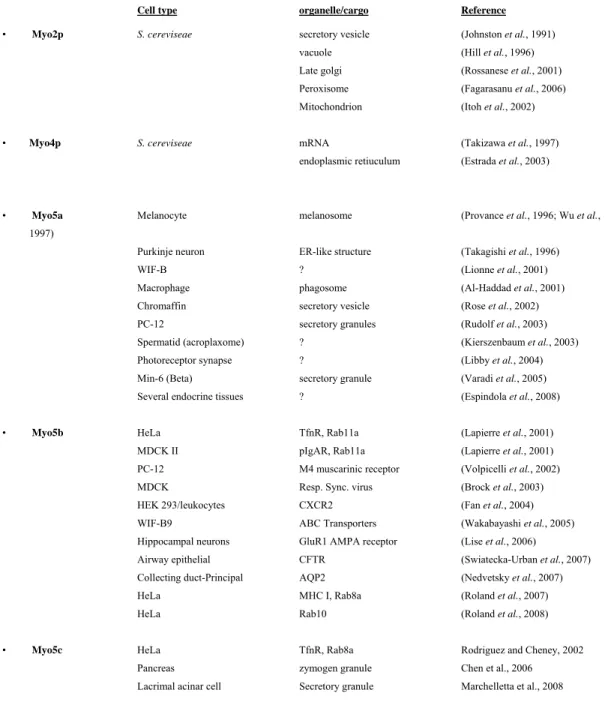
Table 1-1: Class V myosins associate with several organelles
Cell type organelle/cargo Reference
• Myo2p S. cereviseae secretory vesicle (Johnston et al., 1991) vacuole (Hill et al., 1996)
Late golgi (Rossanese et al., 2001) Peroxisome (Fagarasanu et al., 2006) Mitochondrion (Itoh et al., 2002)
• Myo4p S. cereviseae mRNA (Takizawa et al., 1997) endoplasmic retiuculum (Estrada et al., 2003)
• Myo5a Melanocyte melanosome (Provance et al., 1996; Wu et al., 1997)
Purkinje neuron ER-like structure (Takagishi et al., 1996) WIF-B ? (Lionne et al., 2001) Macrophage phagosome (Al-Haddad et al., 2001)
Chromaffin secretory vesicle (Rose et al., 2002) PC-12 secretory granules (Rudolf et al., 2003) Spermatid (acroplaxome) ? (Kierszenbaum et al., 2003) Photoreceptor synapse ? (Libby et al., 2004) Min-6 (Beta) secretory granule (Varadi et al., 2005) Several endocrine tissues ? (Espindola et al., 2008)
• Myo5b HeLa TfnR, Rab11a (Lapierre et al., 2001) MDCK II pIgAR, Rab11a (Lapierre et al., 2001) PC-12 M4 muscarinic receptor (Volpicelli et al., 2002) MDCK Resp. Sync. virus (Brock et al., 2003)
HEK 293/leukocytes CXCR2 (Fan et al., 2004) WIF-B9 ABC Transporters (Wakabayashi et al., 2005) Hippocampal neurons GluR1 AMPA receptor (Lise et al., 2006)
Airway epithelial CFTR (Swiatecka-Urban et al., 2007) Collecting duct-Principal AQP2 (Nedvetsky et al., 2007) HeLa MHC I, Rab8a (Roland et al., 2007)
HeLa Rab10 (Roland et al., 2008)
• Myo5c HeLa TfnR, Rab8a Rodriguez and Cheney, 2002 Pancreas zymogen granule Chen et al., 2006
Lacrimal acinar cell Secretory granule Marchelletta et al., 2008
indicating Myo2p transports and targets secretory vesicles (Johnston et al., 1991).
Myo2p mutants also display synthetic lethality with mutant genes required for actin cable formation (TPM1) and a Rab8 homolog protein important for secretory vesicle fusion (SEC4) (Liu and Bretscher, 1992; Govindan et al., 1995). Much evidence shows that Myo2p is a molecular motor that functions to transport vesicles along actin cables to sites of polarized secretion (Karpova et al., 2000).
Myo2p transports several additional organelles
In addition to secretory vesicle transport, Myo2p is also required for proper transport/localization of several other cellular organelles, including vacuoles,
peroxisomes, late golgi, and mitochondria (Hill et al., 1996; Catlett and Weisman, 1998;
Hoepfner et al., 2001; Rossanese et al., 2001; Itoh et al., 2002; Boldogh et al., 2004;
Fagarasanu et al., 2006; Altmann et al., 2008; Valiathan and Weisman, 2008). Several of these associations appear to be cell cycle dependent and indicate distinct functions in organelle partitioning and inheritance. It is clear that Myo2p interacts with several different organelles and thus mediates several different trafficking functions. As will be discussed below, specific docking complexes have been identified that mediate the attachment of Myo2p to the various organelles.
In addition to organelle trafficking functions, Myo2p also functions to orient the mitotic spindle and to position the spindle pole body by guiding Bim1p/Kar9p-labeled microtubule plus ends along actin cables into the daughter bud tip (Yin et al., 2000;
Gundersen and Bretscher, 2003; Hwang et al., 2003) . Additionally, an exciting recent
study also implicates Myo2p in P-body disassembly and suggests that Myo2p plays a role
in regulating translation of mRNA (Chang et al., 2008). P-bodies are cytoplasmic foci
that contain enzymes important for mRNA turnover and act to inhibit translational machinery by sequestering mRNA for eventual degradation. cRNA hybridization to yeast microarray assays suggest that Myo2p-associated P-bodies contain as many as 2300 mRNA. This study suggests that Myo2p associates with many different mRNAs and is the first report of an association of Myo2p with mRNA turnover.
Myo4p in mRNA and ER transport
Myo4p, the other class V myosin in S. cereviseae (Haarer et al., 1994), along with a complex of She proteins, is required to transport a specific set of 24 mRNAs
(Gonsalvez et al., 2005). This set includes Ash1, which is important in regulating
mating-type switching in the daughter cell (Takizawa et al., 1997). In addition to mRNA transport, Myo4p has also functions during inheritance of cortical endoplasmic reticulum (ER) (Estrada et al., 2003). These observations indicate that Myo4p functions to
transport mRNA’s and cortical ER tubules along actin cables from the mother cell to the bud tip.
Vertebrates
Myo5a and melanosome transport
The role of Myo5a in melanosome transport is one of the best characterized
functions of a vertebrate class V myosin. Melanocytes synthesize and package melanin
(pigment) into membranous organelles (melanosomes) and distribute them to surrounding
hair follicle cells and keratinocytes. In humans, mutations to Myo5a cause Griscelli
Syndrome Type I (Pastural et al., 1997), which is characterized by partial albinism and
retardation. A similar phenotype is observed in dilute-lethal mice (Myo5a-/-) which also display pigmentation defects and a neurological disorder. Studies in wild-type
melanocytes demonstrate that Myo5a localizes on melanosomes that are distributed throughout the cell and are especially enriched in dendritic extensions (Provance et al., 1996; Wu et al., 1997). (Figure 1-2b) In wild-type melanocytes, melanosomes move from the cell center to the periphery via microtubules and microtubule motors. However, in dilute-lethal melanocytes, melanosomes accumulate in the cell center (Provance et al., 1996). These results argue strongly that Myo5a functions to terminate microtubule- based transport and tether the melanosome in the cell cortex (Provance et al., 1996; Wu et al., 1998). Several observations support the hypothesis that Myo5a is required for melanosome transport in melanocytes, including, 1) Myo5a is present on melanosomes (Wu et al., 1997), 2) wild-type melanophores treated with F-actin destabilizers exhibit a similar melanosome distribution as dilute-lethal melanocytes (Rogers and Gelfand, 1998), 3) overexpression of dominant-negative Myo5a tail mimics the dilute-lethal phenotype in wild-type cells (Wu et al., 1998), and 4) in dilute-lethal melanocytes, overexpression of the wild-type Myo5a protein rescues the defect in melanosome distribution (da Silva Bizario et al., 2002). In the melanocyte model system, it is clear that Myo5a is required for maintaining melanosomes in the cell periphery. Whether Myo5a also plays a direct role in transferring the organelles from melanocytes to surrounding keratinocytes is not clear.
Myo5a and trafficking in dendritic spines
Myo5a is expressed most abundantly in brain and neuronal tissues, and dilute-
lethal mice and dilute-opisthotonis rats, which also have mutated Myo5a, suffer from a
serious neurological disorder that is lethal within three weeks of birth (Dekker-Ohno et al., 1996; Takagishi et al., 1996). Although no gross abnormalities in overall brain morphology were detected, mutant animals exhibit severe seizures and ataxia.
Subsequent ultrastructural studies of dendritic spines of Purkinje neurons in the
cerebellum revealed the absence of smooth endoplasmic reticulum (SER), which serves as a calcium storage organelle and labels with inositol triphosphate receptors (IP3R).
These observations suggest that Myo5a functions to transport/tether SER into the actin- rich dendritic spines (Dekker-Ohno et al., 1996; Takagishi et al., 1996). (Figure 2c) Although immunogold labeling of Purkinje neurons in dilute-lethal mice revealed that dendritic spines contained a normal contingent of several protein components essential for synaptic function (Petralia et al., 2001), the Myo5a mutant mice suffered from a loss of long term depression (LTD) (Miyata et al., 2000). These results indicate that loss of Myo5a function inhibits LTD through an inability to transport or tether SER in dendritic spines of Purkinje neurons.
In the dendrites of rat hippocampal neurons, Myo5a appears to associate with organelles containing GluR1 AMPA receptors (Correia et al., 2008). Using a dominant negative approach or RNA interference against Myo5a inhibited GluR1 AMPA receptor trafficking from dendritic shafts into the spine. This transport defect altered activity- induced synaptic potentiation. These results indicate that Myo5a is responsible for short- range transport of organelles containing receptors important for long term potentiation (LTP), thus Myo5a appears to be involved with the regulation of learning and memory.
Myo5a and neuroendocrine secretion
Myo5a is expressed in neuroendocrine cells and exhibits a striking localization on
studies in neuroendocrine cells have demonstrated that filamentous actin is required for efficient exocytosis (Neco et al., 2003; Giner et al., 2005) and that an actin-based molecular motor likely mediates transport of secretory organelles (Neco et al., 2002;
Rose et al., 2002). Several methods demonstrated that Myo5a facilitates a more direct role in secretory granule dynamics in the cell periphery. Both RNA interference against Myo5a and over expression of a dominant negative Myo5a tail alter the distribution of secretory granules and decrease a secretory response in neuroendocrine cells (Rudolf et al., 2003; Varadi et al., 2005; Desnos et al., 2007b), thus supporting a direct association with secretory granules. Although the precise mechanistic role of Myo5a in
neuroendocrine secretion is still not clear, Desnos et al. (Desnos et al., 2007b)
demonstrate that knock down of Myo5a in chromaffin cells appears to decrease the dwell time of individual granules near the plasma membrane, which then decreases the
likelihood of granules becoming docked prior to secretion. These results support the hypothesis that Myo5a tethers secretory granules in the actin-rich cell cortex and may also facilitate short range movements toward sites of secretion (Wu et al., 1998; Desnos et al., 2007b). (Figure 2d)
Espindola et al. (Espindola et al., 2008) have recently surveyed several neuroendocrine tissues from rat and determined that Myo5a is expressed in the pineal gland (pinealocytes), pituitary gland (parenchymal cells), thyroid/parathyroid glands (parafollicular and principal cells), pancreas (almost exclusively endocrine islet cells), adrenal gland (chromaffin cells), testis (spermatogonia), and ovary (follicular and
granulosa cells). These studies indicate a clear association of Myo5a with endocrine cells
and suggest that Myo5a is important in endocrine physiology. Although endocrine
defects have not been detected in dilute-lethal mice thus far, this may be due to their brief lifetimes. For additional information on Myo5a and neuroendocrine secretion, two recent reviews are available (Eichler et al., 2006; Desnos et al., 2007a).
Myo5b and plasma membrane receptor recycling
Myo5b was originally cloned from rat (Zhao et al., 1996), and is about ~60%
identical to Myo5a and ~49% identical to Myo5c by amino acid sequence (Rodriguez and Cheney, 2002). Myo5b has a broad tissue distribution, with the highest levels detected in brain, kidney, liver, and lung (Zhao et al., 1996). Several studies in cultured cells that utilized a dominant negative approach (GFP-Myo5b tail) have indicated that Myo5b associates with endocytic cargoes that undergo recycling to the plasma membrane
(Lapierre et al., 2001; Brock et al., 2003; Nedvetsky et al., 2007; Swiatecka-Urban et al., 2007). Consistent with this, Myo5b interacts with Rab11a, which is an important
component of the recycling endosome (Casanova et al., 1999; Lapierre et al., 2001).
These studies led to the hypothesis that Myo5b is involved in the outbound trafficking of cell surface receptors from an internal recycling compartment back to the plasma
membrane. Overexpression of the Myo5b tail appears to induce a collapse of an
endocytic perinuclear recycling compartment and also inhibit an exocytic arm of a
plasma membrane recycling pathway. In HeLa cells, a non-polarized cell type, Myo5b
tail inhibited the recycling of internalized transferrin and transferrin receptor out of the
perinuclear compartment back to the plasma membrane (Lapierre et al., 2001). Using
this approach, Myo5b appears to be important in the trafficking of several plasma
membrane receptors including the M4 muscarinic receptor in PC12 cells (Volpicelli et
cells, it was recently reported that Myo5b can associate with Rab8a, suggesting that Myo5b functions in an additional trafficking pathway that is distinct from the rab11a recycling pathway (Roland et al., 2007). Yeast two-hybrid screening and Fluorescence Resonance Energy Transfer (FRET), Goldenring and colleagues showed that Myo5b tail can directly interact with wild-type and GTP-bound, (but not GDP-bound), Rab8a
(Roland et al., 2007). In these cells, endogenous Rab8a labels long, membranous tubules and full-length Myo5b (GFP-Myr6) colocalizes at distinct points along the length.
Furthermore, in pulse labeling assays, puncta of Myo5b tail were also shown to accumulate MHCI molecules. Interestingly, MHCI molecules were also shown to associate with long, membranous tubules during recycling assays in HeLa cells (Weigert et al., 2004). These results suggest that Myo5b can form a direct association with Rab8a and/or Rab11a-associated organelles and also demonstrates that Myo5b can interact with at least two distinct compartments in a single cell type.
In polarized epithelial cells, Myo5b appears to specifically associate with cargoes destined for the apical plasma membrane (Lapierre et al., 2001). In fully polarized MDCK cell monolayers, expressed polymeric IgA receptor (pIgAR) was found to localize to Myo5b tail puncta and Rab11a in a subapical recycling endosome.
Interestingly, transferrin receptor was excluded from this compartment. The pIgARs was
inhibited from being expressed at the apical membrane by the dominant negative Myo5b
tail, indicating that Myo5b regulates apically directed transcytotic trafficking in MDCK
cells (Lapierre et al., 2001). In addition to a role in inhibiting transcytosis, the Myo5b
tail also inhibits the polarized distribution of several transmembrane proteins, including
the Na/H+ exchanger in gastric parietal cells (Hales et al., 2001), the GluR1 glutamate
receptor in dendritic spines of hippocampal neurons (Lise et al., 2006), the aquaporin-2 channel in rat kidney (Nedvetsky et al., 2007), the respiratory syncytial virus (Brock et al., 2003), and the cystic fibrosis transmembrane conductance regulator (CFTR) in human airway epithelial cells (Swiatecka-Urban et al., 2007). Importantly, all of these cargoes appear to undergo regulated insertion into the apical plasma membrane.
In these studies, the internalization of receptors from the plasma membrane did not appear to be affected by the dominant negative Myo5b tail, which strongly suggests that Myo5b performs an exocytic trafficking function in these cells. However, an alternative hypothesis suggests that Myo5b functions primarily during the endocytic phase of plasma membrane receptor recycling (Provance et al., 2004; Provance et al., 2008). Mercer and colleagues utilize a full-length Myo5b that has been genetically engineered to bind an oversized ADP analog with specificity, which locks the motor domain into a tight actin-binding state. By locking Myo5b onto actin, uptake of
fluorescent transferrin into a perinuclear recycling compartment was inhibited (Provance et al., 2004). In these studies full length GFP-Myo5b localized to enlarged peripheral puncta and correlated with an apparent increase of transferrin labeling on the plasma membrane. This work suggests that overexpression of full-length Myo5b inhibits inward transport of endocytosed transferrin, and shunts it toward a “fast recycling” pathway, thus explaining the increase in transferrin on the plasma membrane (Provance et al., 2008).
This hypothesis also suggests that the inability of the Myo5b tail to bind actin causes the
peripheral endocytic compartment to collapse into the cell center. In support of an
endocytic function, Yan et al. (Yan et al., 2005) report that Myo5b associates with the
CART complex of proteins, which includes the early endosomal associated proteins, Hrs,
actinin-4, and BERP. Provance et al., (Provance et al., 2008) suggest that Myo5b tethers an endosomal compartment in the cortical actin and functions to oppose inward transport of endocytic cargoes mediated by microtubules to the perinuclear recycling compartment.
Although this is an intriguing hypothesis, further data are required to definitively show whether Myo5b functions to oppose retrograde endocytic transport or support
anterograde exocytic transport, or both.
Myo5b and epithelial polarity
The establishment of cell polarity is vital for epithelial cell function. Polarity establishment requires a segregation of membrane proteins and a complex membrane trafficking system to maintain the polarized distribution of newly synthesized and recycled membrane proteins. Myo5b has been hypothesized to mediate polarity
establishment in several systems. In the hepatocyte-derived WIF-B9 cells, Myo5b and rab11a localized near the centrosome and at the canilicular membrane, indicating Myo5b is associated with apically targeted cargoes (Wakabayashi et al., 2005). Knockdown of Rab11a or expression of dominant-negative forms of Rab11a (S25N) or Myo5b (tail domain) inhibited canilicular formation significantly in these cells. These observations indicate that Myo5b is required for the establishment of polarity and canilicular
formation. Importantly, a genetic screen in humans identified Myo5b as a gene
responsible for microvillus inclusion disease (Muller et al., 2008). Mutations in the
motor domain or a C-terminal truncation appears to cause a loss of microvilli structure on
the luminal surface of mature enterocytes and causes the appearance of villin-labeled,
microvilli-like structures within internal inclusions. These cells also exhibited a defect in
the localization of basolateral markers into a subapical distribution, indicating the polarity
of the enterocyte was compromised. These studies indicate that Myo5b functions to transport organelles that contain cargoes that require a polarized distribution, however more studies are required to determine whether this effect is direct or indirect.
Myo5c: a motor for exocrine secretion?
Myo5c is the third and final member of the class V myosin family to be identified in vertebrates (Berg et al., 2001; Rodriguez and Cheney, 2002). Myo5c was originally cloned from a pancreas cDNA library and the initial characterization of human Myo5c showed that it is expressed most abundantly in exocrine tissues and epithelial cells (Rodriguez and Cheney, 2002). This study also showed that in cultured HeLa cells, expression of a dominant negative, GFP-tagged Myo5c tail construct induced the formation of large, peripheral punctate structures that accumulated Myo5c tail,
exogenous transferrin, and the transferrin receptor. Intriguingly, these puncta also stained positive with antibodies to Rab8, but not rab11a, which suggests that Myo5c associates with one or more organelles that are distinct from the Myo5b-labeled recycling endosome (Rodriguez and Cheney, 2002). Although HeLa cells are a well characterized model for membrane transport, unfortunately the levels of endogenous Myo5c are low, thus making studies in these cells difficult. Future studies are thus required to determine the exact function of Myo5c in recycling of endocytic membranes.
The distribution of Myo5c mRNA in exocrine glands (Rodriguez and Cheney,
2002) and the localization of Myo5c protein in mouse and rat tissues suggested that it is
most abundantly expressed in the acinar cells (Rodriguez and Cheney, 2002; Chen et al.,
2006; Marchelletta et al., 2008). Consistent with this, Myo5c was identified in a
exocrine pancreas (Chen et al., 2006). This approach also identified Rab3D, Rab27b, Rab11a, and Rab8a on the surface of zymogen granules. It is tempting to speculate whether Myo5c may associate with one or more of these Rab proteins to mediate an association with the granule membrane. Consistently, rab8a was shown to function in secretory granule biogenesis in a rat pancreatic acinar cell line (Faust et al., 2008). Using yeast two-hybrid screening, a direct interaction between Myo5c tail and Rab8a was shown using wild-type and GTP-bound Rab8a, but not GDP-bound Rab8a (Roland et al., 2007). Tissue expression and localization data suggests that Myo5c may have a secretory function, and more recent studies have shown that a dominant negative Myo5c tail can partially inhibit stimulated secretion from isolated rabbit lacrimal gland acinar cells (Marchelletta et al., 2008). Although the precise role in acinar cells is not clear, Myo5c likely functions at the late stages of regulated secretion, which may be to transport secretory granules through the F-actin barrier, facilitate fusion of secretory granules with the plasma membrane or with each other), provide force to extrude contents, or provide a cytoskeletal link to membranes targeted for recycling following exocytosis.
Biophysical and biochemical properties of class V myosins
Myo5a has been studied intensively and has biochemical properties that make it
ideal for single molecule studies (Trybus, 2008). The first experiments using purified,
chick brain myosin V showed that it has ATPase activity, does not form filaments, and
was regulated by calcium (Cheney et al., 1993). A distinct characteristic of Myo5a is the
36 nm step size (Mehta et al., 1999; Rief et al., 2000; Sakamoto et al., 2000). Since the
step size is dictated by lever arm length (Sakamoto et al., 2005), all class V myosins are
predicted to have a 36 nm step size. Thirty-six nanometers closely matches the pseudo- helical repeat of F-actin, which allows a class V myosin to walk along an actin filament without following the spiral of the helix (Trybus, 2008). The 36 nm step size may be an advantage for a motor protein that functions to transport large organelles along actin filaments. The average velocity reported for vertebrate Myo5a is ~300 nm/s (Cheney et al., 1993; Watanabe et al., 2007). Similarly, the velocity for Myo5b was reported to be
~220 nm/s (Watanabe et al., 2006) and the reported velocities for Myo5c are 24 nm/s (Takagi et al., 2008) and 160 nm/s (Watanabe et al., 2007).
These studies also revealed that Myo5a is a processive motor, meaning it can undergo multiple rounds of ATP hydrolysis (ie. multiple steps) before dissociating from an actin filament (Mehta et al., 1999; Trybus, 2008). A defining determinant of
processivity for single myosin molecules is the duty ratio, which is defined as the fraction of the ATPase cycle spent in a tight actin-bound state. Myosin Va has a high duty ratio of ~ 0.7, therefore each motor domain spends 70% of its ATPase cycle tightly bound to actin (Watanabe et al., 2006). Similar kinetic studies of human Myo5b showed that it is also a processive motor with a duty ratio of ~0.8 (80%) (Watanabe et al., 2006).
Interestingly, two independent groups showed that human Myo5c does not exhibit
processivity (Watanabe et al., 2007; Takagi et al., 2008). The calculated duty ratios for
Myo5c are ~10% (Takagi et al., 2008) and ~30% (Watanabe et al., 2007), which are low
values, similar to skeletal muscle myosin II (Takagi et al., 2008). This result is intriguing
because it highlights that not all vertebrate class V myosins are processive. Thus, Myo5c
exhibits characteristics similar to Myo2p and Myo4p in yeast (Reck-Peterson et al.,
2001), and also myosin V in Drosophila melanogaster (Toth et al., 2005), which do not
exhibit procesivity. It is interesting to speculate concerning the selective advantage of a non-processive or processive class V myosin. For more detailed information on the biophysics of class V myosins see Trybus (Trybus, 2008).
Regulatory mechanisms of class V myosins
In order for class V myosins to function as motors for organelle transport the motor domain must bind to actin and hydrolyze ATP in a cyclical manner, and the tail domain must provide a stable and specific attachment to the organelle. Hence, regulating motor activity and regulating the association with cargoes provide distinct points to modulate class V myosin function. Recent studies focused on the structure of class V myosins have led to insights on the mechanisms by which the motor activity can be regulated, as well as identifying subdomains important for organelle recognition. In addition, several protein complexes that facilitate the attachment of the motor to the organelle have been described.
How do class V myosins associate with organelles?
Since the globular tail domain of class V myosins has been shown to be important
for cargo association (Wu et al., 1998; Reck-Peterson et al., 1999; Schott et al., 1999),
the tail domain may also provide binding specificity and form a stable attachment to the
organelle. Recent biochemical and structural studies have revealed the presence of
distinct domains important for binding specific cargoes. Here we discuss the solution of
the crystal structure of the globular tail and the growing list of multi-protein organelle
receptors that form specific docking complexes.
Crystal structure of the globular tail
Myo2p associates with several different organelles within the same cell (see Table 1). This observation implies that determinants of organelle specificity may be present in the Myo2p globular tail. To address this, an early mutagenesis screen identified two distinct clusters of residues essential for vacuole (aa 1297-1307) and secretory vesicle (aa 1439-1491) binding (Catlett et al., 2000). The clusters are positioned ~132 amino acids apart, thus indicating that two distinct regions of the globular tail mediate vacuole and secretory vesicle binding (Catlett 2000). Subsequent studies showed that mild proteolysis of the Myo2p globular tail produced two intact globular tail fragments, termed
subdomain I (aa 1131-1345) and subdomain II (aa 1346-1574) (Pashkova et al., 2005).
This study showed that residues essential for vacuole binding reside in subdomain I and many of the residues important for secretory vesicle binding are in subdomain II.
Subdomain I was shown to associate with subdomain II with high affinity, and in living
yeast cells, only simultaneous overexpression of both domains yielded a dominant
negative phenotype (Pashkova et al., 2005). Importantly, a high resolution crystal
structure consisting of subdomain I and subdomain II of the Myo2p globular tail was
obtained (Pashkova et al., 2006). The tertiary structure confirmed the tight association of
the subdomains and that the residues essential for vacuole binding and secretory vesicle
binding appear to be simultaneously exposed ~180° from each other. (Figure 1-3)
Figure 1-3: Crystal structure of the Myo2p globular tail. The Myo2p crystal structure consists of two separate subdomains. Subdomain I (blue) has been shown to be
important for vacuolar binding and subdomain II (red) is important for secretory vesicle binding. Clusters of residues in both subdomain I and subdomain II have been identified that mediate attachment to the yeast vacuole and secretory vesicles, respectively.
Residues were identified through mutagenesis studies in S.cereviseae. Each subdomain
requires a tight association with the other to be functional. Figure reproduced from
Pashkova et al. (2006), and with permission from the Nature Publishing Group.
Crystal structure of the Myo2p globular tail domain
Subdomain I Vacuole binding
Subdomain II
Secretory Vesicle binding
Class V myosins bind organelles through protein complexes
What are the molecular components utilized by class V myosins to attach to an organelle and how is specificity ensured? The answers to these questions are being elucidated and we now are gaining an understanding of the molecular players that determine organelle attachment. The association of Myo5a with the melanosome provides a clear example of how a motor protein attaches to its cargo. It is well established that Myo5a is one member of a tripartite protein docking complex that
includes Rab27a and melanophilin/Slac2-a (Wu et al., 2002b; Wu et al., 2006). (Figure 1- 4a) Rab27a is a member of the Rab family of small Ras-related GTPases that function in organelle identity and vesicle trafficking (Zerial and McBride, 2001) (Figure 1-5).
Melanophilin/Slac2-a is a melanocyte-specific member of the Slac2 family of proteins that appear to function as “adaptor” molecules for Rab27-based organelle transport (Izumi, 2007). The Myo5a motor protein has six exons (exon A-exon F) in the tail domain and three undergo alternative splicing (exons B, D, and F) in a cell type specific manner (Lambert et al., 1998). The melanocyte-specific isoform of Myo5a expresses exon F, which is required for proper melanosome distribution (Wu et al., 2002b).
Biochemical studies of deletion mutants have shown that melanophilin acts as a “linker”
protein by binding to Rab27a at its N-terminus and to exon F of Myo5a at the C-terminus
(Au and Huang, 2002; Wu et al., 2002b; Fukuda and Itoh, 2004). In humans, Griscelli
Syndrome Type I, II, and III, and the equivalent genetic disorders in mice, (Dilute, Ashen,
and Leaden) are the result of mutations to Myo5a, Rab27a, and melanophilin/Slac2-a,
respectively (Mercer et al., 1991; Wilson et al., 2000; Matesic et al., 2001). Each gene,
when mutated, induces a virtually identical coat color phenotype as well as a perinuclear
accumulation of melanosomes in isolated melanocytes (Wu et al., 2006). Recently,
Hammer and colleagues have reconstituted in vitro motility using purified components, thus confirming that Myo5a, melanophilin, and Rab27a are essential to an actin-based transport complex (Wu et al., 2006). These studies convincingly demonstrate that all three proteins are essential to maintaining proper organelle distribution in melanocytes.
Myo5b also associates with a tripartite protein complex that regulates attachment to the recycling endosome. (Figure 1-4b) The Myo5b-associated docking complex is composed of Rab11a and a member of the Rab11-family of interacting proteins (Rab11- FIP), Rab11-FIP2 (Hales et al., 2002). The Rab11 family of Rab GTPases consists of Rab11a, Rab11b, and Rab25, and yeast two-hybrid screens showed the all four Rab11- FIPs are able to bind to each member of the Rab11 family (Hales et al., 2001). However, Rab11-FIP2 bound to Rab11a, regardless of nucleotide state, and also showed a specific interaction with the tail domain of Myo5b. (Figure 1-4b) Of note, in a yeast two-hybrid screen, the Myo5b tail domain also demonstrated a direct interaction with Rab11a (Roland et al., 2007). These results indicate that all three members of this docking complex interact with each other to form a specific and stable interaction with the organelle(s). Also, in Drosophila melanogaster, the single class V myosin (MyoV) expressed in this species forms a ternary complex with Rab11 and dRip11 (D.
melanogaster Rab11-FIP) to facilitate the delivery of organelles important for
rhabdomere biogenesis (Li et al., 2007). Although yeast two hybrid demonstrated that MyoV and dRip11 can each bind Rab11 independently, all three proteins are required for normal organelle transport.
As discussed above, Myo2p functions to transport several different organelles and
our knowledge of Myo2p-associated organelle receptors is growing rapidly. Myo2p
Figure 1-4: Diagram of docking complexes for different organelles. a. Myo5a associates with melanosomes through a tripartite protein docking complex that includes Rab27a and melanophilin. b. Myo5b attaches to endocytic organelles through a tripartite protein adaptor complex that consists of Rab11a and Rab11-FIP2. The Myo5b tail domain interacts directly with Rab11a and Rab11-FIP2 by yeast two-hybrid screen. c.
Although a Myo5c-associated docking complex has not been identified, proteins have
been identified on secretory granules. d. Myo2p interacts with secretory vesicles through
Ypt31/32 (mammalian Rab11). The Myo2p interaction with secretory vesicles is mediate
through the globular tail subdomain II. Sec4p, a rab protein, is known to be present and
is required for secretory vesicle fusion in the bud tip. e. Myo2p attaches to the vacuole
through a tripartite protein organelle receptor that includes Vac8p and Vac17p. Vac8p,
which is not a rab protein, is linked to globular tail subdomain I by Vac17p. Of note,
Vac17p undergoes regulated expression and degradation. f. Myo4p indirectly associates
with mRNA through She3p and She2p, to form an mRNA transport complex. Myo2p
and She3p can also associate with cortical ER and functions during inheritance.
b.
d. e.
Myo2p
Yeast Vacuole
(+) (-)
Vac8p Vac17p
Myo2p
Yeast Secretory
Vesicle
Ypt31/32 (Rab11 family) Sec4p? (Rab8 family)
(-) (+)
f.
c.
(-) (+)
?
Myo4p She2p
She3p
mRNA AAAA
Myo4p
a.
Exocrine Secretory Granule
(-) (+)
Rab?
? ?
Myo5c
Recycling Endosome
(-) (+)
Rab11a Rab11-FIP2
Myo5b
Melanosome
Rab27a
(-) (+)
Melanophilin/Slac2-a