Dichlorido{2-(morpholin-4-yl)-
N
-[1-
(pyridin-2-yl)ethylidene]ethanamine-j
3N
,
N
000,
N
000000}copper(II) monohydrate
Nura Suleiman Gwaram, Hamid Khaledi* and Hapipah Mohd Ali
Department of Chemistry, University of Malaya, 50603 Kuala Lumpur, Malaysia Correspondence e-mail: khaledi@siswa.um.edu.my
Received 7 February 2011; accepted 9 February 2011
Key indicators: single-crystal X-ray study;T= 100 K; mean(C–C) = 0.003 A˚;
Rfactor = 0.025;wRfactor = 0.060; data-to-parameter ratio = 17.0.
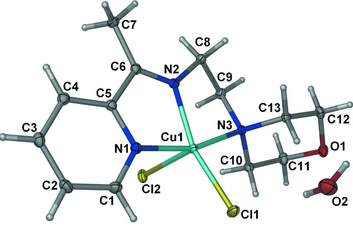
In the title compound, [CuCl2(C13H19N3O)]H2O, the
triden-tate Schiff base ligand and the two Cl atoms complete a distorted square-pyramidal coordination geometry around the CuII ion in which the three N atoms and one Cl atom are located in the basal plane and the other Cl atom is at the apical position. In the crystal, O—H Cl hydrogen bonds link the complex molecules and the uncoordinated water molecules into infinite chains along the a axis. The chains are further connected into a three-dimensional network via C—H O and C—H Cl interactions.
Related literature
For the structures of CuCl2complexes with similar ligands, see:
Saleh Salgaet al.(2010); Wanget al.(2009). For the structure of a CdCl2 complex with the same Schiff base ligand, see:
Ikmal Hishamet al.(2010). For a description of the geometry of complexes with a five-coordinate metal atom, see: Addison
et al.(1984).
Experimental
Crystal data
[CuCl2(C13H19N3O)]H2O
Mr= 385.77
a= 7.9194 (8) A˚ b= 8.5793 (8) A˚
= 91.981 (1)
V= 1556.6 (3) A˚3
Z= 4
MoKradiation
= 1.75 mm1
T= 100 K
0.180.160.09 mm
Data collection
Bruker APEXII CCD diffractometer
Absorption correction: multi-scan (SADABS; Sheldrick, 1996) Tmin= 0.743,Tmax= 0.858
9634 measured reflections 3348 independent reflections 2948 reflections withI> 2(I) Rint= 0.023
Refinement
R[F2> 2(F2)] = 0.025
wR(F2) = 0.060
S= 1.05 3348 reflections 197 parameters 2 restraints
H atoms treated by a mixture of independent and constrained refinement
max= 0.37 e A˚
3
min=0.34 e A˚
[image:1.610.46.259.560.727.2]3
Table 1
Hydrogen-bond geometry (A˚ ,).
D—H A D—H H A D A D—H A
O2—H2A Cl2i 0.84 (2) 2.35 (2) 3.1829 (16) 173 (2) O2—H2B Cl1 0.83 (2) 2.48 (2) 3.2841 (18) 164 (2) C2—H2 O2ii
0.95 2.41 3.307 (2) 156 C3—H3 Cl2iii
0.95 2.82 3.619 (2) 142 C4—H4 O2iv
0.95 2.50 3.445 (2) 172 C7—H7A Cl1v
0.98 2.68 3.6179 (19) 161 C8—H8A O1vi
0.99 2.47 3.336 (2) 146 C10—H10B Cl1 0.99 2.79 3.4496 (19) 124 C10—H10A Cl2 0.99 2.71 3.3566 (19) 123
Symmetry codes: (i)x1;y;z; (ii)xþ1;yþ1;z; (iii)xþ2;yþ2;z; (iv)
xþ1;yþ2;z; (v)x;yþ1;z; (vi)xþ1 2;yþ
1 2;zþ
1 2.
Data collection:APEX2(Bruker, 2007); cell refinement:SAINT (Bruker, 2007); data reduction:SAINT; program(s) used to solve structure:SHELXS97(Sheldrick, 2008); program(s) used to refine structure: SHELXL97 (Sheldrick, 2008); molecular graphics: X-SEED (Barbour, 2001); software used to prepare material for publication:SHELXL97andpublCIF(Westrip, 2010).
The authors thank University of Malaya for funding this study (FRGS grant No. FP004/2010B).
Supplementary data and figures for this paper are available from the IUCr electronic archives (Reference: IS2674).
References
Addison, A. W., Rao, T. N., Reedijk, J., Rijn, V. J. & Verschoor, G. C. (1984).J. Chem. Soc. Dalton Trans.pp. 1349–1356.
Barbour, L. J. (2001).J. Supramol. Chem.1, 189–191.
Bruker (2007).APEX2andSAINT. Bruker AXS Inc., Madison, Wisconsin, USA.
Ikmal Hisham, N., Suleiman Gwaram, N., Khaledi, H. & Mohd Ali, H. (2010). Acta Cryst.E66, m1471.
Saleh Salga, M., Khaledi, H., Mohd Ali, H. & Puteh, R. (2010).Acta Cryst. E66, m508.
Sheldrick, G. M. (1996).SADABS. University of Go¨ttingen, Germany. Sheldrick, G. M. (2008).Acta Cryst.A64, 112–122.
Wang, Q., Bi, C.-F., Wang, D.-Q. & Fan, Y.-H. (2009).Acta Cryst.E65, m439. Westrip, S. P. (2010).J. Appl. Cryst.43, 920–925.
Acta Crystallographica Section E Structure Reports Online
supporting information
Acta Cryst. (2011). E67, m334 [doi:10.1107/S1600536811004892]
Dichlorido{2-(morpholin-4-yl)-
N
-[1-(pyridin-2-yl)ethylidene]ethanamine-κ
3N
,
N
′
,
N
′′
}copper(II) monohydrate
Nura Suleiman Gwaram, Hamid Khaledi and Hapipah Mohd Ali
S1. Comment
The asymmetric unit of the title compound consists of a copper(II) complex and one molecule of water. Like the CdCl2
complex of the Schiff base, 2-morpholino-N-[1-(2-pyridyl)ethylidene]ethanamine, (Ikmal Hisham et al., 2010) the metal ion in the present structure is five-coordinated by the N,N′,N"-tridentate Schiff base ligand and two Cl atoms in a distorted square-pyramidal geometry, the τ value (Addison et al.,1984) being 0.15. The Cu—Cl and Cu—N interatomic distances are comparable to the values reported in the literature (Saleh Salga et al., 2010; Wang et al., 2009). In the crystal, the adjacent metal complexes and water molecules are linked into a three-dimensional network via O—H···Cl, C —H···Cl and C—H···O interactions. In addition, intramolecular C—H···Cl hydrogen bonding is observed.
S2. Experimental
A mixture of 2-acetylpyridine (0.20 g, 1.65 mmol) and 4-(2-aminoethyl)morpholine (0.21 g, 1.65 mmol) in ethanol (20
ml) was refluxed. After 2 hr a solution of copper(II) chloride dihydrate (0.28 g, 1.65 mmol) in a minimum amount of
ethanol was added and the resulting solution was refluxed for 30 min, then set aside at room temperature. The crystals of
the title complex were obtained after a few days.
S3. Refinement
The C-bound hydrogen atoms were placed at calculated positions (C—H 0.95–0.99 Å) and were treated as riding on their
parent atoms. The O-bound H atoms were placed in a difference Fourier map, and were refined with distance restraint of
Figure 1
Displacement ellipsoid plot of the title compound at the 50% probability level. Hydrogen atoms are drawn as spheres of
arbitrary radii.
Dichlorido{2-(morpholin-4-yl)-N-[1-(pyridin-2-yl)ethylidene]ethanamine- κ3N,N′,N′′}copper(II) monohydrate
Crystal data
[CuCl2(C13H19N3O)]·H2O
Mr = 385.77 Monoclinic, P21/n
Hall symbol: -P 2yn
a = 7.9194 (8) Å
b = 8.5793 (8) Å
c = 22.925 (2) Å
β = 91.981 (1)°
V = 1556.6 (3) Å3
Z = 4
F(000) = 796
Dx = 1.646 Mg m−3
Mo Kα radiation, λ = 0.71073 Å Cell parameters from 4229 reflections
θ = 2.5–28.2°
µ = 1.75 mm−1
T = 100 K Block, green
0.18 × 0.16 × 0.09 mm
Data collection
Bruker APEXII CCD diffractometer
Radiation source: fine-focus sealed tube Graphite monochromator
φ and ω scans
Absorption correction: multi-scan (SADABS; Sheldrick, 1996)
Tmin = 0.743, Tmax = 0.858
9634 measured reflections 3348 independent reflections 2948 reflections with I > 2σ(I)
Rint = 0.023
θmax = 27.0°, θmin = 2.5°
h = −10→10
k = −10→10
l = −29→28
Refinement
Refinement on F2
Least-squares matrix: full
R[F2 > 2σ(F2)] = 0.025
wR(F2) = 0.060
3348 reflections 197 parameters 2 restraints
Secondary atom site location: difference Fourier map
Hydrogen site location: inferred from neighbouring sites
H atoms treated by a mixture of independent and constrained refinement
w = 1/[σ2(F
o2) + (0.0258P)2 + 0.893P]
where P = (Fo2 + 2Fc2)/3
(Δ/σ)max = 0.001
Δρmax = 0.37 e Å−3
Δρmin = −0.34 e Å−3
Special details
Geometry. All e.s.d.'s (except the e.s.d. in the dihedral angle between two l.s. planes) are estimated using the full covariance matrix. The cell e.s.d.'s are taken into account individually in the estimation of e.s.d.'s in distances, angles and torsion angles; correlations between e.s.d.'s in cell parameters are only used when they are defined by crystal symmetry. An approximate (isotropic) treatment of cell e.s.d.'s is used for estimating e.s.d.'s involving l.s. planes.
Refinement. Refinement of F2 against ALL reflections. The weighted R-factor wR and goodness of fit S are based on F2,
conventional R-factors R are based on F, with F set to zero for negative F2. The threshold expression of F2 > σ(F2) is used
only for calculating R-factors(gt) etc. and is not relevant to the choice of reflections for refinement. R-factors based on F2
are statistically about twice as large as those based on F, and R- factors based on ALL data will be even larger.
Fractional atomic coordinates and isotropic or equivalent isotropic displacement parameters (Å2)
x y z Uiso*/Ueq
Cu1 0.60251 (3) 0.86379 (2) 0.122328 (9) 0.01099 (7)
Cl1 0.49087 (6) 0.62132 (5) 0.11931 (2) 0.01873 (11)
Cl2 0.90458 (6) 0.85029 (5) 0.15951 (2) 0.01773 (11)
O1 0.32231 (17) 0.82636 (16) 0.30317 (6) 0.0183 (3)
N1 0.68558 (19) 0.86674 (17) 0.03959 (7) 0.0129 (3)
N2 0.60269 (19) 1.09006 (17) 0.10733 (7) 0.0119 (3)
N3 0.51025 (18) 0.92685 (17) 0.20213 (6) 0.0114 (3)
C1 0.7350 (2) 0.7448 (2) 0.00826 (8) 0.0159 (4)
H1 0.7128 0.6427 0.0221 0.019*
C2 0.8177 (2) 0.7617 (2) −0.04381 (8) 0.0167 (4)
H2 0.8513 0.6729 −0.0653 0.020*
C3 0.8501 (2) 0.9106 (2) −0.06373 (8) 0.0156 (4)
H3 0.9077 0.9254 −0.0990 0.019*
C4 0.7975 (2) 1.0386 (2) −0.03157 (8) 0.0147 (4)
H4 0.8168 1.1417 −0.0449 0.018*
C5 0.7169 (2) 1.0126 (2) 0.01989 (8) 0.0121 (4)
C6 0.6645 (2) 1.1396 (2) 0.05987 (8) 0.0127 (4)
C7 0.6910 (2) 1.3055 (2) 0.04320 (8) 0.0172 (4)
H7A 0.6395 1.3737 0.0719 0.026*
H7B 0.6385 1.3247 0.0045 0.026*
H7C 0.8124 1.3271 0.0422 0.026*
C8 0.5483 (2) 1.1856 (2) 0.15617 (8) 0.0127 (4)
H8A 0.4272 1.2127 0.1510 0.015*
H8B 0.6148 1.2832 0.1587 0.015*
C9 0.5779 (2) 1.0881 (2) 0.21117 (8) 0.0132 (4)
H9A 0.7005 1.0830 0.2210 0.016*
H9B 0.5212 1.1379 0.2442 0.016*
C10 0.5778 (2) 0.8179 (2) 0.24813 (8) 0.0139 (4)
H10B 0.5557 0.7094 0.2353 0.017*
C11 0.5017 (2) 0.8419 (2) 0.30720 (8) 0.0174 (4)
H11A 0.5492 0.7642 0.3352 0.021*
H11B 0.5316 0.9471 0.3221 0.021*
C12 0.2547 (2) 0.9419 (2) 0.26393 (8) 0.0171 (4)
H12A 0.2853 1.0468 0.2789 0.021*
H12B 0.1299 0.9340 0.2620 0.021*
C13 0.3217 (2) 0.9220 (2) 0.20293 (8) 0.0130 (4)
H13A 0.2818 0.8210 0.1867 0.016*
H13B 0.2749 1.0057 0.1774 0.016*
O2 0.0878 (2) 0.59912 (18) 0.08146 (7) 0.0273 (3)
H2A 0.035 (3) 0.659 (3) 0.1032 (10) 0.033*
H2B 0.183 (2) 0.602 (3) 0.0978 (11) 0.033*
Atomic displacement parameters (Å2)
U11 U22 U33 U12 U13 U23
Cu1 0.01348 (12) 0.00927 (11) 0.01044 (12) 0.00023 (8) 0.00364 (8) 0.00007 (8) Cl1 0.0256 (3) 0.0112 (2) 0.0200 (2) −0.00334 (17) 0.00966 (19) −0.00245 (17) Cl2 0.0123 (2) 0.0245 (2) 0.0165 (2) 0.00191 (17) 0.00273 (17) 0.00437 (18) O1 0.0157 (7) 0.0231 (7) 0.0165 (7) −0.0002 (5) 0.0050 (5) 0.0047 (6) N1 0.0131 (7) 0.0133 (7) 0.0124 (8) 0.0007 (6) 0.0013 (6) 0.0005 (6) N2 0.0124 (7) 0.0112 (7) 0.0120 (8) 0.0006 (6) 0.0008 (6) −0.0010 (6) N3 0.0109 (7) 0.0114 (7) 0.0120 (8) −0.0005 (6) 0.0013 (6) −0.0006 (6) C1 0.0190 (10) 0.0125 (9) 0.0162 (10) 0.0001 (7) 0.0019 (8) −0.0010 (7) C2 0.0175 (9) 0.0177 (9) 0.0151 (10) 0.0032 (7) 0.0029 (7) −0.0031 (7) C3 0.0142 (9) 0.0215 (10) 0.0113 (9) 0.0014 (7) 0.0017 (7) 0.0002 (7) C4 0.0145 (9) 0.0155 (9) 0.0142 (9) 0.0001 (7) 0.0004 (7) 0.0017 (7) C5 0.0127 (9) 0.0130 (8) 0.0107 (9) 0.0010 (7) 0.0003 (7) −0.0004 (7) C6 0.0108 (8) 0.0141 (9) 0.0130 (9) 0.0002 (7) 0.0003 (7) 0.0008 (7) C7 0.0229 (10) 0.0124 (9) 0.0169 (10) 0.0016 (7) 0.0070 (8) 0.0016 (7) C8 0.0150 (9) 0.0109 (8) 0.0124 (9) −0.0008 (7) 0.0031 (7) −0.0010 (7) C9 0.0145 (9) 0.0126 (9) 0.0126 (9) −0.0025 (7) 0.0007 (7) −0.0029 (7) C10 0.0127 (9) 0.0155 (9) 0.0134 (9) 0.0024 (7) 0.0007 (7) 0.0033 (7) C11 0.0160 (9) 0.0228 (10) 0.0134 (9) 0.0002 (8) 0.0021 (7) 0.0025 (8) C12 0.0152 (9) 0.0194 (9) 0.0171 (10) 0.0013 (7) 0.0056 (8) −0.0004 (8) C13 0.0099 (8) 0.0144 (9) 0.0148 (9) −0.0005 (7) 0.0009 (7) 0.0000 (7) O2 0.0307 (9) 0.0231 (8) 0.0285 (9) 0.0035 (7) 0.0053 (7) −0.0013 (7)
Geometric parameters (Å, º)
Cu1—N2 1.9715 (15) C5—C6 1.491 (2)
Cu1—N1 2.0290 (15) C6—C7 1.490 (2)
Cu1—N3 2.0654 (15) C7—H7A 0.9800
Cu1—Cl1 2.2604 (5) C7—H7B 0.9800
Cu1—Cl2 2.5143 (5) C7—H7C 0.9800
N1—C1 1.336 (2) C8—H8B 0.9900
N1—C5 1.356 (2) C9—H9A 0.9900
N2—C6 1.281 (2) C9—H9B 0.9900
N2—C8 1.464 (2) C10—C11 1.515 (3)
N3—C10 1.494 (2) C10—H10A 0.9900
N3—C13 1.495 (2) C10—H10B 0.9900
N3—C9 1.495 (2) C11—H11A 0.9900
C1—C2 1.389 (3) C11—H11B 0.9900
C1—H1 0.9500 C12—C13 1.522 (3)
C2—C3 1.383 (3) C12—H12A 0.9900
C2—H2 0.9500 C12—H12B 0.9900
C3—C4 1.395 (3) C13—H13A 0.9900
C3—H3 0.9500 C13—H13B 0.9900
C4—C5 1.379 (3) O2—H2A 0.837 (16)
C4—H4 0.9500 O2—H2B 0.831 (16)
N2—Cu1—N1 79.77 (6) C6—C7—H7B 109.5
N2—Cu1—N3 84.20 (6) H7A—C7—H7B 109.5
N1—Cu1—N3 163.90 (6) C6—C7—H7C 109.5
N2—Cu1—Cl1 154.62 (5) H7A—C7—H7C 109.5
N1—Cu1—Cl1 97.01 (4) H7B—C7—H7C 109.5
N3—Cu1—Cl1 96.79 (4) N2—C8—C9 106.53 (14)
N2—Cu1—Cl2 95.63 (5) N2—C8—H8A 110.4
N1—Cu1—Cl2 88.97 (5) C9—C8—H8A 110.4
N3—Cu1—Cl2 94.20 (4) N2—C8—H8B 110.4
Cl1—Cu1—Cl2 109.544 (19) C9—C8—H8B 110.4
C11—O1—C12 109.02 (14) H8A—C8—H8B 108.6
C1—N1—C5 118.92 (16) N3—C9—C8 110.42 (14)
C1—N1—Cu1 127.12 (12) N3—C9—H9A 109.6
C5—N1—Cu1 113.06 (12) C8—C9—H9A 109.6
C6—N2—C8 126.51 (15) N3—C9—H9B 109.6
C6—N2—Cu1 118.51 (13) C8—C9—H9B 109.6
C8—N2—Cu1 114.57 (11) H9A—C9—H9B 108.1
C10—N3—C13 107.88 (14) N3—C10—C11 113.72 (15)
C10—N3—C9 111.29 (14) N3—C10—H10A 108.8
C13—N3—C9 112.19 (14) C11—C10—H10A 108.8
C10—N3—Cu1 109.42 (11) N3—C10—H10B 108.8
C13—N3—Cu1 112.81 (11) C11—C10—H10B 108.8
C9—N3—Cu1 103.24 (10) H10A—C10—H10B 107.7
N1—C1—C2 122.40 (17) O1—C11—C10 110.79 (15)
N1—C1—H1 118.8 O1—C11—H11A 109.5
C2—C1—H1 118.8 C10—C11—H11A 109.5
C3—C2—C1 118.62 (17) O1—C11—H11B 109.5
C3—C2—H2 120.7 C10—C11—H11B 109.5
C1—C2—H2 120.7 H11A—C11—H11B 108.1
C2—C3—C4 119.35 (17) O1—C12—C13 111.41 (15)
C2—C3—H3 120.3 O1—C12—H12A 109.3
C5—C4—C3 118.75 (17) O1—C12—H12B 109.3
C5—C4—H4 120.6 C13—C12—H12B 109.3
C3—C4—H4 120.6 H12A—C12—H12B 108.0
N1—C5—C4 121.95 (16) N3—C13—C12 112.83 (15)
N1—C5—C6 114.30 (15) N3—C13—H13A 109.0
C4—C5—C6 123.69 (16) C12—C13—H13A 109.0
N2—C6—C7 126.57 (17) N3—C13—H13B 109.0
N2—C6—C5 113.71 (16) C12—C13—H13B 109.0
C7—C6—C5 119.70 (16) H13A—C13—H13B 107.8
C6—C7—H7A 109.5 H2A—O2—H2B 101 (2)
Hydrogen-bond geometry (Å, º)
D—H···A D—H H···A D···A D—H···A
O2—H2A···Cl2i 0.84 (2) 2.35 (2) 3.1829 (16) 173 (2)
O2—H2B···Cl1 0.83 (2) 2.48 (2) 3.2841 (18) 164 (2)
C2—H2···O2ii 0.95 2.41 3.307 (2) 156
C3—H3···Cl2iii 0.95 2.82 3.619 (2) 142
C4—H4···O2iv 0.95 2.50 3.445 (2) 172
C7—H7A···Cl1v 0.98 2.68 3.6179 (19) 161
C8—H8A···O1vi 0.99 2.47 3.336 (2) 146
C10—H10B···Cl1 0.99 2.79 3.4496 (19) 124
C10—H10A···Cl2 0.99 2.71 3.3566 (19) 123

![(Azido κN){(E) 2 [1 (pyridin 2 yl)ethylideneamino]phenolato κ3N,N′,O}copper(II)](data:image/gif;base64,R0lGODlhAQABAIAAAP///wAAACH5BAEAAAAALAAAAAABAAEAAAICRAEAOw==)